And now spit!

Did you know that human saliva is 99.5% water?

1369155421_drool

But a recent set of studies have suggested that the remaining 0.5% holds some interesting insights into Parkinson’s disease.


Interesting fact about saliva – while there is a lot of debate as to how much saliva we produce on a daily basis (anywhere between 0.75 to 1.5 litres per day), it is generally accepted that during sleep the amount of saliva produced drops to almost nothing. Why? Big shrug.


Saliva is a solution produced by three main sets of glands in our mouth: the parotid, Sublingual, and Submandibular glands:

h9991831_001

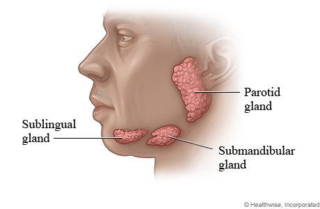
The human salivary glands. Source: WebMD 

The solution produced serves several important functions, namely:

  • beginning the process of digestion by breaking down food particles.
  • protecting teeth from bacterial decay.
  • Moisturising food to aid in the initiation of swallowing.

As we mentioned above, 99.5% of saliva is water. The remaining 0.5% is made up of enzymes and antimicrobial agents. There is also a number of cells in each millilitre of saliva (as many as 8 million human and 500 million bacterial cells per millilitre).

By analysing those human cells, scientists can learn a lot about a person. For example, they can conduct genetic analysis and determine if a person has a particular mutation.

So what has this got to do with Parkinson’s disease?

Well, recently several research groups have been looking at saliva with the hope that biomarkers – chemicals that may allow for early detection or better monitoring of Parkinson’s disease – could be found.

And recently, some of that research has seemingly paid big dividends:

Spit3-title

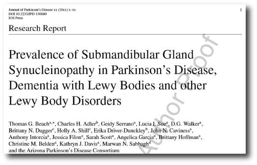
Title: Prevalence of Submandibular Gland Synucleinopathy in Parkinson’s Disease, Dementia with Lewy Bodies and other Lewy Body Disorders.
Authors: Beach TG, Adler CH, Serrano G, Sue LI, Walker DG, Dugger BN, Shill HA, Driver-Dunckley E, Caviness JN, Intorcia A, Filon J, Scott S, Garcia A, Hoffman B, Belden CM, Davis KJ, Sabbagh MN.
Journal: J Parkinsons Dis. 2016 [Epub ahead of print]
PMID: 26756744

In this study, published in January of this year, the researchers collected small biopsies of the submandibular gland (one of the three primary producers of saliva) from the bodies of people who died with various conditions (including Parkinson’s disease). They analysed the biopsies for alpha synuclein – the chemical in the brain associated with Parkinson’s disease. We have previously written about alpha synuclein, a chemical in the brain that is associated with Parkinson’s disease (for a primer on alpha synuclein, click here). They found that alpha synuclein was present in the saliva gland of 89% of the subjects who died with Parkinson’s disease, but none of the 110 control samples.

This result led the same research groups to attempt a similar study on live subjects and they published the results of that study in February of this year:

Spit2--title

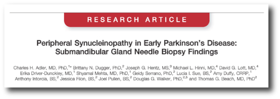
Title: Peripheral Synucleinopathy in Early Parkinson’s Disease: Submandibular Gland Needle Biopsy Findings.
Authors: Adler CH, Dugger BN, Hentz JG, Hinni ML, Lott DG, Driver-Dunckley E, Mehta S, Serrano G, Sue LI, Duffy A, Intorcia A, Filon J, Pullen J, Walker DG, Beach TG.
Journal: Mov Disord. 2016 Feb;31(2):250-6.
PMID: 26799362

The researchers enrolled 25 people with early-stage Parkinson’s disease (less than  5 years since diagnosis) and 10 control subjects. All of these subjects underwent a small biopsy of the submandibular gland. Those biopsies were then analysed for alpha synuclein and the researchers found that 74% of the Parkinsonian biopsies and 22% control biopsies had alpha synuclein present in the submandibular gland.

And remarkably, this report was followed up this last week by a group in Italy, who published some very interesting data:

Spit1-title

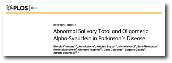
Title: Abnormal Salivary Total and Oligomeric Alpha-Synuclein in Parkinson’s Disease.
Authors: Vivacqua G, Latorre A, Suppa A, Nardi M, Pietracupa S, Mancinelli R, Fabbrini G, Colosimo C, Gaudio E, Berardelli A.
Journal: PLoS One. 2016 Mar 24;11(3):e0151156.
PMID: 27011009    (this report is OPEN-ACCESS if you would like to read it)

The researchers collected salivary samples – actual spit – from 60 people with Parkinson’s disease and 40 age/sex matched control subjects. They then measured the saliva for different types of alpha synuclein. In this study, the researchers measured both the total amount of alpha synuclein in the saliva and also special forms of alpha synuclein.

Alpha synuclein initially starts out in the brain in a monomeric form – as a single version of alpha synuclein. This form of alpha synuclein is believed to be safe. A more mature form of alpha synuclein, called oligomeric, is believed to be the seed of the aggregations found in the Parkinsonian brain, Lewy bodies.

Curiously, in this study the researchers found that the total amount of alpha synuclein in the salivary of people with Parkinson’s disease was lower than that of the control subjects. But – and it’s a big ‘but’ – the amount of alpha synuclein oligomers was higher in the people with Parkinson’s disease than normal healthy controls.

The researchers proposed that the decreased concentration of total alpha-synuclein may reflect the formation of lewy bodies in the brain, and that this test might help the early diagnosis of Parkinson’s disease.


Here at the Science of Parkinson’s we are approaching this research cautiously. Previous attempts at measuring saliva in Parkinson’s disease have not had such significant results when comparing people with Parkinson’s disease and controls (click here for more about that study). The need for better biomarkers of Parkinson’s disease provides the reasons for this research, but the variability between the results different groups are getting leaves one wondering about the viability of the approach. It would indeed make for a very easy, non-invasive testing platform for Parkinson’s disease (‘Please spit into this tube for me’), but more research is needed before it can be applied on the large scale.

We’ll keep watching and hoping.

Herpes Simplex virus and Alpha-Synuclein

The theory of universal gravitation is not cast-iron. No theory is, and there is always room for improvement – Isaac Asimov


Previously we have discussed the possibility that a virus could be one of the causative agents in Parkinson’s disease (Click here for that post).

Well, recently an interesting research report was published that offers evidence to support that idea and proposes an interesting new idea:

Caggiu1

Title: Humoral cross reactivity between α-synuclein and herpes simplex-1 epitope in Parkinson’s disease, a triggering role in the disease?
Authors: Caggiu E, Paulus K, Arru G, Piredda R, Sechi GP, Sechi LA.
Journal: J Neuroimmunol. 2016 Feb 15;291:110-4.
PMID: 26857504

In this study, the researchers began with a simple hypothesis:

Pathogens that resemble normal proteins in the body may cause the immune system to attack cells that have those normal proteins.

A pathogen is a biological entity that can cause damage or disease in our bodies. Our immune system’s response is to generate antibodies – small proteins that label the pathogen as foreign (or ‘not self’). The removal cells in the immune system then know which stuff in the body to attack and which to leave alone.

But what happens if parts of a particular pathogen look very similar to a normal protein in the body? Causing an immune response through a process of ‘molecular mimicry’. This is the question that the scientists behind today’s study were asking.

To test their hypothesis, the scientists looked at the herpes simplex virus 1 and compared it with the Parkinson’s disease-related protein, alpha synuclein. They found that there were two regions on the herpes simplex virus 1 that exhibited the same appearance as regions on the alpha synuclein protein.

Importantly, when they analysed the blood of people with Parkinson’s disease and some age-matched control, they found a statistically significant difference in the levels of antibodies generated against of those one regions of the Herpes simplex virus 1 (that regions was Ul4222–36) in people with Parkinson’s disease. Those antibodies were also reactive to a site on the alpha synuclein protein (that region was 100–114).

The researchers suggested that the results may implicate the involvement of Herpes simplex virus 1 in stimulating immune cells against the alpha synuclein resulting in neurons that have a lot of alpha synuclein in the brain being attacked (especially those that have alpha synuclein containing Lewy bodies).

What is Herpes simplex virus 1?

Herpes_simplex_virus_TEM_B82-0474_lores

Herpes simplex virus. Source: Wikipedia

Herpes simplex virus 1 and 2 are members of the herpesvirus (Herpesviridae) family of viruses that infect humans. The two viruses should not be confused – Herpes simplex virus 1 produces cold sores, while herpes simplex virus 2 is associated with genital herpes.

Both herpes simplex virus 1 & 2 are not only able to infect neurons in the brain, but they can able to become dormant and hide in neurons, away from the immune system. They can remain in that state until suddenly/spontaneously becoming reactivated for reasons unknown.

Is there any association between Parkinson’s disease and Herpes simplex virus 1?

There is one paper published in 1993, that found an association between previous exposure to Herpes and developing Parkinson’s disease:

Title: Infections as a risk factor for Parkinson’s disease: a case-control study.
Authors: Vlajinac H, Dzoljic E, Maksimovic J, Marinkovic J, Sipetic S, Kostic V.
Journal: Int J Neurosci. 2013 May;123(5):329-32. doi: 10.3109/00207454.2012.760560. Epub 2013 Feb 4.
PMID: 23270425

In this study, the researchers found that Parkinson’s Disease was also significantly associated to mumps, scarlet fever, influenza, and whooping cough as well as herpes simplex 1 infections. They found no association between Parkinson’s disease and Tuberculosis, measles or chickenpox though.

This result raises the tantalizing possibility that other viruses may be stimulating the immune system by ‘molecular mimicry’. Obviously this still needs to be tested. Plus that study was based only 110 people with Parkinson’s (compared with 220 controls) in one particular geographical location (Belgrade, Serbia).

Other than making the immune system attack cells, could the antibodies to the virus be having other effects?

In the discussion of their paper, the authors of the Herpes simplex virus 1 study point out that alpha synuclein can be divided in three parts:

  1. the N-terminal region (which contains several of the point mutations related to early onset Parkinson’s disease)
  2. the central region (which appears to promote aggregation)
  3. the C-terminal portion (which tends to decrease protein aggregation)

The segment of alpha synuclein (100–114) that cross reacts with antibodies for Herpes simplex virus 1 (Ul4222–36) is part of the C-terminal region. Given that antibodies are binding to and removing to that non-aggregating section of alpha synuclein, are the remaining segments of α-synuclein left in place to foster aggregation (and perhaps forming Lewy bodies)?

Interesting research report that leaves us with new questions to explore.

A sense of (and the science of) smell

Losing the sense of smell is a common feature associated with Parkinson’s disease. But this feature of the condition may help us to better understand the condition. Some autopsy studies have suggested that the olfactory system is one of the first structures in the brain to be affected by the disease.

bad-smell-001

Source: Guardian

How do we smell?

Bad.

That’s both a pathetic attempt at humour and a serious answer. Compared with fellow members of the mammalian family, human beings have a pretty poor sense of smell.

The process of smelling stuff is conducted through structures called the olfactory bulbs. The human olfactory bulbs lie on the base of our brains, protruding forward towards our nose (and nasal cavity).

olfactory_bulb_(inferior_view)1321478082612

A view of the human brain from below (olfactory bulbs are in yellow). Source: StudyBlue

olfactorybulbq1318387403476

A view of the human brain from in front (olfactory bulbs are in yellow). Source: StudyBlue

Inside your nose there is an area of smell sensitive cells that lies on the roof of the nasal cavity (about 7 cm behind your nostrils). That area is called the olfactory epithelium, and it plays a critical role in our sense of smell.

The size of the human olfactory epithelium is rather small and reflects our poor sense of smell, especially when compared, for example, to a dog  (humans have about 10 cm2 (1.6 sq in) of olfactory epithelium, while some dogs have 170 cm2 (26 sq in)).

odorant_eng

The human olfactory system. Source: Biology junction.com

When you inhale an odor (or odorant molecules) through your nose, there are tiny receptors (called olfactory receptors) on the  olfactory epithelium that are the first step in detecting the smell. Every single olfactory receptor cell presents just one (and only one) type type of odorant receptor. When they detect that odor, the olfactory receptor cell reacts by sending an electrical signal along its branch (called an axon) to the olfactory bulbs in the brain.

As the axon of olfactory receptor cell enters the olfactory bulb it forms clusters with other olfactory receptor cell axons, and these clusters are called glomeruli. Inside the glomerulus (singular), the axons make contact the branches of a type of brain cell called a mitral cell. Mitral cells send their axons to many different areas of the brain, including the anterior olfactory nucleus, piriform cortex, the amygdaloid complex, the entorhinal cortex, and the olfactory tubercle.

fulllengths-olfaction-2

Source: YaleScientific

From here our understanding of olfactory processing is less well understood. The piriform cortex is considered the area most likely associated with identifying particular odor. The amygdala is involved in emotional and social functions (eg. mating and recognition), while the entorhinal cortex (and connected hippocampus) is associated with memory – this area is probably activated when a particular smell reminds us of something in our childhood.

What is known about our sense of smell in Parkinson’s disease?

In 1975, two researchers in Minnesota noticed that many of their people with Parkinson’s disease that they were assessing had reduced olfactory abilities. They decided to test this observation:

olfactory-title

Title: Olfactory function in patients with Parkinson’s disease.
Authors: Ansari KA, Johnson A.
Journal: J Chronic Dis. 1975 Oct;28(9):493-7.
PMID: 1176578

The researchers took 22 people with Parkinson’s disease and 37 age/sex-matched controls and repeatedly tested them in a double blind study to determine their olfactory acuity. In each test, the subjects were given five test tubes. Two of the tubes in each set contained 0.5 ml of diluted amyl acetate (which has a distinct smell). The other three tubes contained just water. The subjects were asked to inhale through their nose and then identify which two tubes in each set contained the amyl acetate. The highest dilution (the weakest smelling solution) at which the subject could correctly identify the two amyl acetate containing tubes was designated as their olfactory threshold.

The researchers found that people with Parkinson’s disease had a significantly reduced olfactory acuity (a lower olfactory threshold than compared to control subjects). They also noted that subjects with more progressive forms of the disease exhibited a worse performance on the test. Numerous studies have now replicated this overall result, including a recent study that indicated that smoking may have a protective role on the olfactory ability (Click here and here for more on this).


EDITORIAL NOTE: Please understand that the loss of smell in Parkinson’s disease does not immediately mean that you will have a more progressive form of the condition. There is simply a trend in the data that suggests the loss of smell is a risk factor for having a more progressive version of the condition. 

We would also like to discourage any thoughts of taking up smoking in order to protect your sense of smell.


So what is actually happening in the Parkinson’s disease brain?

This is Prof Heiko Braak:

heiko-braak-01

He’s a dude. We’ve mentioned him before in a previous post.

Many years ago, he and his colleagues were intrigued with the hyposmia (reduction in olfactory ability) in Parkinson’s disease. They conducted a series of autopsy studies, looking at 413 brains! Specifically, they were looking for deposits of the Parkinson’s disease-related protein, alpha synuclein, in the brains and where the protein was accumulating. The accumulation of alpha synuclein is believed to be associated with the loss of cells in the brain.

In total they found 30 brains that exhibited accumulation of alpha synuclein. Of interest, they found that 16 of those brains had accumulation of alpha synuclein in the olfactory bulb. And in one particular case, the olfactory bulb was the only affected part of the brain, except for a tiny region of the brain stem.

The researchers were curious about the possibility that the olfactory system could be a potential starting point for Parkinson’s disease, but they were quick to point out that only half the cases they analysed (16/30) had accumulation of alpha synuclein in the olfactory bulb. Thus, while the olfactory system may be involved, it seems unlikely that the nose is the sole induction site of Parkinson’s disease.

After this study was published, however, Braak and his colleagues went on to analyse the accumulation of alpha synuclein in the lining of the gut and their results suggested this as another possible site of induction (we have written about this in a previous post). They have subsequently proposed a model of disease spread based on entry to the brain via the nose and gut:

nrneurol.2012.80-f1

The Braak stages of Parkinson’s disease. Source: Nature Reviews Neurology.

It is interesting to observe that studies by other scientists have indicated that the nasal epithelium of people with Parkinson’s disease (both with and without the loss of olfactory abilities) is not damaged or presenting an accumulation of alpha synuclein (Click here for more on this).

So what happens to the olfactory bulbs in Parkinson’s disease?

A recent review of the previous studies investigating olfactory bulb volume in people with Parkinson’s disease was published in the Open Access journal PlosOne:

Olfactory_title

Title: Changes in Olfactory Bulb Volume in Parkinson’s Disease: A Systematic Review and Meta-Analysis.
Authors: Li J, Gu CZ, Su JB, Zhu LH, Zhou Y, Huang HY, Liu CF.
Journal: PLoS One. 2016 Feb 22;11(2):e0149286.
PMID: 26900958  (this report is OPEN ACCESS if you would like to read it)

The authors of the study conducted a systematic review (or meta-analysis) of all of the previous studies (six in total) that have measured the size of the olfactory bulb in the brains of people with Parkinson’s disease (using brain imaging techniques). They found that in all of the 6 studies (collectively 216 PD patients and 175 healthy controls) there was a significant reduction in the size of the olfactory bulbs of people with Parkinson’s disease. Strangely, they authors also found the right olfactory bulb was larger than the left in subjects with Parkinson’s disease across all of the studies, and this effect was not found in the healthy controls.

The motor features of Parkinson’s disease usually begin asymmetrically – by this we mean that the left arm is affected before the right, or the right leg has tremor before the left. This is different for each person, as the disease has no particular preference for either side of the body. So why on earth is the right olfactory bulb more affected than the left?

There is your homework question for tonight!

I’ll expect your answers tomorrow.


Cleaning up with Ambroxol

Exciting news recently with the announcement of the Ambroxol study starting.

Exciting for two reasons:

  1. Ambroxol has the potential to make a major impact in the lives of some people with Parkinson’s disease.
  2. It illustrates how FAST things are moving in the world of Parkinson’s disease!


Inside each and every cell, there are millions of tiny actions taking place. Minute processes all working in a collective manner allowing the cell to function normally. There are lots of proteins helping to make other proteins, lots of proteins helping other proteins to get to where they need to be, and lots of proteins helping to break down other proteins after they have done their job.

All this activity generates a lot of waste. And a fundamental part of the activity in any cell is waste disposal. If that does not function properly, the cell is in serious trouble.

One of the most common genetic mutations associated with Parkinson’s disease – called GBA – results in cells having trouble getting rid of waste.

GBA-cartoon

Adapted from a cartoon by Dr Jing Pu. Source: The Nichd connection

What is GBA?

Glucocerebrosidase (or GBA) is an enzyme that helps with the recycling of waste. It is active in inside ‘lysosomes‘.

What are Lysosomes?

Lysosomes are small structures inside cells that act like recycling centers. Waste gets put inside the lysosome where enzymes like GBA help to break it down into useful parts. Mutations in the GBA gene can result in an abnormally short, non-functioning version of the enzyme. And in those cases the breaking down of waste inside the lysosome because inhibited.

What is the connection between GBA and Parkinson’s disease?

GBA mutations are the most common genetic anomaly associated with Parkinson’s disease. People with a mutation in their GBA gene are at higher risk of developing Parkinson’s disease than the general population. And people with Parkinson’s are approximately five times more likely to carry a GBA mutation than healthy control subjects.

So what is Ambroxol?

Ambroxol is a commonly used treatment for respiratory diseases. It promotes mucus clearance and eases coughing. Ambroxol is also anti-inflammatory, reducing redness in a sore throat.

Ok, but why the excitement for Parkinson’s disease?

In May of 2014 – less than 2 years ago – this study was published:

McNeil1

Title: Ambroxol improves lysosomal biochemistry in glucocerebrosidase mutation-linked Parkinson disease cells.
Authors: McNeill A, Magalhaes J, Shen C, Chau KY, Hughes D, Mehta A, Foltynie T, Cooper JM, Abramov AY, Gegg M, Schapira AH.
Journal: Brain. 2014 May;137(Pt 5):1481-95.
PMID: 24574503    (This report is OPEN ACCESS if you want to read it)

It was the first time that Ambroxol – a commercially available drug – had been tested in a Parkinson’s disease related context.

In this study the researchers collected skin cells (called fibroblasts) from eleven people with GBA mutations (some had been diagnosed with Parkinson’s disease). They measured the amount of glucocerebrosidase protein and enzyme activity in these cells, and they found that glucocerebrosidase enzyme activity was significantly reduced in fibroblasts from GBA mutations (on average just the enzyme was acting at just 5% of normal levels). They found that ambroxol increased glucosylceramidase activity in fibroblasts from people with GBA mutations AND in fibroblasts from healthy controls. Ambroxol treatment also reduced markers of oxidative stress in GBA mutant cells.

Given the increase in glucocerebrosidase activity after ambroxol treatment, the researchers wondered whether the drug would reduce alpha-synuclein levels in cells that were over-expressing this protein. Amazingly, after 5 days of ambroxol treatment, levels of alpha-synuclein had decreased significantly (15% on average 15%).

You can understand why the researchers were a little bit excited by these results. Here was a drug that re-activated the recycling unit in the cell and reduced levels of one of the main proteins associated with Parkinson’s disease. If the drug can reduce the levels of alpha synuclein in the brains of people with Parkinson’s disease, maybe the researchers will be able to slow down (or even halt) the disease!

Additional studies have now been reported which have confirmed the initial results.

And now the clinical trial?

Funded by the Cure Parkinson’s Trust and the Van Andel Research Institute (USA), it was announced this week that they had started recruiting subjects to be involved in a clinical trial at the Royal Free Hospital in London. The trial is a phase 1 study which will test the safety of Ambroxol in Parkinson’s disease. The researchers will also look to see if Ambroxol can increase levels of glucocerebrosidase and whether this has any beneficial effects in the subjects. The study will be conducted on 20 people with Parkinson’s disease who also have GBA mutations. They will be given the drug and followed over the next 24 months.

These are exciting times for the world of Parkinson’s disease as these drugs are no longer simply reducing the motor features of the condition, but actually attempting to slow/halt the disease.

And as we suggested at the start of the post the pace of these developments is becoming hard to keep up with.

New research – the disorder of Alpha Synuclein

A couple of interesting scientific papers were published this week dealing with the Parkinson’s disease-related protein, Alpha Synuclein. If you are not familiar with it, we suggest that you check out our primer page on Alpha Synuclein before reading any further.

So, what’s new in the world of Alpha Synuclein?

Two studies.

One in the prestigious journal Nature and the other in her sister Nature Communications. Both studies came from the same lab (good job guys!)

The first study :

Theillet-title

Title: Structural disorder of monomeric α-synuclein persists in mammalian cells.
Authors: Theillet FX, Binolfi A, Bekei B, Martorana A, Rose HM, Stuiver M, Verzini S, Lorenz D, van Rossum M, Goldfarb D, Selenko P.
Journal: Nature. 2016 Jan 25.
PMID: 26808899

This first study presented a very detailed analysis of the structure of alpha synuclein – at the atomic level – inside living cells.

Interestingly, when the researchers injected alpha synuclein (at concentrations that have been observed in normal neurons) into 5 different types of cells (both neuron and others types), they found that the protein remains extremely disordered – it changed shape rapidly. They determined this by using nuclear magnetic resonance spectroscopy (try saying that 3 times really fast!), which provides a shallow peak readout for stable proteins and a sharp peak for disordered proteins (see image below).

nature16871-f1

The researchers found a lot of sharp peaks in cells that they injected Alpha Synuclein into. Source: Nature

Rather remarkably, despite the fact that disordered proteins are usually removed from cells by enzymatic degradation, the alpha synuclein that was injected by these researchers appears to have remained intact in the cells for several days (50+ hours). And the cells did not seem to be adversely affected by this.

The second Alpha Synuclein study published this week illustrated an equally interesting result:

Binolfi-title

Title: Intracellular repair of oxidation-damaged α-synuclein fails to target C-terminal modification sites.
Authors: Binolfi A, Limatola A, Verzini S, Kosten J, Theillet FX, May Rose H, Bekei B, Stuiver M, van Rossum M, Selenko P.
Journal: Nature Communications, 2016 Jan 25;7:10251.
PMID: 26807843

In this study, the researchers injected damaged alpha synuclein into cells and then watched the cells try to repair that damaged protein. There are specific enzymes that help to maintain/repair proteins like Alpha Synuclein inside each cell. This is a normal recycling process for cells, but something interesting happened with this damaged version of alpha synuclein: only one end of the protein was repaired. The other end (called the C-terminus) was left damaged and this end failed to function correctly.

fnins-09-00059-g001

The structure of Alpha Synuclein. The c-terminus is the area in red. Source: Frontiers in Neuroscience

This led the authors to conclude that damage can cause the accumulation of chemically and functionally altered Alpha Synuclein in cells.

What does this mean for Parkinson’s disease?

The results are very interesting and the researchers should be congratulated on the complexity of their work. The findings add to our understanding of Alpha Synuclein, but both of these results need to be replicated and expanded on before we can fully appreciate their impact.

One possible implications of the results is that designing drugs to target Alpha Synuclein may be more complicated than originally thought. If the protein remains as disordered as the first study suggests, it could be difficult to target. Further investigations, however, focused on the c-terminus end of Alpha synuclein may offer novel targets for therapies looking to clear damaged proteins from cells.

If Alpha Synuclein is the big, bad enemy in Parkinson’s disease, we now know a lot more about him and we can focus on his weaknesses.

Vaccination for Parkinson’s disease

There has never been a more exciting time in Parkinson’s disease research. At no point in the past has the progress been made at such a frenetic pace. New week, new discoveries. And it has to be said that none of this would be possible without the advocacy and fundraising efforts of groups such as the Michael J Fox foundation, the Cure PD Trust, and Parkinson’s UK.

In addition to learning a great deal about the basic science of Parkinson’s disease – a better understanding of the biology underlying the disease – we are also making tremendous gains in new areas of treatment. Until now, the basic treatment has been dopamine replacement with L-dopa. But now, like never before, novel therapeutic approaches are being tested in the clinic.

One of these new approaches, however, is based on a very old idea: Vaccination.

220px-Edward_Jenner_by_James_Northcote

Edward Jenner (1749 – 1823). Source: Wikipedia

While Edward Jenner is considered to be the pioneer of the world’s first vaccine (for Smallpox), the idea of vaccination/inoculation actually originated in India in 1000 BC, where it was briefly mentioned in Sact’eya Grantham, an Ayurvedic text. The first really credible mention of inoculation, however, was in China where it was described in the book Yuyi cao (寓意草 or Notes on My Judgment) by Yu Chang, published in 1643.

getty_rf_photo_of_senior_man_getting_pneumonia_vaccination

Vaccination. Source: WebMD

The basic idea of vaccination is to deliberately introduce an individual to a small component of a disease-causing agent so that the body can build up an immune response to the disease prior to being attacked by the full disease.  That fragment of the disease-causing agent becomes what is known as an an ‘antigen’ (this comes from a French word, antigène, derived from the Greek anti- or “against”, and the word-forming suffix -gen, “thing that produces or causes”), and it will serve as the target for the immune system. In response to the antigen, the body produces beacons that bind to the antigen for the immune system to look out for  – these beacons are called ‘antibodies’, and they tell the immune system that what they have bound to is ‘not of this body – get rid of it’!

Vaccines will sometimes be made of an empty virus – the surface of the virus will be present, but the internal disease-causing mechanisms have been destroyed or removed. Think of it as training the immune system for some big event. In this way, by exposing and thus priming the body against a particular part of s disease-causing agent, if the body is ever attacked by the full agent, the immune system will be ready to deal with it.

So what does this have to do with Parkinson’s disease?

Good question!

In July of 2014, a small Austrian company called “AFFiRiS” announced the results of one of their first clinical trials for Parkinson’s disease.

imgres

The AFFiRiS drug (called PD01A in the AFF008 trial)  is an vaccine that targets the Parkinson’s disease-related protein ‘Alpha-synuclein’. The vaccine causes the body to produce Alpha-synuclein-specific antibodies. These antibodies allow the immune system to then attack and remove this protein from the blood and fluid surrounding the brain. Any loose alpha-synuclein floating around should be removed.

Alpha-synuclein is a very common protein in the brain – it makes up about 1% of the material in neurons. It is also one of the proteins that is present in the ‘Lewy bodies’ that are associated with Parkinson’s disease.

shutterstock_227273575

A cartoon of a neuron, with the Lewy body indicated within the cell body. Source: Alzheimer’s news

Lewy bodies are one of the defining characteristic features of the Parkinsonian brain (having said that, it is interesting to note that approx. 30% of the population over the age of 70 will have Lewy bodies but no clinical symptoms/problems). They are densely packed, spherical shaped, clusters of protein inside the cell body. We are not entirely sure if they are causing cells to die, but they should not be there so it is assumed that if we get rid of them, the cells will be healthier.

url

An actual photo of a Lewy body (brown) within a neuron. Source: Medicalia

Given that Alpha-synuclein is one of the major components of Lewy bodies, it is the first protein to be targeted by a vaccine for Parkinson’s disease. Some researchers believe that the passing of Alpha synuclein from one cell to another may be the mechanism by which the disease spreads. By removing any Alpha-synuclein that floating around outside of cells, companies like Affiris hope that they will be able to slow down or even halt the spread of Parkinson’s disease within the brain.

The results from the first Affiris trial look rather promising.

The phase one trial run by Affiris was very small (just 12 subjects received the vaccine) and lasted only 12 months. The primary endpoint of any phase one trial is ‘safety and tolerability’ – that is to say, the study is a test of whether the drug is ok for humans use and can be well tolerated (e.g. it has no hidden/unknown side effects). Two different doses of the PD01A vaccine were given in the study and both were well tolerated by the participants in the study.

The Affiris researchers, however, were also looking at a second endpoint in their trial: whether the vaccine caused Alpha Synuclein-specific antibodies to be produced. Thankfully, Affiris found measurable levels of alpha-synuclein-specific antibodies in serum samples (a component of blood) and cerebrospinal fluid (the liquid surrounding the brain) collected from their participants, suggesting that the vaccine is doing it’s job and causing the immune system to react to the antigen being introduced.

Obviously a larger study is now required to determine if the vaccine will actually slow or halt Parkinson’s disease, but when the Affiris researchers compared the subjects in their first trial that received the vaccine with a group of control subjects at the end of the 12 months, they claim that they found PD01A subjects ‘functionally stabilised compared to the control group’.

And Affiris is not the only biotech company trialling the vaccine approach for Parkinson’s disease. In March 2015, an Irish company called ‘Prothena‘ announced that their vaccine reduced Alpha synuclein levels in the serum by 96%! And again the vaccine was well tolerated, with few side effects. 40 subjects were used in the Prothena study and the company will continue to follow them. They expect to release follow-up data – with clinical and imaging results – in early to mid 2016.

We will be watching this area of research very closely. Fingers crossed!