“Red hair, sir, in my opinion, is dangerous”

Henna-Hair-Red-Hair-Ginger-Hair-Benefits-Beauty

The quote entitling this post is from a PG Wodehouse book ‘Very Good, Jeeves!’.


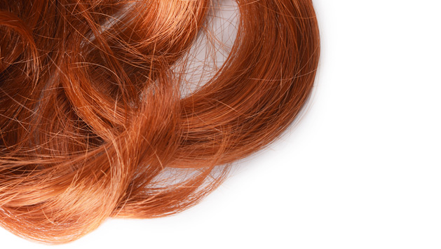
We have previously discussed the curious connection between melanoma and Parkinson’s disease. There is also a well known connection between melanoma and red hair. And believe it or not, there is another really strange relationship between Parkinson’s disease and red hair.

Redhair

Title: Genetic determinants of hair color and Parkinson’s disease risk.
Authors: Gao X, Simon KC, Han J, Schwarzschild MA, Ascherio A.
Journal: Ann Neurol. 2009 Jan;65(1):76-82.
PMID: 19194882

In 2009, researchers from Harvard University found a relationship between hair color and risk of Parkinson’s disease, when they examined the records of 131,821 US men and women who participated in the two large longitudinal studies, the Health Professionals Follow-up Study (HPFS) and the Nurses’ Health Study (NHS).


The HPFS, which started in 1986, sends questionnaires to US health professionals (dentists, optometrists, etc) – aged 40-75.  Every couple of years, members of the study receive questionnaires dealing with diseases and health-related issues (e.g. smoking, physical activity, etc). The questionnaire is supplemented by another questionnaires which is sent every four years, that deals with dietary information.

The NHS study – which was established in 1976 and then expanded in 1989 – has also collected questionnaire-based information from 238,000 registered nurses. Similar to the HPFS, every two years the study participants receive a questionnaire dealing with diseases and health-related topics.


In their study, the investigators found 264 of the male and 275 of the female responders to the HPFS and NHS questionnaires had been diagnosed with Parkinson’s disease. Of these individuals, 33 were black haired, 418 had brown hair, 62 were blond and 26 were redheads. Given that redheads make up just 1% of the general population but 5% of the people who were diagnosed with Parkinson’s disease in their study, the authors suggested that red haired people have a higher risk of developing Parkinson’s disease. Interestingly, they found a stronger association between hair color and Parkinson’s disease in younger-onset of PD (that is being diagnosed before 70 years of age) than those with age of onset greater than 70 years. When they took health and age related matters into account, the authors concluded that people with red hair are almost four times more likely to develop Parkinson’s disease than people with black hair.

NOTE: This result does not mean that people with red hair are definitely going to develop Parkinson’s disease, it simply suggests that they may be more vulnerable to the condition. And we should add that this result have never been replicated and we are not sure if anyone has ever attempted to reproduce it with a different database.

So how does (or could) this work?

The short answer is: we really don’t know.

The long answer involves explaining where there are no connections:

Red hair results from a genetic mutation. 80% of people with red hair have a mutation in a gene called MC1R – full name: melanocortin-1 receptor. Another gene associated with red hair is called HCL2 – ‘Hair colour 2’. We know that the connection between red hair and Parkinson’s disease is not genetic, as there is no association between MC1R mutations and Parkinson’s disease (for more on this, click here). We are not sure about HCL2, but this gene has never been associated with any disease.

What we do know is that redheads:

  • are more sensitive to cold (for more on this, click here)
  • are less responsive to subcutaneously (under the skin) administered anaesthetics (for more on this, click here)
  • suffer more from toothaches (for more on on this, click here)
  • are more sensitive to painkillers (for more on this, click here)
  • require more anesthetic for surgery (for more on this, click here)

Common myths associated with red hair include:

  • redheads bled more than others (this is not true – click here)…but they do bruise easier!
  • redheads are at greater risk of developing endometriosis (this is not true – click here)
  • redheads are more frequently left-handed (I can find no evidence for this, so I’ll put it in the myth basket until corrected).

There is also a strange link between red hair and multiple sclerosis, but it is too complicated to understand at the moment (women with red hair are more vulnerable to multiple sclerosis than men with red hair, for more on this, click here).

How any of these findings relates to Parkinson’s disease is unclear – we provide them here for those who are interested in following up this curious relationship.

One important caveat regarding this study is that incidence rates of Parkinson’s disease in countries with very high levels of red hair do not support the relationship (PD & red hair). In Scotland, approx. 10% of the population have red hair (source), and yet the England has a higher incidence of Parkinson’s disease (28.0/10,000 in England vs 23.9/10,000 in Scotland – source).

It may well be, however, that there is no direct connection between red hair and Parkinson’s disease. And until the results of the 2009 study mentioned above are replicated or supported by further findings, we here at the ‘Science of Parkinson’s disease’ shall consider this simply as a curious correlation.


The difference between men and women

At the bottom of our previous post, we mentioned that Japan is the only country where women have a higher incidence of Parkinson’s disease than men.

JapanPanorama_top

We also suggested that we have no idea why this difference exists. Well, a study presented at the Cardiovascular, Renal and Metabolic Diseases conference in Annapolis City (Maryland) last week may now be able to explain why this is.


The prevalence of Alzheimer’s disease is significantly higher in women compared to men. One recent estimate suggested that almost two-thirds of individuals diagnosed with Alzheimer’s disease are women (More information here). One possible reason for this is that Alzheimer’s disease is a condition of the elderly and women live longer.

So why is it then is the exact opposite true in Parkinson’s disease???


elderly-cake_2165089b

Source: The Telegraph Newspaper

Men are approximately twice as likely to develop Parkinson’s disease as females (More information here)

In addition, women are on average diagnosed 2 years later than men (More information here)

This gender difference has long puzzled the Parkinson’s research community. But now a group from the University of North Texas Health Science Center think that they may have the answer.

UNTHSC-copy

The researchers – lead by Shaletha Holmes from Dr Rebecca Cunningham’s lab – observed that when they stressed dopamine neurons, adding the male hormone testosterone made the damage worse. Interestingly, they found that testosterone was doing this by acting on a protein called cyclooxygenase 2 (or COX2). When they blocked the actions of COX2 while stressing dopamine neurons, they found that they also blocked the damaging effect of testosterone. The researchers concluded that testosterone may exacerbate the damage (and death) in dopamine neurons that occurs in Parkinson’s disease, thus possibly explaining the sex differences described above.

Now, there are several interesting aspects to this finding:

Firstly, the use of Ibuprofen, the nonsteroidal anti-inflammatory drug used for relieving pain, has long been associated with reducing the risk of Parkinson’s disease (More information here).

Ibuprofen is a COX2 inhibitor.

But more importantly, several years ago it was shown that Japanese men have lower levels of testosterone than their Western equivalents. Here is the study:

Japan1

Title: Evidence for geographical and racial variation in serum sex steroid levels in older men.
Authors: Orwoll ES, Nielson CM, Labrie F, Barrett-Connor E, Cauley JA, Cummings SR, Ensrud K, Karlsson M, Lau E, Leung PC, Lunggren O, Mellström D, Patrick AL, Stefanick ML, Nakamura K, Yoshimura N, Zmuda J, Vandenput L, Ohlsson C; Osteoporotic Fractures in Men (MrOS) Research Group.
Journal: Journal of Clinical Endocrinol. Metab. 2010 Oct;95(10):E151-60.
PMID: 20668046

The study suggested that total testosterone levels (while similar in men from Sweden, Tobago and the US) were 16 per cent higher in men from Hong Kong and Japan. BUT – and here’s the catch – Japanese men also had higher levels of a testosterone-binding hormone (Sex hormone-binding globulin or SHBG), so there is less of the testosterone floating around free to act. As a result, Japanese men had the lowest levels of active testosterone in the study.

Intriguingly, the researchers found that Japanese men who emigrated to the US had similar testosterone levels to men of European descent, suggesting that environmental influences may be having an effect of testosterone levels. Diet perhaps?

If testosterone is found to play a role in the gender difference found in Parkinson’s disease, the lower levels of free testosterone observed in Japanese men may explain why women in Japan have a higher risk of Parkinson’s disease than men.


EDITOR’S NOTE: WHILE WE HAVE NO DOUBTS REGARDING THE RESEARCH OF DR CUNNINGHAM AND HER GROUP, WE ARE TAKING A LEAP IN THIS POST BY APPLYING THE TESTOSTERONE RESULTS TO THE GENDER DIFFERENCE IN JAPAN. THIS IS PURE SPECULATION ON OUR PART. WE HAVE SIMPLY SAT DOWN AND TRIED TO NUT OUT POSSIBLE REASONS AS TO WHY THERE IS A REVERSED GENDER DIFFERENCE FOR PARKINSON’S DISEASE IN JAPAN. OUR THEORY IS YET TO BE TESTED, AND MAY BE COMPLETELY BONKERS. WE PRESENT IT HERE PURELY FOR DISCUSSION SAKE AND WELCOME YOUR THOUGHTS.

The Honolulu Heart Study

In 1950, Dr Tavia Gordon noticed that while the overall mortality rates for men in the USA and Japan were very similar, the incidence of heart disease was significantly lower in Japan. This observation resulted in three longitudinal studies – one of which became known as the Honolulu Heart Study.

Dr Travis Gordon. Source: JSTOR


The original purpose of the study was to determine whether there was a difference in heart disease incidence between Japanese people living in Japan and individuals of Japanese ancestry living in Hawaii.

The subjects recruited for the study were “non-institutionalized men of Japanese ancestry, born 1900-1919, resident on the island of Oahu.” In all, 12,417 men were identified as meeting the criteria. Of those contacted, 1,269 questionnaires were ‘return to sender’, 2,962 men declined to participate in the study, and 180 died before the study commenced. That left 8,006 participants who would be studied and followed for the rest of their lives.

From October 1965 onwards, the participants were interviewed and given physical examinations every few years. The interview processed asked for:

  • Family and personal history of illness
  • Sociological history
  • Smoking status
  • Physical activity level

The physical examination was very thorough, looking at:

  • ECG (Electrocardiography – electrical activity of the heart)
  • Urine analysis
  • Measurements of weight, height, skinfold thickness, etc.
  • Blood pressure and serum cholesterol

As a result, the study built up a HUGE amount of epidemiological information regarding these 8,006 individuals.

So, what does this have to do with Parkinson’s disease????

Given the enormous number of individuals involved in the study and the length of time that they were followed, it was inevitable that a certain percentage of them would develop Parkinson’s disease as the study progressed. As a result, the Honolulu Heart Study represents one of the largest epidemiological study of Parkinson’s to date. In 1994, a group of research involved in the study, published some very interesting findings relating to Parkinson’s disease. That published article was:

Morens


Title: Epidemiologic observations on Parkinson’s disease: incidence and mortality in a prospective study of middle-aged men.
Authors: Morens DM, Davis JW, Grandinetti A, Ross GW, Popper JS, White LR.
Journal: Neurology, 1996 Apr;46(4):1044-50.
PMID: 8780088

In total, 92 of the 8006 individuals enrolled in the study developed Parkinson’s disease. The incidence of Parkinson’s cases was registered between 1965 and November 30th 1994. The majority of the cases were diagnosed between 55 and 79 years of age (n=80). Diagnosis after the age of 80 was very rare. It is interesting to note that when the researchers divided the group into those ‘born before 1910’ and those ‘born after 1910’, the older group (born before 1910) had a lower risk of Parkinson’s disease.

In another study, the same group of investigators noted


Smoking


Title: Prospective study of cigarette smoking and the risk of developing idiopathic Parkinson’s disease.
Authors:  Grandinetti A, Morens DM, Reed D, MacEachern D.
Journal: American Journal of Epidemiology 1994 Jun 15;139(12):1129-38.
PMID: 8209872

In this study the authors found that men who had smoked cigarettes at any time prior to their enrollment in the study in 1965, had a reduced risk of developing idiopathic Parkinson’s disease (relative risk = 0.39). That is to say, smoking reduced the chance of developing Parkinson’s disease. And a few years later the authors published a follow up paper which rejected the possibility that smoking was killing people before they could develop Parkinson’s disease (selective mortality representing a false positive). That follow up report can be found here.

EDITOR’S NOTE: THIS DOES NOT MEAN THAT EVERYONE SHOULD RUSH OUT AND START SMOKING. THERE DOES, HOWEVER, APPEAR TO BE SOME INGREDIENT IN CIGARETTES THAT REDUCES THE INCIDENCE OF PARKINSON’S DISEASE. A LOT OF RESEARCH IS CURRENTLY TRYING TO IDENTIFY THAT INGREDIENT.

This finding was made alongside other interesting correlations (Note: coffee and alcohol reduce the risk of Parkinson’s disease):

Smoking-table

From Grandinetti et al (1994).

It should be noted that many of these associations (smoking in particular) had been reported before, but the Honolulu Heart Study was the first epidemiological study providing definitive proof. And it should be noted that subsequent epidemiological studies have found similar results.

INTERESTING FACTS ABOUT THE JAPANESE:

  1. The Japanese as a population have a lower incidence of Parkinson’s disease (much like most of the Asian nations) than their western equivalents, despite living longer.
  2. Japan is the only country in the world where females have a higher incidence of Parkinson’s disease than men (and we have no idea why!). Look here for more on this.

Alzheimer’s news – and how it relates to Parkinson’s disease

It all began with a 51 year old woman named Auguste Deter.

320px-Auguste_D_aus_Marktbreit

Auguste Deter. Source: Wikipedia

She was admitted by her husband to the Institution for the Mentally Ill and for Epileptics in Frankfurt, Germany on the 25th November, 1901. Her husband complained that she suffering memory loss and having delusions.

The attending doctor was Dr Alois Alzheimer.

Over the next year, Alois continued to examine Auguste – and what he began calling the “Disease of Forgetfulness” – until he left the institute to take up a position in Munich. He made regular visits back to Frankfurt, however, to follow up on Auguste.

Auguste dies on the 8th April, 1906. She had become completely demented and had existed in a vegatative state. When he examined the brain, Alois found the hall marks of what we today call ‘Alzheimer’s disease’ (namely neurofibrillary tangles and plaques).

Now, almost 110 years later, Alzheimer’s disease is the most common neurodegenerative condition – Parkinson’s disease is the second most common. Alzheimer’s affects 850,000 people in the UK alone (Source: Alzheimer’s Society). Huge efforts have been made in researching this condition and last week some interesting new data was published about the disease that may also have implications for Parkinson’s disease.

Jaunmuktane

Title: Evidence for human transmission of amyloid-β pathology and cerebral amyloid angiopathy.
Authors: Jaunmuktane Z, Mead S, Ellis M, Wadsworth JD, Nicoll AJ, Kenny J, Launchbury F, Linehan J, Richard-Loendt A, Walker AS, Rudge P, Collinge J, Brandner S.
Journal: Nature. 2015 Sep 10;525(7568):247-50.
PMID: 26354483

Published in the prestigious science journal, Nature, the article found signs of Alzheimer’s disease in the autopsied brains of people who had died from Creutzfeldt-Jakob disease (CJD) – the prion induced neurodegenerative condition.

What’s a prion?

Good question! A prion is a small infectious particle – usually composed of an abnormally-folded version of a normal bodily protein – that causes progressive neurodegenerative conditions. The first prion discovered in mammals was Prion protein (PRP): this is the prion that causes CJD.

PrP is considered the only known prion in mammals, but recently other proteins have exhibited prion-like behaviour. One such protein is Amyloid-β protein – the protein that is found clustered in clumps in the brains of people with Alzheimer’s disease.

The brains that were analysed in the study from the journal Nature were collected at death from people who had received human growth-hormone earlier in their lives. The growth-hormone had been extracted from human cadavers and it was injected into people with growth problems (this was a common practise during the 1950s to mid 1980s). Unfortunately, some of the growth-hormone appears to have been contaminated with PrP (possibly one of the cadavers used had undiagnosed CJD) and numerous people were injected with it (65 cases in Britain alone). Many of these individuals have been followed and we have learned a great deal from them regarding CJD. Some of these individuals have also donated their brains to science and it was some of these brains that were analysed in the study being discussed here.

What the authors of the study were expecting to see when they analysed these brains was lots of clusters of PrP. What the authors were not expecting to see was the clustering of Amyloid-β protein in these brains.

Fig1H

Amyloid-β protein (brown) in a section of brain tissue. Source: Nature

Of the eight brains (from people who received PrP infected growth-hormone) the authors analysed, six of them had clustering of Amyloid-β protein present in the brain (in four of those cases it was wide-spread). These brains came from people aged between aged 36–51 years – in such cases it is very rare to see large accumulations of Amyloid-β protein. The researchers also analysed the DNA of the individuals involved in the study and found that none of them were genetically susceptible to Alzheimer’s disease.

The researchers then compared these six brain with the brains of people who died from CJD caused by other means – 119 brains in total and none of them had Amyloid-β protein present in the brain. From these and other experiments, the authors suggested that this was the first human evidence of transmission of Alzheimer’s related pathology.

It is very important to note several details in the study:
1. None of the people whose brains were used in the study exhibited the clinical signs of Alzheimer’s.

2. None of the brains with Amyloid-β pathology had what is called ‘hyperphosphorylated tau neurofibrillary tangles’ – SImilar clumps of Amyloid-β protein, tau neurofibrillary tangles are another characteristic feature of Alzheimer’s disease brains. Their absence is curious.

3. The authors can not dismiss the possibility that the Amyloid-β was not present in the growth-hormone solution. In this case, the Amyloid-β accumulation in the brains could have been caused by some other unknown agent that was present in the injected solution.


A rare editorial note here: The Science of PD is disappointed with the way that this study has been handled by the wider media. While the results are interesting and the authors can be congratulated on their work, a correct interpretation of the results requires further study. This study has simply demonstrated was that Amyloid-β protein may be transmissible in a similar fashion to PrP. 


So why are we discussing this Alzheimer’s research here at the Science of Parkinson’s Disease?

Well, for a long time now Parkinson’s researchers have suspected that similar mechanisms may underlying what is happening in PD. That is to say, a prion-like protein may be transmitted between cells in the body (possibly from the gut to the brain – see previous posts) allowing the disease to progress. One protein in particular, Alpha Synuclein, which is present in Lewy bodies – the neurological features associated with Parkinson’s disease, has been implicated in this regards. Recent evidence from lab-based studies suggest that this is possible in cell cultures and in rodents, but whether it is possible in humans is yet to be determined.


NOTE:  Since publishing this post, we contacted the authors of the study regarding the presence of Alpha Synuclein and they told us that they were currently conducted a large study investigating what other proteins are also present. Thus far they have not seen any Alpha Synuclein accumulation. Interesting….

Melanoma and Parkinson’s – interesting connection

FACT: People with Alzheimer’s have a reduced chance of developing all known cancers (Source).

FACT: People with Parkinson’s disease have a reduced chance of developing all known cancers (Source)……except for one.

Melanoma

Melanoma. Source: Wikipedia

It is a curious fact, but people with Parkinson’s disease are 2-8 times more likely to develop melanoma than people without Parkinson’s (Source: Olsen et al, 2005; Olsen et al, 2006; Driver et al 2007; Gao et al 2009; Lo et al 2010; Bertoni et al 2010; Schwid et al 2010; Ferreira et al, 2010Inzelberg et al, 2011; Liu et al 2011; Kareus et al 2012; Wirdefeldt et al 2014; Catalá-López et al 2014; Constantinescu et al 2014; Ong et al 2014).

PLEASE NOTE: This is NOT to say that people with Parkinson’s disease are going to develop melanoma, it is simply making people with PD (and their carers) more aware that they should be keeping an eye out for it.

And there is another interesting connection between Parkinson’s disease and melanoma – if you have melanoma you are almost 3 times more likely to develop Parkinson’s disease than someone without melanoma (Source: Baade et al 2007; Gao et al 2009).

PLEASE NOTE AGAIN: This is NOT to say that if you have a melanoma you are automatically going to develop Parkinson’s disease. It is just something to be aware of. 

So, what’s going on here?

The simple answer is: we don’t know.

A lot of people are now looking at this issue though and we know that the connection is probably NOT genetic: approx. 10% of all cases of Parkinson’s disease can be associated with genetic mutants passed down through families. But none of the known Parkinson’s mutations make you more susceptible to melanoma. Equally, genetic susceptibility has been associated with 4-8% of all melanoma cases, but none of those genetic mutations makes one vulnerable to Parkinson’s disease (Meng et al 2012; Dong et al 2014; Elincx-Benizri et al 2014).

While we’re not sure what is happening between Melanoma and Parkinson’s disease, we are definitely very interested in the connection, and we will be watching this space closely. We’ll be sure to report any new discoveries relating to this in the future.

A gut feeling

New Parkinson’s Research this week:

Vagotomy

Title: Vagotomy and Subsequent Risk of Parkinson’s Disease.
Authors: Svensson E, Horváth-Puhó E, Thomsen RW, Djurhuus JC, Pedersen L, Borghammer P, Sørensen HT.

Journal: Annals of Neurology, 2015, May 29. doi: 10.1002/ana.24448.
PMID: 26031848

What’s it all about?

This is Prof Heiko Braak:

heiko-braak-01

Source – Memim.com

Many years ago, Prof Braak – a German neuroanatomist – sat down and examined hundreds of postmortem brains from people with Parkinson’s disease.

He had collected brains from people at different stages of Parkinson’s disease and was looking for any kind of pattern that might explain where and how the disease starts. His research led to what is referred to as the Braak stages of Parkinson’s disease – a six step explanation of how the disease spreads up from the brain stem and into the rest of the brain (see Braak et al, 2003).

nrneurol.2012.80-f1

The Braak stages of PD. Source: Doty RL (2012) Nature Reviews Neurology 8, 329-339.

Braak’s results also led him to propose that Parkinson’s disease may actually begin in the gut and then spread to the central nervous system (the brain). He based this on the observation that many brains that exhibited the very early stages of Parkinson’s disease had disease-related pathology in a population of neurons called the dorsal motor nucleus of the vagal nerve. This population of neurons connects to different organs in the body, such as the lungs, heart, kidneys and the gastrointestinal system (or the gut).

gut_aid_in_PD
A diagram illustrating the vagal nerve connection with the enteric nervous system which lines the gastric system. Source: Braak et al (2006) Nature Reviews Neurology 8, 329-339

Braak and his colleagues went on to examine the nerves fibres around the gastrointestinal system and in those fibres he found large deposits of a Parkinson’s disease-related substance: a protein called alpha synuclein. These deposits were present even at very early stages of the disease, which supported his theory that maybe the disease was starting in the gut.

This ‘gut to brain’ theory was supported by the fact that people with Parkinson’s disease often complain of gastrointestinal problems (eg. constipation) and some of these issues may predate the onset of other Parkinson’s disease symptoms. In addition, a couple of years ago, a group of scientists in the USA found alpha-synuclein present in bowel biopsies taken from people years before they were actually diagnosed with Parkinson’s disease (that study can be found here).

The ‘gut to brain’ theory received a further boost recently with the publication of a paper by a Danish group, who retrospectively looked at all the people in Denmark that received a vagotomy between 1975 and 1995.

So what’s a vagotomy?

Good question.

A vagotomy is a surgical procedure in which the vagus nerve are cut. It is typically due to help treat stomach ulcers. A vagotomy can be ‘truncal‘ (in which the main nerve is cut) or ‘superselective’ (in which specific branches of the nerve are cut, which the main nerve is left in tact).

Vagotomy

A schematic demonstrating the vagal nerve surrounding the stomach. Image A. indicates a ‘truncal’ vagotomy, where the main vagus nerves are cut above the stomach; while image B. illustrates the ‘superselective’ vagotomy, cutting specific branches of the vagus nerve connecting with the stomach. Source: Score

And what did the Danish scientists find?

The Danish researcher found that between 1975 and 1995, 5339 individuals had a truncal vagotomy and 5870 had superselective vagotomy. Using the Danish National registry (which which stores everyone’s medical information), they then looked for how many of these individuals went on to be diagnosed with Parkinson’s disease. They compared these vagotomy subjects with more than 60,000 randomly-selected, age-matched controls.

They found that subjects who had a superselective vagotomy had the same chance of developing Parkinson’s disease as anyone else in the general public (a hazard ratio (or HR) of 1 or very close to 1). But when they looked at the number of people in the truncal vagotomy group who were later diagnosed with Parkinson’s disease, the risk had dropped by 35%. Further, when they followed up the Truncal group 20 years later, checking to see who had been diagnosed with Parkinson’s in 2012, they found that they rate was half that of both the superselective group and the control group (see table below – HR=0.53). The authors concluded that a truncal vagotomy reduces the risk of developing Parkinson’s disease.

Svensson_Table2

Source: Svensson et al (2015) Annals of Neurology – Table 2.

So what does it all mean?

The study is an extremely novel approach to investigating the ‘gut to brain’ theory of Parkinson’s disease and the authors can be congratulated on some excellent work. It adds further weight to the idea that something is happening in the gut very early in Parkinson’s disease. It suggests that by cutting one of the main nerves connecting to the stomach, the disease is slowed down if not avoided all together. It might also suggest that the disease is slower and strikes earlier than previously thought (given that some people with truncal vagotomies still developed Parkinson’s disease – maybe the condition started before the nerve was cut).

But there are a couple of important details that should be considered about the paper before everyone rushes out to get a vagotomy:

  1. The number of people that received a truncal vagotomy (total = 5339) who went on develop Parkinson’s disease 20 years later was just 10 (compared with 29 in the superselective group). There may be some individuals who got lost in the system, but the number is still very low and caution should be used before we get too excited about a result based on a low number of subjects. It is important to determine whether this result can be replicated (in other countries).
  2. The gut may not be the only avenue for the disease. There has also been theories regarding an environmental aspect to the cause of Parkinson’s disease, and there have been studies conducted looking at the nasal/olfactory system of people with Parkinson’s disease to determine if this is another point of entry for the disease (for a recent review on this, see this paper here).

In summary, very interesting study and fascinating result, but please don’t rush to your doctor and ask for your vagus nerve to be cut!

Beginnings

Welcome to the Science of Parkinson’s Disease – a blog that has been set up by scientists to provide information and understanding about the neurodegenerative condition known as Parkinson’s disease.

Over the last 10 years, the advocacy for Parkinson’s disease has been tremendous and real awareness has been created by groups such as the Michael J Fox foundation, the Cure PD Trust, and Parkinson’s UK. They have generated enormous amounts of funding for scientific research and provided hope for disease halting therapies, while supporting and improving the general welfare of people suffering from this condition.

The media regularly announces new breakthroughs in the medical and scientific world, but there are few forums available for the general public to ask questions related to the science being conducted.  The Science of Parkinson’s disease has been set up for this purpose.

The Science of Parkinson’s disease is run by research scientists working in the field, and it was begun with several goals in mind:

  • Try to answer any questions you may have about Parkinson’s disease.
  • Report each week on interesting/exciting research in the world of Parkinson’s disease.
  • Interview Parkinson’s disease researchers, providing a face to the people doing the work.
  • Help you to understand this disease better.

We look forward to hearing from you.

The Team