An update on the connection between Melanoma and Parkinson’s disease

We have previously discussed the strange connection between Melanoma and Parkinson’s disease (click here to read that post).

Melanoma

That post included the curious observations that:

  • People with Parkinson’s disease are 2-8 times more likely to develop melanoma than people without Parkinson’s.
  • People with melanoma are almost 3 times more likely to develop Parkinson’s disease than someone without melanoma.

And we have no idea why (there is no shared genetic predisposition for the two conditions).

Research published this week, however, may begin to explain part of the connection:

Melanoma-title

Title: Parkinson disease (PARK) genes are somatically mutated in cutaneous melanoma.
Authors: Inzelberg R, Samuels Y, Azizi E, Qutob N, Inzelberg L, Domany E, Schechtman E, Friedman E.
Journal: Neurol Genet. 2016 Apr 13;2(3):e70.
PMID: 27123489     (This research article is OPEN ACCESS if you would like to read it)

In this study, the scientists looked at somatic mutations in cells from 246 tissue samples of melanoma.

What are somatic mutations?

Somatic mutations are genetic alteration that have been acquired by a cell that can then be passed to the progeny of that mutated cell (via cell division). These somatic mutations are different from ‘germline’ mutations, which are inherited genetic alterations that are present in the sperm and egg that were used in making each of us.

germlinesomatic1

Somatic vs Germline mutations. Source: AutismScienceFoundation

In the 246 samples analysed, the researchers found 315,914 somatic mutations in 18,758 genes. Yes, that is a lot, but what was very interesting was their discovery of somatic mutations in many of the PARK genes.

What are PARK genes?

There are a number (approx. 20) genes that are now recognised as conferring vulnerability to developing Parkinson’s disease. These genes are referred to as PARK genes. They include the gene that makes the protein Alpha synuclein ( SNCA ) and many others with interesting names (like PINK1 and LRRK2). Approximately 15% of cases of Parkinson’s are believed to occur because of a mutation in one (or more) of the  PARK genes. As a result there is a lot of research being conducted on the PARK genes.

Were all of PARK genes mutated in the Melanoma samples?

Somatic mutation in 14 of the 15 PARK genes (that the researchers analysed) were present in the melanoma samples. This means that after the skin cells turned into melanoma cancer cells, they acquired mutations in some of the PARK genes. Overall, 48% of the analysed samples had a mutation in at least 1 PARK gene, and 25% had mutations in multiple PARK genes (2–8 mutated genes). One PARK gene in particular, PARK 8, was more significantly present in the melanoma cells than the others. PARK8 is also known as Leucine-rich repeat kinase 2 or LRRK2 (we have previously discussed Lrrk2 – click here to read that post). Three additional PARK genes (PARK2, PARK18, and PARK20) were also significantly present, but not as significant as Lrrk2.

So what does it all mean?

The researchers speculate in the discussion of their report about what the findings could mean, but it is interesting to note that many of the PARK genes are susceptible to acquiring mutations (particularly  Lrrk2). And this is important to consider when thinking about our development as individual human beings – even though you may not born with a particular mutation for Parkinson’s disease (you haven’t inherited it from our parents), somewhere along the developmental pathway (from egg fusing with sperm to full grown adult) you could acquire some of these mutations which would make you vulnerable to Parkinson’s disease.And here we should note that skin and brain share the same developmental source (called the ectoderm). A mutation in a PARK gene could occur during your development and you would never know.

We thought this was a very interesting study – certainly worthy of reporting here.

Older siblings and Parkinson’s disease

Curious new research results out of Sweden this weekend…

To all of our readers who have older siblings that you grew up fighting with – you should  go and give them a hug today, because apparently they have lowered your risk of Parkinson’s disease.

Like I said ‘curious’.


enhanced-873-1401832891-14

Source: Buzzfeed

Older siblings. Nothing but trouble (a bit like younger siblings now that I think about it).

Who needs them.

Well, according to a massive new epidemiological study from the Karolinska Institutet, Stockholm (Sweden), we all do!

Siblings-title

Title: Early-Life Factors and Risk of Parkinson’s Disease: A Register-Based Cohort Study.
Authors: Liu B, Chen H, Fang F, Tillander A, Wirdefeldt K.
Journal: PLoS One. 2016 Apr 15;11(4):e0152841.
PMID: 27082111      (This article is OPEN ACCESS if you would like to read it)

This is a fascinating study, not only in its size and scale, but for the interesting details in the results.

The investigators collected a huge amount of information from multiple nationwide Swedish registers that are cross-linked thanks to the national personal identification number system that is used in Sweden (each Swedish resident is assigned a unique number).

sweden_stockholm

Stockholm, the capital of Sweden. Source: Budgetyourtrip

The information was collected from:

  • The Swedish Multi-Generation Register (MGR) – which holds information about the biological and adoptive parents for all residents born in 1932 or later, and were alive or lived in Sweden in 1961. This database covers over 95% of Swedish-born residents, plus more than 22% of foreign-born residents in Sweden.
  • The Swedish Patient Register – established in 1964/1965, this databases collects inpatient discharge records. It became nationwide in 1987, and since 2001, the Patient Register has recorded information on every inpatient visit and vast majority of the outpatient visits for all Swedish residents. 
  • They also linked their data to the Migration Register and Swedish Population and Housing Censuses from 1960, 1970, 1980, and 1990 for information on socio-economic status.

Like I said, ‘a huge amount of information’. They next set up a selection criteria. Within their pool of people for analysis, individuals had to:

  • be born in Sweden between 1932 and 1970
  • have information available regarding maternal links in the MGR
  • be alive and free of Parkinson’s disease on January 1, 2002,
  • 40 years or older on January 1, 2002 or turned 40 years during the study period

3 545 612 people fulfilled this criteria. 8779 cases of Parkinson’s disease were identified within that population of people (a further 2658 people were identified as having Parkinson’s disease, but since they were diagnosed before 2002, they were excluded). When looking at the findings of the analysis of this study:

Unsurprisingly:

  • the average age of diagnosis was 65.1 years of age
  • males had a higher risk than females (1.5 times more men than women)
  • parental occupation as farmers increased risk of Parkinson’s
  • a family history of the condition results in a higher risk of Parkinson’s disease.
  • No difference between blue or white collar occupations, or self employed roles
  • No difference between month/season of birth
  • No association with early life factors, including flu burden in the year of birth.

Surprisingly:

Compared to those without older siblings, the risk of developing Parkinson’s disease was 7% lower among participants with older siblings (HR = 0.93, 95% CI: 0.89, 0.98). The number of people with no older siblings was 1.68 million, of which 5384 had Parkinson’s disease. But of those with older siblings (1.86 million) only 3395 had Parkinson’s disease. Curiously, however, there was no further associations (eg. the number of older siblings or the interval length between the individual and their older siblings).

The effect (7%) is small, but the number of cases is very large, so we can assume that the finding is real. But how to explain it?!?

Even more surprisingly:

This is not the first time we’ve seen something like this:

Fang-title

Title: Maternal age, exposure to siblings, and risk of amyotrophic lateral sclerosis.
Authors: Fang F, Kamel F, Sandler DP, Sparén P, Ye W.
Journal: Am J Epidemiol. 2008 Jun 1;167(11):1281-6.
PMID: 18367467

In a similar sort of study published in 2008 (also from researchers at the Karolinska Institute in Sweden), it was reported that the risk of amyotrophic lateral sclerosis (ALS, also called Lou Gehrig’s disease; another neurodegenerative condition) increased with the number of younger siblings, and children whose first younger sibling was born after the age of 6 years had the highest risk of ALS. In contrast to the Parkinson’s research above, however, exposure to older siblings was not associated with an increased risk of ALS.

And a similar sort of result has also been observed in cases of Schizophrenia:

Westergaard-title

Title: Exposure to prenatal and childhood infections and the risk of schizophrenia: suggestions from a study of sibship characteristics and influenza prevalence.
Authors: Westergaard T, Mortensen PB, Pedersen CB, Wohlfahrt J, Melbye M.
Journal: Arch Gen Psychiatry. 1999 Nov;56(11):993-8.
PMID: 10565498

This research came from a different Scandinavian capital (Copenhagen), and involved only 1.74 million people, but it suggested that larger sibships were associated with an increased risk of developing schizophrenia. This result was independent of birth order or interval length between siblings. 

Why these effects exist is a question yet to be answered. In each of these studies, the authors propose elaborate possibilities (eg. developmental theories involving the immune system, etc), but there is no evidence (yet) to support them. Given that the effects are small (just a 7% reduction in risk in the case of Parkinson’s), it would be interesting to investigate differences between subjects within the Parkinson’s population, to determine if there is a subset of individuals more affected than others by this sibling phenomenon. By comparing which commonalities they may share (genetic, environmental or otherwise) we could identify patterns of risk factors for specific individuals.

So while the Parkinson’s connection is an interesting finding, obviously more research is required to better understand what is going on.

Curious result though, right?


The Parkinson’s UK 2016 Gretschen Amphlet Memorial Lecture

Gretschen Amphlet was a long-time resident of Cambridge (UK) who suffered from Parkinsons’s disease. Every year she is remembered in a memorial lecture in April.

This year, Prof Roger Barker of Cambridge University was asked to give the talk.

roger_barker

He is a Professor of Clinical Neuroscience at the University of Cambridge and an Honorary Consultant Neurologist at Addenbrooke’s Hospital in Cambridge. Prof |Barker conducts both lab-based and clinical research on Parkinson’s disease, co-ordinating large clinical studies such as the Transeuro cell transplantation trial currently being conducted.

His lecture was titled: Can stem cells deliver on their promise for Parkinson’s?

The event is organised by Parkinson’s UK.

logo

It was a beautiful evening outside the auditorium at FitzWilliams college in Cambridge…

FitzCollege

…and we were present in the lecture hall for the event. Parkinson’s UK filmed the lecture and that footage is available online (click here to watch it). We also offer a transcript of the lecture – to read the transcript, please click here.


Chromosome 22 and Parkinson’s disease

A wise man once told me:

“When trying to understand genetics, think of DNA as the stream of words in a book. The nucleotides (A, G, T and C) are the individual letters. These ‘letters’ collect together to make up the genes (the sentences) which give the  book meaning and convey information. And the chromosomes are the chapters in that book.

Some of these ‘books’ are short reads – the fly has only 139.5 million nucleotides (‘words’) and 15,682 genes (sentences) spread across just 4 chromosomes (‘chapters’), while others are long books – humans = 3 billion words, divided into 22,000 sentences, and 23 chapters.

They were helpful words – putting things in perspective – and I hope that they might aid you dear reader as we tackle the topic of this post – a genetic mutation in a particular location of chromosome 22 and its relationship with Parkinson’s disease.

Oh, and do not be fooled into thinking that size matters when it comes to chromosomes. The mighty hedgehog and faultless pigeon have almost twice as many chromosomes as we do (45 and 40 pairs, respectively), and yet…


As most of you will be aware, human beings have 23 pairs of chromosomes.

Chromosomes are a concept that many people are aware of (a pub quiz type of topic), but what are they?

What exactly is a chromosome?

In a nutshell, a chromosome is a very efficient way of packing a lot of DNA into a cell.

Within most of the cells in your body, DNA is densely coiled into discrete packages called chromosomes. Without such packaging, the stringy DNA molecules would be too long to fit inside the cell. In fact, if you uncoiled all of the DNA molecules in a single human cell and placed them end-to-end, they would stretch for at least 6 feet. And that’s just for one cell – remember that the humans have approx. 40 trillion cells in their body!

CDR761781 A schematic demonstrating the arrangement of DNA- Genes-Chromosomes. Source: cancergenome.nih.gov

When a cell is not dividing, the chromosomes usually sit in the nucleus of the cell in loose strands called chromatin. When the cell decides to divide, the chromatin condenses and wrap up very tightly, becoming chromosomes. Both loose chromatin and tightly wound chromosomes are very difficult to see, even with a microscope.

Chromosomes come in pairs – one set of 23 chromosomes from each parent, giving us a total of 46 chromosomes per cell. All of these pairs reside inside the nucleus of each cell, where their DNA is read and instructions (RNA) are sent off to be made into proteins which performs functions within the cell.

Within the DNA in the chromosomes there are sometimes mistakes (think of them as spelling mistakes in the book example we mentioned above). The mistakes are called ‘mutations’ or variants. They can involve sections of DNA being absent or sections of DNA being replicated multiple times.

This week new research was published dealing with Parkinson’s disease and a mutation in chromosome 22.

What do we know about Chromosome 22?

Chromosome 22 is the second smallest human chromosome, being only slightly larger than chromosome 21. Chromosome 22 spans approximately 50 million DNA base pairs and represents 1.5-2% of the total DNA in each cell.

Human_male_karyotpe_high_resolution_-_Chromosome_22

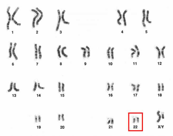
The 23 chromosomes of humans (this set is from a male). Chromosome 22 is highlighted. Source: Wikipedia

There are approx. 1000 genes on chromosome 22. The functions of many of these genes (what they tell the cell/body to do) is still being determined. Mutations in some of those genes, however, are associated with certain diseases. One particular disease associated with Chromosome 22 is called chromosome 22q11.2 deletion syndrome.

What is 22q11.2 deletion syndrome?
Chromosome 22q11.2 deletion syndrome (also known as DiGeorge syndrome) is a condition that arises from a section of chromosome 22 being absent. The ’22q11.2′ code part of the name relates to the specific location on chromosome 22 where the missing sections become apparent. About 87% of deletions occur within a 3 million base pair (nucleotides) region in the middle of one copy of chromosome 22 in each cell (remember that chromosomes come in pairs). The region contains at least 52 known genes.

Given the number of possible gene affected, there are numerous clinical features associated with 22q11.2 deletion syndrome, including heart defects, an opening in the roof of the mouth (a cleft palate), subtle facial features, learning issues, and low calcium levels.

Small ‘micro deletions’ within chromosome 22 are some of the most frequent known deletions found in human beings, occurring in about 25 in 100 000 people. These micro deletions are inherited from an affected parent in 5–10% of cases, while the rest occur spontaneously.


So what does Chromosome 22 have to do with Parkinson’s disease?

In 2009, this research report was published:

Zaleski-title

Title: The co-occurrence of early onset Parkinson disease and 22q11.2 deletion syndrome.
Authors: Zaleski C, Bassett AS, Tam K, Shugar AL, Chow EW, McPherson E.
Journal: Am J Med Genet A. 2009 Mar;149A(3):525-8.
PMID: 19208384

In this report the researchers described two patients, both with chromosome 22q11.2 deletion syndrome and early onset Parkinson’s disease (diagnosed before 45 years of age). The researchers suggested that this co-occurrence of chromosome 22q11.2 deletion syndrome and Parkinson’s disease in two unrelated patients was unlikely to be coincidence (given the low frequency of the conditions).

That first study was followed up by a second study:

Butcher-title

Title: Association between early-onset Parkinson disease and 22q11.2 deletion syndrome: identification of a novel genetic form of Parkinson disease and its clinical implications.
Authors: Butcher NJ, Kiehl TR, Hazrati LN, Chow EW, Rogaeva E, Lang AE, Bassett AS.
Journal: JAMA Neurol. 2013 Nov;70(11):1359-66.
PMID: 24018986

In this report, the scientists conducted an observational study of the occurrence of Parkinson’s disease in the world’s largest cohort of well-characterized adults with a chromosome 22q11.2 deletion syndrome (n = 159; age range = 18.1-68.6 years). They found that people with chromosome 22q11.2 deletion syndrome had a significantly elevated occurrence of Parkinson’s disease compared with standard population estimates.

Curiously, they suggested that the common use of antipsychotics in patients with chromosome 22q11.2 deletion syndrome (to manage associated psychiatric symptoms) delayed diagnosis of Parkinson’s disease by up to 10 years. And in postmortem analysis of the brains of people with both conditions, they found the loss of dopamine neurons and the occurrence of Lewy bodies – characteristic features of Parkinson’s disease.

This was proof that people with chromosome 22q11.2 deletion syndrome were more vulnerable to developing Parkinson’s disease. But what about people with Parkinson’s disease? Do they have deletions with chromosome 22q11.2?

This week we got the answer to that question:

Mok-title

Title: Deletions at 22q11.2 in idiopathic Parkinson’s disease: a combined analysis of genome-wide association data.
Authors: Mok KY, Sheerin U, Simón-Sánchez J, Salaka A, Chester L, Escott-Price V, Mantripragada K, Doherty KM, Noyce AJ, Mencacci NE, Lubbe SJ; International Parkinson’s Disease Genomics Consortium (IPDGC), Williams-Gray CH, Barker RA, van Dijk KD, Berendse HW, Heutink P, Corvol JC, Cormier F, Lesage S, Brice A, Brockmann K, Schulte C, Gasser T, Foltynie T, Limousin P, Morrison KE, Clarke CE, Sawcer S, Warner TT, Lees AJ, Morris HR, Nalls MA, Singleton AB, Hardy J, Abramov AY, Plagnol V, Williams NM, Wood NW.
Journal: Lancet Neurol. 2016 Mar 23. [Epub ahead of print]
PMID: 27017469

The researchers analysed the DNA of 9387 people with Parkinson’s disease and 13 863 controls. They identified eight unrelated people with Parkinson’s disease who carried the chromosome 22q11.2 deletions. None of the controls had any of these deletions. Those people with Parkinson’s disease who had chromosome 22q11.2 deletions had earlier ages of onset (average age of diagnosis = 41 years old) than people with Parkinson’s disease who did not have the deletions (average age of diagnosis = 60.3 years). The researchers concluded that chromosome 22q11.2 deletions are associated with early onset Parkinson’s disease.

Some concluding thoughts

While the results of the Lancet Neurology study are very interesting, there are several important aspects to consider.

Firstly, the researchers noted that the estimated prevalence of 22q11.2 deletion syndrome in the general population is 0·024% or 24 in every 100,000 people. More importantly, as the study indicated the frequency of a 22q deletion among people with early-onset Parkinson’s disease is also very low (0·49% or 5 in every 1000 people with early-onset Parkinson’s disease). In fact, the number of people with the 22q11.2 deletion syndrome mutation and Parkinson’s disease is less than 20. So obviously this is a very low frequency event.

It is also interesting to consider that only 3% of patients with 22q11.2 deletion syndrome go on to develop Parkinson’s disease. Also a low frequency event. But studying this small population of people with a very specific genetic circumstance may enlighten us to some of the biological mechanisms causing this low frequency occurrence. And that may further aid us in better understanding other forms of Parkinson’s disease.

And that really is the take home message from this study:  we are gradually building a map of the connections between genetics and Parkinson’s disease, and while genetics will not explain every case of this condition, the knowledge we gain from this process will allow us to better target the disease in the long run.

And now spit!

Did you know that human saliva is 99.5% water?

1369155421_drool

But a recent set of studies have suggested that the remaining 0.5% holds some interesting insights into Parkinson’s disease.


Interesting fact about saliva – while there is a lot of debate as to how much saliva we produce on a daily basis (anywhere between 0.75 to 1.5 litres per day), it is generally accepted that during sleep the amount of saliva produced drops to almost nothing. Why? Big shrug.


Saliva is a solution produced by three main sets of glands in our mouth: the parotid, Sublingual, and Submandibular glands:

h9991831_001

The human salivary glands. Source: WebMD 

The solution produced serves several important functions, namely:

  • beginning the process of digestion by breaking down food particles.
  • protecting teeth from bacterial decay.
  • Moisturising food to aid in the initiation of swallowing.

As we mentioned above, 99.5% of saliva is water. The remaining 0.5% is made up of enzymes and antimicrobial agents. There is also a number of cells in each millilitre of saliva (as many as 8 million human and 500 million bacterial cells per millilitre).

By analysing those human cells, scientists can learn a lot about a person. For example, they can conduct genetic analysis and determine if a person has a particular mutation.

So what has this got to do with Parkinson’s disease?

Well, recently several research groups have been looking at saliva with the hope that biomarkers – chemicals that may allow for early detection or better monitoring of Parkinson’s disease – could be found.

And recently, some of that research has seemingly paid big dividends:

Spit3-title

Title: Prevalence of Submandibular Gland Synucleinopathy in Parkinson’s Disease, Dementia with Lewy Bodies and other Lewy Body Disorders.
Authors: Beach TG, Adler CH, Serrano G, Sue LI, Walker DG, Dugger BN, Shill HA, Driver-Dunckley E, Caviness JN, Intorcia A, Filon J, Scott S, Garcia A, Hoffman B, Belden CM, Davis KJ, Sabbagh MN.
Journal: J Parkinsons Dis. 2016 [Epub ahead of print]
PMID: 26756744

In this study, published in January of this year, the researchers collected small biopsies of the submandibular gland (one of the three primary producers of saliva) from the bodies of people who died with various conditions (including Parkinson’s disease). They analysed the biopsies for alpha synuclein – the chemical in the brain associated with Parkinson’s disease. We have previously written about alpha synuclein, a chemical in the brain that is associated with Parkinson’s disease (for a primer on alpha synuclein, click here). They found that alpha synuclein was present in the saliva gland of 89% of the subjects who died with Parkinson’s disease, but none of the 110 control samples.

This result led the same research groups to attempt a similar study on live subjects and they published the results of that study in February of this year:

Spit2--title

Title: Peripheral Synucleinopathy in Early Parkinson’s Disease: Submandibular Gland Needle Biopsy Findings.
Authors: Adler CH, Dugger BN, Hentz JG, Hinni ML, Lott DG, Driver-Dunckley E, Mehta S, Serrano G, Sue LI, Duffy A, Intorcia A, Filon J, Pullen J, Walker DG, Beach TG.
Journal: Mov Disord. 2016 Feb;31(2):250-6.
PMID: 26799362

The researchers enrolled 25 people with early-stage Parkinson’s disease (less than  5 years since diagnosis) and 10 control subjects. All of these subjects underwent a small biopsy of the submandibular gland. Those biopsies were then analysed for alpha synuclein and the researchers found that 74% of the Parkinsonian biopsies and 22% control biopsies had alpha synuclein present in the submandibular gland.

And remarkably, this report was followed up this last week by a group in Italy, who published some very interesting data:

Spit1-title

Title: Abnormal Salivary Total and Oligomeric Alpha-Synuclein in Parkinson’s Disease.
Authors: Vivacqua G, Latorre A, Suppa A, Nardi M, Pietracupa S, Mancinelli R, Fabbrini G, Colosimo C, Gaudio E, Berardelli A.
Journal: PLoS One. 2016 Mar 24;11(3):e0151156.
PMID: 27011009    (this report is OPEN-ACCESS if you would like to read it)

The researchers collected salivary samples – actual spit – from 60 people with Parkinson’s disease and 40 age/sex matched control subjects. They then measured the saliva for different types of alpha synuclein. In this study, the researchers measured both the total amount of alpha synuclein in the saliva and also special forms of alpha synuclein.

Alpha synuclein initially starts out in the brain in a monomeric form – as a single version of alpha synuclein. This form of alpha synuclein is believed to be safe. A more mature form of alpha synuclein, called oligomeric, is believed to be the seed of the aggregations found in the Parkinsonian brain, Lewy bodies.

Curiously, in this study the researchers found that the total amount of alpha synuclein in the salivary of people with Parkinson’s disease was lower than that of the control subjects. But – and it’s a big ‘but’ – the amount of alpha synuclein oligomers was higher in the people with Parkinson’s disease than normal healthy controls.

The researchers proposed that the decreased concentration of total alpha-synuclein may reflect the formation of lewy bodies in the brain, and that this test might help the early diagnosis of Parkinson’s disease.


Here at the Science of Parkinson’s we are approaching this research cautiously. Previous attempts at measuring saliva in Parkinson’s disease have not had such significant results when comparing people with Parkinson’s disease and controls (click here for more about that study). The need for better biomarkers of Parkinson’s disease provides the reasons for this research, but the variability between the results different groups are getting leaves one wondering about the viability of the approach. It would indeed make for a very easy, non-invasive testing platform for Parkinson’s disease (‘Please spit into this tube for me’), but more research is needed before it can be applied on the large scale.

We’ll keep watching and hoping.

Another connection between skin and Parkinson’s disease

This is very interesting.

We have previously written blog posts dealing with the connection between melanoma and Parkinson’s disease. And now, there is new research providing a new link between another skin condition and Parkinson’s disease.


What is Rosacea?

Rosacea is a chronic skin condition, classically characterized by a redness of the face. This is the result of dilation of blood vessels in the facial skin, and is usually accompanied by pustules and swelling. Rosacea is indiscriminate in which age group it afflicts and there are four subtypes: three specifically affecting the skin and another affecting the eyes (ocular rosacea).

ps_150331_rosacea_800x600

An example of Rosacea.Source: Medscape

Rosacea is diagnosed in women almost three times more than men. It is also more common in people between the ages of 30 and 50, and appears to have a preference for Caucasians of northwestern European descent (hence it’s nickname: the “curse of the Celts”).

What has this skin condition got to do with Parkinson’s disease?

Well, back in 2001 this study was published:

Fischer-title

Title: Skin function and skin disorders in Parkinson’s disease.
Authors: Fischer M, Gemende I, Marsch WC, Fischer PA.
Journal: J Neural Transm. 2001;108(2):205-13.
PMID: 11314773

In this study, the researchers were investigating seborrheic dermatitis (similar to rosacea, this is an inflammation condition that presents itself as flaky, itchy, and red skin) and hyperhidrosis (abnormal increase in sweating) in Parkinson’s disease. They measured these afflictions in  70 people with Parkinson’s disease and 22 matched control subjects. Almost 20% of the people with Parkinson’s disease had seborrheic dermatitis and half of the Parkinson’s population had hyperhidrosis. The researchers also found that half of the Parkinson’s group also had abnormal sebum levels – sebum being a waxy substance produced by the skin (interestingly, we have previously mentioned sebum in a post about a lady who can smell Parkinson’s disease).

This was an interesting result, but it was never really followed up…until this last week, when another study was published:

Egeberg-title

Title: Exploring the Association Between Rosacea and Parkinson Disease: A Danish Nationwide Cohort Study.
Authors: Egeberg A, Hansen PR, Gislason GH, Thyssen JP.
Journal: JAMA Neurol. 2016 Mar 21. [Epub ahead of print]
PMID: 26999031

The size of this new study is amazing: the researchers looked at data from an national database which includes all Danish citizens 18 years or older from January 1, 1997 to December 31, 2011. That is a reference population of 5.4 million individuals!

Of these, 22 387 individuals (43.8% women) received a diagnosis of Parkinson disease between 1997 -2011, and 68 053 individuals (67.2% women) had a history of the skin condition rosacea.

The general population rate of Parkinson disease was 3.5 cases per 10 000 person. But in the population that had a history of rosacea the rate of Parkinson’s disease was 7.6 cases per 10 000 people – almost twice as high as the general population. Interestingly, when they looked at the subtypes of rosacea, the researchers found that there was a more than 2-fold increase in the risk of Parkinson disease in patients who had a history of ocular rosacea.

Even more interesting: treatment with tetracycline – an antibiotic – appears to have reduced the risk of Parkinson’s disease. The researchers also noted that people with severe rosacea have the same risk of developing Parkinson’s disease as do those who have more moderate rosacea.

This is an interesting study, further indicating a connection between the skin and Parkinson’s disease. Whether the relationship indicates anything causal or simply occurring in parallel is yet to be determined. But given similar previous association, we obviously need to take a closer look at skin.

Does the age of onset make a difference?

This is Dr Henri Huchard (1844-1910; a French neurologist and cardiologist):

Henri_Huchard

Source: Wikipedia

In 1875, he described the case was of a child who, at just 3 years of age, presented with all the clinical features of Parkinson’s disease. Since that report, there have been many studies detailing the condition that has come to be known as ‘juvenile Parkinsonism‘. Currently the youngest person diagnosed with Parkinson’s disease is a young lady from Oklahoma. She was diagnosed at 10-years of age, but had shown symptoms since the age of 2.

We are going to explore juvenile Parkinson’s disease in future post, but today’s post will review some new research that looks at the differences in Parkinson’s disease features of people diagnosed at different ages.


For most members of the general public, Parkinson’s disease is considered a condition of the elderly. And this is a fair line of thinking given what they probably observe out there in the big wide world.


A few years ago though, Parkinson’s UK commissioned and published a report of the statistics/demographics of Parkinson’s disease in the UK (Click here for a copy of that report). In that report, they present this table:

Table1

It illustrates the estimated number of people in each age bracket that have Parkinson’s disease. As you can see, the bulk of the people affected are over the age of 60. But this does not mean that Parkinson’s disease is simply a condition of the aged. It is believed that worldwide at least 5% of diagnoses are to people below the age of 50 – this is considered early onset Parkinson’s disease.

There are many people – actor Michael J Fox among them – who have been diagnosed below the age of 40.

leadership-fox-m-img_2

Actor Michael J Fox was diagnosed with Parkinson’s disease at age 30.
Source: MJFox foundation

Given this wide spectrum of age of onset, it is curious that more research has not been conducted comparing the differences in features of the condition between the different age groups. This situation, however was remedied recently:

Age-of-onset-PD

Title: Age at onset and Parkinson disease phenotype.
Authors: Pagano G, Ferrara N, Brooks DJ, Pavese N.
Journal: Neurology. 2016 Feb 10.
PMID: 26865518

The investigators in this study took 422 people who had recently been diagnosed with Parkinson’s disease (none of them were on any anti-Parkinson’s medication, eg. L-dopa). The subjects were divided into 4 groups according to their age at diagnosis:

  1. younger than 50 years (58 subjects)
  2. 50-59 years (117 subjects)
  3. 60-69 years (168 subjects)
  4. older than 70 years (79 subjects)

The researchers then investigated differences in:

  • side of onset (left or right; dominant or non-dominant side of the body)
  • type of symptoms (rigidity or tremor, etc)
  • localization of symptoms occurrence (eg. arms, legs, etc)
  • severity of motor features (rigidity, tremor,…)
  • severity of nonmotor features (memory, attention,…)
  • severity nigrostriatal function (brain imaging of the dopamine system)
  • CSF biomarkers (Chemicals in the cerebrospinal fluid which surrounds the brain)

Curiously in all of the four groups, a quarter of the people had a family history of Parkinson’s disease. Familial history could suggest a genetic connection, and the genetic aspect of Parkinson’s disease has generally been associated with the early onset group. But this does not appear to be the case in this  study – there was no bias towards the younger onset group.

Asymmetry of motor features onset (eg. tremor, etc) was apparent in 97.8% of the total population, with 55% of those subjects having symptoms on their dominant side. It is interesting to note here, however, that the young onset group were the only group in which the non-dominant side was more affected than the dominant. Similarly, almost all of the symmetrical onset individuals (exhibiting no asymmetry in their motor features) were in the oldest group.

In all four groups, the arm was the more likely site of motor features (this was the case in approx. 85% in all groups). When considering other sites of onset, the head was more frequent in the older groups than the younger group, while the leg was more common in the younger group than the older group.

The older the age at onset the more severe the motor (eg. resting tremor, and postural instability scores) and nonmotor features (including autonomic, olfactory, and cognitive functions). This was accompanied by a greater dopaminergic dysfunction on the brain scan, and a reduction of alpha synuclein floating around in the cerebrospinal fluid.

Rigidity was more common in the young-onset group.

There were no differences between the groups in terms of issues associated with activities of daily living, measures of depression and anxiety, impulsive control, or sleep problems.

What does it all mean?

Why these difference exist and what they might tell us about the condition is yet to be determined. The results are interesting when one considers that the subjects had similar disease duration (they had all just been diagnosed; within 6 months of diagnosis). This suggests that the observed differences may be specific to the different age groups. A direct contribution of the aging process, however, has to be considered when assessing the older group.

This kind of analysis is necessary as it is the only way small details about the disease can be determined. We thought this was an interesting study

A sense of (and the science of) smell

Losing the sense of smell is a common feature associated with Parkinson’s disease. But this feature of the condition may help us to better understand the condition. Some autopsy studies have suggested that the olfactory system is one of the first structures in the brain to be affected by the disease.

bad-smell-001

Source: Guardian

How do we smell?

Bad.

That’s both a pathetic attempt at humour and a serious answer. Compared with fellow members of the mammalian family, human beings have a pretty poor sense of smell.

The process of smelling stuff is conducted through structures called the olfactory bulbs. The human olfactory bulbs lie on the base of our brains, protruding forward towards our nose (and nasal cavity).

olfactory_bulb_(inferior_view)1321478082612

A view of the human brain from below (olfactory bulbs are in yellow). Source: StudyBlue

olfactorybulbq1318387403476

A view of the human brain from in front (olfactory bulbs are in yellow). Source: StudyBlue

Inside your nose there is an area of smell sensitive cells that lies on the roof of the nasal cavity (about 7 cm behind your nostrils). That area is called the olfactory epithelium, and it plays a critical role in our sense of smell.

The size of the human olfactory epithelium is rather small and reflects our poor sense of smell, especially when compared, for example, to a dog  (humans have about 10 cm2 (1.6 sq in) of olfactory epithelium, while some dogs have 170 cm2 (26 sq in)).

odorant_eng

The human olfactory system. Source: Biology junction.com

When you inhale an odor (or odorant molecules) through your nose, there are tiny receptors (called olfactory receptors) on the  olfactory epithelium that are the first step in detecting the smell. Every single olfactory receptor cell presents just one (and only one) type type of odorant receptor. When they detect that odor, the olfactory receptor cell reacts by sending an electrical signal along its branch (called an axon) to the olfactory bulbs in the brain.

As the axon of olfactory receptor cell enters the olfactory bulb it forms clusters with other olfactory receptor cell axons, and these clusters are called glomeruli. Inside the glomerulus (singular), the axons make contact the branches of a type of brain cell called a mitral cell. Mitral cells send their axons to many different areas of the brain, including the anterior olfactory nucleus, piriform cortex, the amygdaloid complex, the entorhinal cortex, and the olfactory tubercle.

fulllengths-olfaction-2

Source: YaleScientific

From here our understanding of olfactory processing is less well understood. The piriform cortex is considered the area most likely associated with identifying particular odor. The amygdala is involved in emotional and social functions (eg. mating and recognition), while the entorhinal cortex (and connected hippocampus) is associated with memory – this area is probably activated when a particular smell reminds us of something in our childhood.

What is known about our sense of smell in Parkinson’s disease?

In 1975, two researchers in Minnesota noticed that many of their people with Parkinson’s disease that they were assessing had reduced olfactory abilities. They decided to test this observation:

olfactory-title

Title: Olfactory function in patients with Parkinson’s disease.
Authors: Ansari KA, Johnson A.
Journal: J Chronic Dis. 1975 Oct;28(9):493-7.
PMID: 1176578

The researchers took 22 people with Parkinson’s disease and 37 age/sex-matched controls and repeatedly tested them in a double blind study to determine their olfactory acuity. In each test, the subjects were given five test tubes. Two of the tubes in each set contained 0.5 ml of diluted amyl acetate (which has a distinct smell). The other three tubes contained just water. The subjects were asked to inhale through their nose and then identify which two tubes in each set contained the amyl acetate. The highest dilution (the weakest smelling solution) at which the subject could correctly identify the two amyl acetate containing tubes was designated as their olfactory threshold.

The researchers found that people with Parkinson’s disease had a significantly reduced olfactory acuity (a lower olfactory threshold than compared to control subjects). They also noted that subjects with more progressive forms of the disease exhibited a worse performance on the test. Numerous studies have now replicated this overall result, including a recent study that indicated that smoking may have a protective role on the olfactory ability (Click here and here for more on this).


EDITORIAL NOTE: Please understand that the loss of smell in Parkinson’s disease does not immediately mean that you will have a more progressive form of the condition. There is simply a trend in the data that suggests the loss of smell is a risk factor for having a more progressive version of the condition. 

We would also like to discourage any thoughts of taking up smoking in order to protect your sense of smell.


So what is actually happening in the Parkinson’s disease brain?

This is Prof Heiko Braak:

heiko-braak-01

He’s a dude. We’ve mentioned him before in a previous post.

Many years ago, he and his colleagues were intrigued with the hyposmia (reduction in olfactory ability) in Parkinson’s disease. They conducted a series of autopsy studies, looking at 413 brains! Specifically, they were looking for deposits of the Parkinson’s disease-related protein, alpha synuclein, in the brains and where the protein was accumulating. The accumulation of alpha synuclein is believed to be associated with the loss of cells in the brain.

In total they found 30 brains that exhibited accumulation of alpha synuclein. Of interest, they found that 16 of those brains had accumulation of alpha synuclein in the olfactory bulb. And in one particular case, the olfactory bulb was the only affected part of the brain, except for a tiny region of the brain stem.

The researchers were curious about the possibility that the olfactory system could be a potential starting point for Parkinson’s disease, but they were quick to point out that only half the cases they analysed (16/30) had accumulation of alpha synuclein in the olfactory bulb. Thus, while the olfactory system may be involved, it seems unlikely that the nose is the sole induction site of Parkinson’s disease.

After this study was published, however, Braak and his colleagues went on to analyse the accumulation of alpha synuclein in the lining of the gut and their results suggested this as another possible site of induction (we have written about this in a previous post). They have subsequently proposed a model of disease spread based on entry to the brain via the nose and gut:

nrneurol.2012.80-f1

The Braak stages of Parkinson’s disease. Source: Nature Reviews Neurology.

It is interesting to observe that studies by other scientists have indicated that the nasal epithelium of people with Parkinson’s disease (both with and without the loss of olfactory abilities) is not damaged or presenting an accumulation of alpha synuclein (Click here for more on this).

So what happens to the olfactory bulbs in Parkinson’s disease?

A recent review of the previous studies investigating olfactory bulb volume in people with Parkinson’s disease was published in the Open Access journal PlosOne:

Olfactory_title

Title: Changes in Olfactory Bulb Volume in Parkinson’s Disease: A Systematic Review and Meta-Analysis.
Authors: Li J, Gu CZ, Su JB, Zhu LH, Zhou Y, Huang HY, Liu CF.
Journal: PLoS One. 2016 Feb 22;11(2):e0149286.
PMID: 26900958  (this report is OPEN ACCESS if you would like to read it)

The authors of the study conducted a systematic review (or meta-analysis) of all of the previous studies (six in total) that have measured the size of the olfactory bulb in the brains of people with Parkinson’s disease (using brain imaging techniques). They found that in all of the 6 studies (collectively 216 PD patients and 175 healthy controls) there was a significant reduction in the size of the olfactory bulbs of people with Parkinson’s disease. Strangely, they authors also found the right olfactory bulb was larger than the left in subjects with Parkinson’s disease across all of the studies, and this effect was not found in the healthy controls.

The motor features of Parkinson’s disease usually begin asymmetrically – by this we mean that the left arm is affected before the right, or the right leg has tremor before the left. This is different for each person, as the disease has no particular preference for either side of the body. So why on earth is the right olfactory bulb more affected than the left?

There is your homework question for tonight!

I’ll expect your answers tomorrow.


Cleaning up with Ambroxol

Exciting news recently with the announcement of the Ambroxol study starting.

Exciting for two reasons:

  1. Ambroxol has the potential to make a major impact in the lives of some people with Parkinson’s disease.
  2. It illustrates how FAST things are moving in the world of Parkinson’s disease!


Inside each and every cell, there are millions of tiny actions taking place. Minute processes all working in a collective manner allowing the cell to function normally. There are lots of proteins helping to make other proteins, lots of proteins helping other proteins to get to where they need to be, and lots of proteins helping to break down other proteins after they have done their job.

All this activity generates a lot of waste. And a fundamental part of the activity in any cell is waste disposal. If that does not function properly, the cell is in serious trouble.

One of the most common genetic mutations associated with Parkinson’s disease – called GBA – results in cells having trouble getting rid of waste.

GBA-cartoon

Adapted from a cartoon by Dr Jing Pu. Source: The Nichd connection

What is GBA?

Glucocerebrosidase (or GBA) is an enzyme that helps with the recycling of waste. It is active in inside ‘lysosomes‘.

What are Lysosomes?

Lysosomes are small structures inside cells that act like recycling centers. Waste gets put inside the lysosome where enzymes like GBA help to break it down into useful parts. Mutations in the GBA gene can result in an abnormally short, non-functioning version of the enzyme. And in those cases the breaking down of waste inside the lysosome because inhibited.

What is the connection between GBA and Parkinson’s disease?

GBA mutations are the most common genetic anomaly associated with Parkinson’s disease. People with a mutation in their GBA gene are at higher risk of developing Parkinson’s disease than the general population. And people with Parkinson’s are approximately five times more likely to carry a GBA mutation than healthy control subjects.

So what is Ambroxol?

Ambroxol is a commonly used treatment for respiratory diseases. It promotes mucus clearance and eases coughing. Ambroxol is also anti-inflammatory, reducing redness in a sore throat.

Ok, but why the excitement for Parkinson’s disease?

In May of 2014 – less than 2 years ago – this study was published:

McNeil1

Title: Ambroxol improves lysosomal biochemistry in glucocerebrosidase mutation-linked Parkinson disease cells.
Authors: McNeill A, Magalhaes J, Shen C, Chau KY, Hughes D, Mehta A, Foltynie T, Cooper JM, Abramov AY, Gegg M, Schapira AH.
Journal: Brain. 2014 May;137(Pt 5):1481-95.
PMID: 24574503    (This report is OPEN ACCESS if you want to read it)

It was the first time that Ambroxol – a commercially available drug – had been tested in a Parkinson’s disease related context.

In this study the researchers collected skin cells (called fibroblasts) from eleven people with GBA mutations (some had been diagnosed with Parkinson’s disease). They measured the amount of glucocerebrosidase protein and enzyme activity in these cells, and they found that glucocerebrosidase enzyme activity was significantly reduced in fibroblasts from GBA mutations (on average just the enzyme was acting at just 5% of normal levels). They found that ambroxol increased glucosylceramidase activity in fibroblasts from people with GBA mutations AND in fibroblasts from healthy controls. Ambroxol treatment also reduced markers of oxidative stress in GBA mutant cells.

Given the increase in glucocerebrosidase activity after ambroxol treatment, the researchers wondered whether the drug would reduce alpha-synuclein levels in cells that were over-expressing this protein. Amazingly, after 5 days of ambroxol treatment, levels of alpha-synuclein had decreased significantly (15% on average 15%).

You can understand why the researchers were a little bit excited by these results. Here was a drug that re-activated the recycling unit in the cell and reduced levels of one of the main proteins associated with Parkinson’s disease. If the drug can reduce the levels of alpha synuclein in the brains of people with Parkinson’s disease, maybe the researchers will be able to slow down (or even halt) the disease!

Additional studies have now been reported which have confirmed the initial results.

And now the clinical trial?

Funded by the Cure Parkinson’s Trust and the Van Andel Research Institute (USA), it was announced this week that they had started recruiting subjects to be involved in a clinical trial at the Royal Free Hospital in London. The trial is a phase 1 study which will test the safety of Ambroxol in Parkinson’s disease. The researchers will also look to see if Ambroxol can increase levels of glucocerebrosidase and whether this has any beneficial effects in the subjects. The study will be conducted on 20 people with Parkinson’s disease who also have GBA mutations. They will be given the drug and followed over the next 24 months.

These are exciting times for the world of Parkinson’s disease as these drugs are no longer simply reducing the motor features of the condition, but actually attempting to slow/halt the disease.

And as we suggested at the start of the post the pace of these developments is becoming hard to keep up with.

Something lrrk-ing in the water…


Before you read any further, I feel it only fair to warn the squeamish amongst you that todays post is going to deal with the topic of urine. I myself have a little ‘three-nager’ who is potty training at the moment, so I am rather intimately familiar with the stuff. But consider yourselves fair warned.


Warning out of the way, let’s begin:

What is urine?

Urine is a liquid excression from our body, consisting of water, salts and a substance called urea. It is made in the kidneys, temporarily stored in the bladder, and eventually released through the urethra. Pretty simple right.

On a good day approximately 90-95%  of your urine will be water. Within the remaining 5%, however, there is a lot of solids that have been removed from the blood system by the kidneys. Those solids may be considered waste by our bodies, but they can tell us a lot about what is happening inside us.

Last week some researchers from the University of Alabama and Columbia University (NY) published a study that analysed some of those solids – looking at one enzyme in particular – being excreted in urine. They wanted to determine whether there were any differences between normal healthy individuals and people with Parkinson’s disease.

Their results are really interesting:


Urine-title

Title: Urinary LRRK2 phosphorylation predicts parkinsonian phenotypes in G2019S LRRK2 carriers.
Authors: Fraser KB, Moehle MS, Alcalay RN, West AB; LRRK2 Cohort Consortium.
Journal: Neurology. 2016 Feb 10.
PMID: 26865512

We have previously discussed Lrrk2 (and you can find that post here). It is a gene that is particularly interesting with regards to Parkinson’s disease because mutations in that gene are associated with susceptibility to the condition.

The Lrrk2 gene gives rise to an enzyme that has different functions in our cells. The researchers in the current study extracted the lrrk2 enzyme from the solid waste of urine and started analysing the “phosphorylation status of the enzyme”.

Ok, um,…what is Phosphorylation?

Phosphorylation is the process by which a phosphoryl group is added to a molecule.

And what is a phosphoryl group?!?

Oh, never you mind. Just remember that phosphorylation is basically the way in which many enzymes – like Lrrk2 – are turned on (and off when they are dephosphorylated). Through phosphorylation the function/activity of an enzyme is changed. They can go from dormant to active through this process. And this addition of the phosphoryl group to the molecule can occur at different places on that molecule, affecting the resulting activity in different ways.

So what did the researchers find?

The scientists found that people with Parkinson’s disease who also have a particular mutation in the Lrrk2 gene (that mutation is called p.G2019S) had almost 5 times more phosphorylation at a particular part of the Lrrk2 enzyme than normal healthy control subjects. Interestingly, those levels were also 4.5 times higher than those of people with PD, but who did not have the Lrrk2 mutation.

This means that the researchers have found a potential biomarker of the Lrrk2 mutation (independent of Parkinson’s disease itself). This finding could offers us a means of determining people with the Lrrk2 mutation – who may be susceptible to Parkinson’s disease – with a simple urine test.

But the researchers also noticed that among all of the study participants who have the Lrrk2 mutation,those who also had Parkinson’s disease had levels of phosphorylation twice as high as those who did not have Parkinson’s disease. Thus the overall results suggest that regardless of mutation status, higher levels of Lrrk2 phosphorylation are associated with a greater risk or the presence of Parkinson’s disease.

Lrrk-Urine1

A diagram graph illustrating the findings of the Lrrk2 study. Source: Neurology

What does this mean?

Firstly, we need to point out that the study was conducted on a small population of men (two studies actually – the first had 14 subjects, and the second had 62 subjects). The results need to be independently replicated in larger groups (ideally also containing some female participants).

The results are very exciting, however, as they may point towards potential therapeutic pathways. It could also provide a means of monitoring clinical trials – a feature that the University of Alabama researchers are currently testing in another clinical trial. They are investigating if a LRRK2 inhibitor drug, called Sunitinib, results in lower leaves of phosphorylated Lrrk2 in the urine.

The research is also encouraging with regards to the search for biomarkers in Parkinson’s disease – a quest that has struggled somewhat until recently. Novel biomarkers provide useful tools in our fight against this terrible disease.