Brain (not Heart) warming research

The great Isaac Asimov once said:

“The most exciting phrase to hear in science, the one that heralds new discoveries, is not ‘Eureka!’ but ‘huh, that’s funny’…”

Here at the Science of Parkinson’s disease we suspect that this was the situation when some Italian scientists made a curious discovery in some early-onset Parkinson’s disease subjects.

Brain-scan

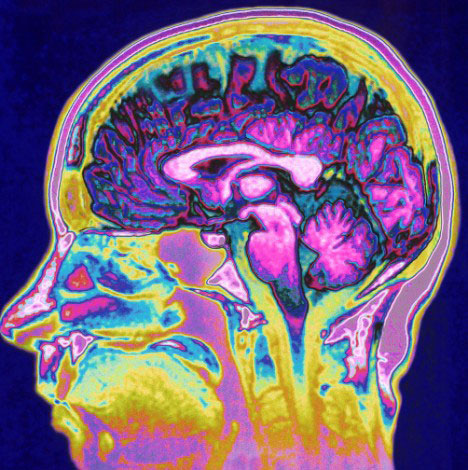
An image of a brain scan. Source: DailyMail

Last week they published their observation in the journal Movement Disorders:

temp-title

Title: Abnormal Brain Temperature in Early-Onset Parkinson’s Disease
Authors: Rango M, Piatti M, Di Fonzo A, Ardolino G, Airaghi L, Biondetti P, & Bresolin N.
Journal: Movement Disorders, 2016 Mar;31(3):425-6.
PMID: 26873586

The researchers were conducting brain scans of 5 people with early onset Parkinson’s disease (3 men and 2 women, with an average age of 41±6 years) and 10 control/normal subjects (6 men and 4 women with an average age of 43±7 years). The study was a follow on from a smaller previous study conducted by the same researchers. There was absolutely no difference in the average body temperature of all the subjects (36.7±0.48°C) and healthy subjects (36.5±0.84 °C).

But when the researchers began looking at different brains regions, they found there were substantial increases in temperature in the early-onset Parkinson’s patients when compared to the control subjects.

The areas of the brain where significant temperature differences were observed included:

  • the hypothalamus (38.50±0.20 vs. 37.01±0.60 °C; PD subjects vs controls)
  • the posterior cingulate gyrus (37.60±0.20 vs. 36.70±0.40 °C)
  • the centrum semiovale (38.00±0.60 vs. 36.60±0.60 °C)
  • the lenticulate nucleus (38.80±0.80 vs. 37.40±0.60 °C)

There was also a slight difference in the visual cortex in the patient group, but this was not statistically significant (37.20±0.20 vs. 36.80±0.40 °C).

Dysfunction in the hypothalamus is known to occur in Parkinson’s disease (click here for more information on this). Changes in the posterior cingulate gyrus (an area involved with emotion) have also been reported (click here for more information on this). But our knowledge of the role of the centrum semiovale and lenticulate nucleus in Parkinson’s disease requires further investigation.

Please remember that all things being equal, there should be no difference whatsoever in brain temperatures. The brain is an extremely sensitive organ and its temperature is rigidly controlled.

So why is there a difference?

Basically the researchers have no idea and, to their credit, they admit as much.

They also point out to the reader that any temperature change in the hypothalamus – an area of the brain that regulates temperature in the body – should result in a corrective response to restore proper temperature in the brain. But apparently in the early-onset Parkinsonian brain it doesn’t. They also note that dopamine-based Parkinson’s treatments (such as levodopa) should decrease overall brain temperature because they increase cerebral blood flow (a natural cooling system for the brain). But again, this doesn’t appear to be happening.

They speculate that maybe these temperature differences are the result of ongoing disease-related activities in the brain, and they offer some well considered ideas as to why this maybe. But there are many other areas of the brain that are affected by Parkinson’s disease – why is there no change in temperature in those regions?

The researchers also ask whether cooling the brain may be considered as a therapeutic option. An interesting idea but this still needs to be tested. And the results of the current study also need to be replicated – independently validated by other groups.

In those replication studies it would be interesting to conduct the same experiment on people with Parkinson’s disease at different stages of the disease to see the effect is consistent or changing over time.

A curious result. Opening up new areas of research. And further evidence that it’s the ‘huh, that’s funny’ results that ultimately lead to the  ‘Eureka!’ moments.

New Research – on how movement is controlled


A couple of very interesting studies were published a week ago that help us to better understand how we move. They are particularly important with respects to Parkinson’s disease.


The parts of the brain involved in movement

Movement is largely controlled by the activity in a specific collection of brain regions, collectively known as the ‘Basal ganglia‘.

B9780702040627000115_f11-01-9780702040627

The location of the basal ganglia structures (blue) in the human brain. Source: iKnowledge

The basal ganglia receives signals from the overlying cortex, processes that information before sending the signal on down the spinal cord to the muscles that are going to perform the movement.

There is also another important participant in the regulation of movement: the thalamus.

Brain_chrischan_thalamus

A brainscan illustrating the location of the thalamus in the human brain. Source: Wikipedia

The thalamus is a structure deep inside the brain that acts like the central control unit of the brain. Everything coming into the brain from the spinal cord, passes through the thalamus. And everything leaving the brain, passes through the thalamus. It is aware of most everything that is going on and it plays an important role in the regulation of movement.

The direct/indirect pathways

The processing of movement in the basal ganglia involves a direct pathway and an indirect pathway. In simple terms, the direct pathway encourages movement, while the indirect pathway does the opposite (inhibits it). The two pathways work together like a carefully choreographed symphony.

The motor features of Parkinson’s disease (slowness of movement and resting tremor) are associated with a breakdown in the processing of those two pathways, which results in a stronger signal coming from the indirect pathway – thus inhibiting/slowing movement.

Pathways

Excitatory signals (green) and inhibitory signals (red) in the basal ganglia, in both a normal brain and one with Parkinson’s disease. Source: Animal Physiology 3rd Edition

Both the direct and indirect pathways finish in the thalamus, but their effects on the thalamus are very different. The direct pathway leaves the thalamus excited and active, while the indirect pathway causes the thalamus to be inhibited.

The thalamus will receive signals from the two pathways and then decide – based on those signals – whether to send an excitatory or inhibitory message to the cortex, telling it what to do (‘get excited and movement’ or ‘don’t get excited and don’t move’, respectively).

Where does dopamine come into the picture?

In Parkinson’s disease, the cells in the brain that produce the chemical dopamine are lost. These cells reside in a structure called the substantia nigra (or SNc in the figure above). What effect does this cell loss have on the direct and indirect pathways? Under normal circumstances the dopamine neurons excite the direct pathway and inhibit the indirect pathway.

In Parkinson’s disease the loss of dopamine neurons results in increased activity in the indirect pathway. As a result, the thalamus is kept inhibited. With the thalamus subdued, the overlying motor cortex has trouble getting excited, and thus the motor system is unable to work properly.

So what was published last week?

Two papers.

Both from the same lab (Well done!)

One in the prestigious scientific journal, Cell and the other in her sister journal, Neuron:

Roseberry-title

Title: Cell-Type-Specific Control of Brainstem Locomotor Circuits by Basal Ganglia.
Authors: Roseberry TK, Lee AM, Lalive AL, Wilbrecht L, Bonci A, Kreitzer AC.
Journal: Cell, 2016 Jan 28;164(3):526-537.
PMID: 26824660

The researchers in this study discovered signal sent from the basal ganglia that selectively activates a group of neurons an area of the brainstem called the ‘mesencephalic locomotor region’. Some of the neurons in this area release a chemical called glutamate. Glutamate is a neurotransmitter that excites the cells it comes into contact with. The researchers who conducted this study found that these glutamate-releasing cells in the mesencephalic locomotor region are responsible for initiating movement.

Print

The researchers used a new technique called ‘optogenetics’ that allows light to activate or inhibit specific cells in the brain. By using this technique on the cells in the direct (dMSN in the figure above) or indirect pathways (iMSN) of the basal ganglia, the researchers were able to control the glutamate-releasing neurons in the mesencephalic locomotor region of mice -initiating or inhibiting their movement, respectively.

The researchers then took the study one step further and used the optogenetics approach directly on the glutamate-releasing neurons in the mesencephalic locomotor region, and they were able to control the initiation of movement in the mice irrespective of the signal being generated by the direct or indirect pathways. That is to say, when the glutamate-releasing neurons in the mesencephalic locomotor region were activated, the mouse would move even when the basal ganglia was sending an inhibitory signal.

So what does it all mean?

While some of the findings of the study were already known, the researchers here have elegantly linked the workings of the basal ganglia and the mesencephalic locomotor region, helping us to better understand the neurological functioning of movement. Deep brain stimulation of the mesencephalic locomotor region has already been attempted and it has demonstrated mixed results in people with Parkinson’s disease (it does appear to help with regards to reducing falls – click here and here for more on this).

It will be interesting to follow the research resulting from this current study.


Parker-title
Title: Pathway-specific remodeling of thalamostriatal synapses in Parkinsonian mice
Authors: Parker PRL, Lalive AL, Kreitzer AC.
Journal: Neuron, 2016
PMID: 26833136

In the second study, the researchers (the same folks who gave us the first paper!) found that the basal ganglia is biased towards the direct pathway. The signal coming from the neurons involved in the direct pathway are stronger than those in the indirect pathway. When dopamine is removed however (as in the case of Parkinson’s disease), the system swings in the opposite direction and becomes biased toward the indirect pathway – the neurons in the direct pathway begin to produce a weaker signal than their counters in the indirect pathway which increase the strength of their signal.

Given that both pathways influence the activity of the thalamus, the researchers next turned their attention to that structure. Again using the ‘optogenics‘ (light-activation) technique, the investigators reduced the inhibitory signal coming from the thalamus and were able to reversibly correct the motor impairs observed in the mice with Parkinson’s-like features.

What does this mean for Parkinson’s disease?
This study turns our attention away from what is happening in the basal ganglia and focuses it on the thalamus, which has not receive the same amount of attention with regards to Parkinson’s disease.

There is a lot already known about changes in the thalamus in Parkinson’s disease (click here for more on this), and deep brain stimulation of structures in neighbouring regions is a regular therapy for Parkinson’s disease (targeting the subthalamic nuclei). But this new paper further breaks down the circuitry of movement for us and offers novel directions for future therapeutic approaches for Parkinson’s disease.

We can be sure that a lot of Parkinson’s disease research is now going to focus on the thalamus.


New research – new targets of Lrrk2

This is Sergey Brin.

sergey_brin

He’s a dude.

Having brought us ‘Google’, he is now turning his attention to other projects.

One of these other projects is close to our hearts: Parkinson’s disease.


In 1996, Sergey’s mother started experiencing numbness in her hands. Initially it was believed to be RSI, but then her left leg started to drag. In 1999, following a series of tests, Sergey’s mother was diagnosed with Parkinson’s disease. It was not the first time the family had been affected by the condition: Sergey’s late aunt had also had Parkinson’s disease.

Both Sergey and his mother have had their genome scanned for mutations that increase the risk of Parkinson’s disease. And both of them discovered that they were carrying a mutation on the 12th chromosome, in a gene called Leucine-rich repeat kinase 2 or Lrrk2.

Not everyone with this particular mutation will go on to develop Parkinson’s disease, but Sergey has decided that his chances are 50:50. Being one of the founders of a large company like Google, however, has left Sergey with resources at his disposal. And he has chosen to focus some of those resources on Lrrk2 research (call it an insurance  policy).

Today, the fruits of some of that research has been published and the results are really interesting:

Elife


Title: Phosphoproteomics reveals that Parkinson’s disease kinase LRRK2 regulates a subset of Rab GTPases
Authors: Martin Steger, Francesca Tonelli, Genta Ito, Paul Davies, Matthias Trost, Melanie Vetter, Stefanie Wachter, Esben Lorentzen, Graham Duddy, Stephen Wilson, Marco AS Baptista, Brian K Fiske, Matthew J Fell, John A Morrow, Alastair D Reith, Dario R Alessi, Matthias Mann
Journal: Elife 2016;10.7554/eLife.12813
PMID: 26824392  (This report is openly available for reading on the Elife website)

So what is Lrrk2?

Also known as dardarin (Basque for ‘trembling‘), Lrrk2 is a gene in our DNA that is responsible for making an enzyme. That Lrrk2 enzyme is involved in many different aspects of cell biology. From cellular remodeling and moving (‘trafficking’) various proteins around in the cell, to protein degradation and stabilization, Lrrk2 has numerous roles.

Discovered in 2004, Lrrk2 was quickly associated with Parkinson’s disease because mutations in this gene are amongst the most common in ‘familial Parkinson’s‘ (where an inherited genetic mutation is present in the sufferer; accounting for about 10-20% of all cases of Parkinson’s disease). The most common mutation of LRRK2 gene is G2019S, which is present in 5–6% of all familial cases of Parkinson’s disease, and is also present in 1–2% of all sporadic cases.

Curiously, mutations in Lrrk2 are also associated with increased risk of Crohn’s disease and cancer.

image1

The structure of Lrrk2 and where various mutations lie. Source: Intech

Given the association with Parkinson’s disease, there have been attempts to develop inhibitors of Lrrk2 as a means of treating the condition. These efforts, however, have been hampered by a poor agreement as to which proteins are interacting with Lrrk2.

The goal of the current study was to identify the key proteins that Lrrk2 acts upon.

What did they discover?

Using various techniques to accomplish their task, the scientists began with 30,000 possible targets and gradually whittled that number down to a small group of Lrrk2 targets.

Most importantly, they found that Lrrk2 is deactivating certain proteins that are called ‘Rabs’. The Rab family are heavily involved with trafficking (and that’s not the mafia drug variety!). Trafficking in cells in moving proteins around within the cell itself. And Lrrk2 was found to deactivate 4 Rab family members (3, 8, 10 and 12).

This is a very important result as not only does it provide us with novel Lrrk2 targets, but it also offers us an excellent tool with which we can determine if Lrrk2 inhibitors are actually working  – a functioning Lrrk2 inhibitor will lower the activity of Rab 3, 8 10 & 12 and this can be measured.

The results represent a major leap forward in our understanding of Lrrk2 and a significant return on investment for one Mr Sergey Brin.


A gut feeling

New Parkinson’s Research this week:

Vagotomy

Title: Vagotomy and Subsequent Risk of Parkinson’s Disease.
Authors: Svensson E, Horváth-Puhó E, Thomsen RW, Djurhuus JC, Pedersen L, Borghammer P, Sørensen HT.

Journal: Annals of Neurology, 2015, May 29. doi: 10.1002/ana.24448.
PMID: 26031848

What’s it all about?

This is Prof Heiko Braak:

heiko-braak-01

Source – Memim.com

Many years ago, Prof Braak – a German neuroanatomist – sat down and examined hundreds of postmortem brains from people with Parkinson’s disease.

He had collected brains from people at different stages of Parkinson’s disease and was looking for any kind of pattern that might explain where and how the disease starts. His research led to what is referred to as the Braak stages of Parkinson’s disease – a six step explanation of how the disease spreads up from the brain stem and into the rest of the brain (see Braak et al, 2003).

nrneurol.2012.80-f1

The Braak stages of PD. Source: Doty RL (2012) Nature Reviews Neurology 8, 329-339.

Braak’s results also led him to propose that Parkinson’s disease may actually begin in the gut and then spread to the central nervous system (the brain). He based this on the observation that many brains that exhibited the very early stages of Parkinson’s disease had disease-related pathology in a population of neurons called the dorsal motor nucleus of the vagal nerve. This population of neurons connects to different organs in the body, such as the lungs, heart, kidneys and the gastrointestinal system (or the gut).

gut_aid_in_PD
A diagram illustrating the vagal nerve connection with the enteric nervous system which lines the gastric system. Source: Braak et al (2006) Nature Reviews Neurology 8, 329-339

Braak and his colleagues went on to examine the nerves fibres around the gastrointestinal system and in those fibres he found large deposits of a Parkinson’s disease-related substance: a protein called alpha synuclein. These deposits were present even at very early stages of the disease, which supported his theory that maybe the disease was starting in the gut.

This ‘gut to brain’ theory was supported by the fact that people with Parkinson’s disease often complain of gastrointestinal problems (eg. constipation) and some of these issues may predate the onset of other Parkinson’s disease symptoms. In addition, a couple of years ago, a group of scientists in the USA found alpha-synuclein present in bowel biopsies taken from people years before they were actually diagnosed with Parkinson’s disease (that study can be found here).

The ‘gut to brain’ theory received a further boost recently with the publication of a paper by a Danish group, who retrospectively looked at all the people in Denmark that received a vagotomy between 1975 and 1995.

So what’s a vagotomy?

Good question.

A vagotomy is a surgical procedure in which the vagus nerve are cut. It is typically due to help treat stomach ulcers. A vagotomy can be ‘truncal‘ (in which the main nerve is cut) or ‘superselective’ (in which specific branches of the nerve are cut, which the main nerve is left in tact).

Vagotomy

A schematic demonstrating the vagal nerve surrounding the stomach. Image A. indicates a ‘truncal’ vagotomy, where the main vagus nerves are cut above the stomach; while image B. illustrates the ‘superselective’ vagotomy, cutting specific branches of the vagus nerve connecting with the stomach. Source: Score

And what did the Danish scientists find?

The Danish researcher found that between 1975 and 1995, 5339 individuals had a truncal vagotomy and 5870 had superselective vagotomy. Using the Danish National registry (which which stores everyone’s medical information), they then looked for how many of these individuals went on to be diagnosed with Parkinson’s disease. They compared these vagotomy subjects with more than 60,000 randomly-selected, age-matched controls.

They found that subjects who had a superselective vagotomy had the same chance of developing Parkinson’s disease as anyone else in the general public (a hazard ratio (or HR) of 1 or very close to 1). But when they looked at the number of people in the truncal vagotomy group who were later diagnosed with Parkinson’s disease, the risk had dropped by 35%. Further, when they followed up the Truncal group 20 years later, checking to see who had been diagnosed with Parkinson’s in 2012, they found that they rate was half that of both the superselective group and the control group (see table below – HR=0.53). The authors concluded that a truncal vagotomy reduces the risk of developing Parkinson’s disease.

Svensson_Table2

Source: Svensson et al (2015) Annals of Neurology – Table 2.

So what does it all mean?

The study is an extremely novel approach to investigating the ‘gut to brain’ theory of Parkinson’s disease and the authors can be congratulated on some excellent work. It adds further weight to the idea that something is happening in the gut very early in Parkinson’s disease. It suggests that by cutting one of the main nerves connecting to the stomach, the disease is slowed down if not avoided all together. It might also suggest that the disease is slower and strikes earlier than previously thought (given that some people with truncal vagotomies still developed Parkinson’s disease – maybe the condition started before the nerve was cut).

But there are a couple of important details that should be considered about the paper before everyone rushes out to get a vagotomy:

  1. The number of people that received a truncal vagotomy (total = 5339) who went on develop Parkinson’s disease 20 years later was just 10 (compared with 29 in the superselective group). There may be some individuals who got lost in the system, but the number is still very low and caution should be used before we get too excited about a result based on a low number of subjects. It is important to determine whether this result can be replicated (in other countries).
  2. The gut may not be the only avenue for the disease. There has also been theories regarding an environmental aspect to the cause of Parkinson’s disease, and there have been studies conducted looking at the nasal/olfactory system of people with Parkinson’s disease to determine if this is another point of entry for the disease (for a recent review on this, see this paper here).

In summary, very interesting study and fascinating result, but please don’t rush to your doctor and ask for your vagus nerve to be cut!

Beginnings

Welcome to the Science of Parkinson’s Disease – a blog that has been set up by scientists to provide information and understanding about the neurodegenerative condition known as Parkinson’s disease.

Over the last 10 years, the advocacy for Parkinson’s disease has been tremendous and real awareness has been created by groups such as the Michael J Fox foundation, the Cure PD Trust, and Parkinson’s UK. They have generated enormous amounts of funding for scientific research and provided hope for disease halting therapies, while supporting and improving the general welfare of people suffering from this condition.

The media regularly announces new breakthroughs in the medical and scientific world, but there are few forums available for the general public to ask questions related to the science being conducted.  The Science of Parkinson’s disease has been set up for this purpose.

The Science of Parkinson’s disease is run by research scientists working in the field, and it was begun with several goals in mind:

  • Try to answer any questions you may have about Parkinson’s disease.
  • Report each week on interesting/exciting research in the world of Parkinson’s disease.
  • Interview Parkinson’s disease researchers, providing a face to the people doing the work.
  • Help you to understand this disease better.

We look forward to hearing from you.

The Team