Older siblings and Parkinson’s disease

Curious new research results out of Sweden this weekend…

To all of our readers who have older siblings that you grew up fighting with – you should  go and give them a hug today, because apparently they have lowered your risk of Parkinson’s disease.

Like I said ‘curious’.


enhanced-873-1401832891-14

Source: Buzzfeed

Older siblings. Nothing but trouble (a bit like younger siblings now that I think about it).

Who needs them.

Well, according to a massive new epidemiological study from the Karolinska Institutet, Stockholm (Sweden), we all do!

Siblings-title

Title: Early-Life Factors and Risk of Parkinson’s Disease: A Register-Based Cohort Study.
Authors: Liu B, Chen H, Fang F, Tillander A, Wirdefeldt K.
Journal: PLoS One. 2016 Apr 15;11(4):e0152841.
PMID: 27082111      (This article is OPEN ACCESS if you would like to read it)

This is a fascinating study, not only in its size and scale, but for the interesting details in the results.

The investigators collected a huge amount of information from multiple nationwide Swedish registers that are cross-linked thanks to the national personal identification number system that is used in Sweden (each Swedish resident is assigned a unique number).

sweden_stockholm

Stockholm, the capital of Sweden. Source: Budgetyourtrip

The information was collected from:

  • The Swedish Multi-Generation Register (MGR) – which holds information about the biological and adoptive parents for all residents born in 1932 or later, and were alive or lived in Sweden in 1961. This database covers over 95% of Swedish-born residents, plus more than 22% of foreign-born residents in Sweden.
  • The Swedish Patient Register – established in 1964/1965, this databases collects inpatient discharge records. It became nationwide in 1987, and since 2001, the Patient Register has recorded information on every inpatient visit and vast majority of the outpatient visits for all Swedish residents. 
  • They also linked their data to the Migration Register and Swedish Population and Housing Censuses from 1960, 1970, 1980, and 1990 for information on socio-economic status.

Like I said, ‘a huge amount of information’. They next set up a selection criteria. Within their pool of people for analysis, individuals had to:

  • be born in Sweden between 1932 and 1970
  • have information available regarding maternal links in the MGR
  • be alive and free of Parkinson’s disease on January 1, 2002,
  • 40 years or older on January 1, 2002 or turned 40 years during the study period

3 545 612 people fulfilled this criteria. 8779 cases of Parkinson’s disease were identified within that population of people (a further 2658 people were identified as having Parkinson’s disease, but since they were diagnosed before 2002, they were excluded). When looking at the findings of the analysis of this study:

Unsurprisingly:

  • the average age of diagnosis was 65.1 years of age
  • males had a higher risk than females (1.5 times more men than women)
  • parental occupation as farmers increased risk of Parkinson’s
  • a family history of the condition results in a higher risk of Parkinson’s disease.
  • No difference between blue or white collar occupations, or self employed roles
  • No difference between month/season of birth
  • No association with early life factors, including flu burden in the year of birth.

Surprisingly:

Compared to those without older siblings, the risk of developing Parkinson’s disease was 7% lower among participants with older siblings (HR = 0.93, 95% CI: 0.89, 0.98). The number of people with no older siblings was 1.68 million, of which 5384 had Parkinson’s disease. But of those with older siblings (1.86 million) only 3395 had Parkinson’s disease. Curiously, however, there was no further associations (eg. the number of older siblings or the interval length between the individual and their older siblings).

The effect (7%) is small, but the number of cases is very large, so we can assume that the finding is real. But how to explain it?!?

Even more surprisingly:

This is not the first time we’ve seen something like this:

Fang-title

Title: Maternal age, exposure to siblings, and risk of amyotrophic lateral sclerosis.
Authors: Fang F, Kamel F, Sandler DP, Sparén P, Ye W.
Journal: Am J Epidemiol. 2008 Jun 1;167(11):1281-6.
PMID: 18367467

In a similar sort of study published in 2008 (also from researchers at the Karolinska Institute in Sweden), it was reported that the risk of amyotrophic lateral sclerosis (ALS, also called Lou Gehrig’s disease; another neurodegenerative condition) increased with the number of younger siblings, and children whose first younger sibling was born after the age of 6 years had the highest risk of ALS. In contrast to the Parkinson’s research above, however, exposure to older siblings was not associated with an increased risk of ALS.

And a similar sort of result has also been observed in cases of Schizophrenia:

Westergaard-title

Title: Exposure to prenatal and childhood infections and the risk of schizophrenia: suggestions from a study of sibship characteristics and influenza prevalence.
Authors: Westergaard T, Mortensen PB, Pedersen CB, Wohlfahrt J, Melbye M.
Journal: Arch Gen Psychiatry. 1999 Nov;56(11):993-8.
PMID: 10565498

This research came from a different Scandinavian capital (Copenhagen), and involved only 1.74 million people, but it suggested that larger sibships were associated with an increased risk of developing schizophrenia. This result was independent of birth order or interval length between siblings. 

Why these effects exist is a question yet to be answered. In each of these studies, the authors propose elaborate possibilities (eg. developmental theories involving the immune system, etc), but there is no evidence (yet) to support them. Given that the effects are small (just a 7% reduction in risk in the case of Parkinson’s), it would be interesting to investigate differences between subjects within the Parkinson’s population, to determine if there is a subset of individuals more affected than others by this sibling phenomenon. By comparing which commonalities they may share (genetic, environmental or otherwise) we could identify patterns of risk factors for specific individuals.

So while the Parkinson’s connection is an interesting finding, obviously more research is required to better understand what is going on.

Curious result though, right?


Chromosome 22 and Parkinson’s disease

A wise man once told me:

“When trying to understand genetics, think of DNA as the stream of words in a book. The nucleotides (A, G, T and C) are the individual letters. These ‘letters’ collect together to make up the genes (the sentences) which give the  book meaning and convey information. And the chromosomes are the chapters in that book.

Some of these ‘books’ are short reads – the fly has only 139.5 million nucleotides (‘words’) and 15,682 genes (sentences) spread across just 4 chromosomes (‘chapters’), while others are long books – humans = 3 billion words, divided into 22,000 sentences, and 23 chapters.

They were helpful words – putting things in perspective – and I hope that they might aid you dear reader as we tackle the topic of this post – a genetic mutation in a particular location of chromosome 22 and its relationship with Parkinson’s disease.

Oh, and do not be fooled into thinking that size matters when it comes to chromosomes. The mighty hedgehog and faultless pigeon have almost twice as many chromosomes as we do (45 and 40 pairs, respectively), and yet…


As most of you will be aware, human beings have 23 pairs of chromosomes.

Chromosomes are a concept that many people are aware of (a pub quiz type of topic), but what are they?

What exactly is a chromosome?

In a nutshell, a chromosome is a very efficient way of packing a lot of DNA into a cell.

Within most of the cells in your body, DNA is densely coiled into discrete packages called chromosomes. Without such packaging, the stringy DNA molecules would be too long to fit inside the cell. In fact, if you uncoiled all of the DNA molecules in a single human cell and placed them end-to-end, they would stretch for at least 6 feet. And that’s just for one cell – remember that the humans have approx. 40 trillion cells in their body!

CDR761781 A schematic demonstrating the arrangement of DNA- Genes-Chromosomes. Source: cancergenome.nih.gov

When a cell is not dividing, the chromosomes usually sit in the nucleus of the cell in loose strands called chromatin. When the cell decides to divide, the chromatin condenses and wrap up very tightly, becoming chromosomes. Both loose chromatin and tightly wound chromosomes are very difficult to see, even with a microscope.

Chromosomes come in pairs – one set of 23 chromosomes from each parent, giving us a total of 46 chromosomes per cell. All of these pairs reside inside the nucleus of each cell, where their DNA is read and instructions (RNA) are sent off to be made into proteins which performs functions within the cell.

Within the DNA in the chromosomes there are sometimes mistakes (think of them as spelling mistakes in the book example we mentioned above). The mistakes are called ‘mutations’ or variants. They can involve sections of DNA being absent or sections of DNA being replicated multiple times.

This week new research was published dealing with Parkinson’s disease and a mutation in chromosome 22.

What do we know about Chromosome 22?

Chromosome 22 is the second smallest human chromosome, being only slightly larger than chromosome 21. Chromosome 22 spans approximately 50 million DNA base pairs and represents 1.5-2% of the total DNA in each cell.

Human_male_karyotpe_high_resolution_-_Chromosome_22

The 23 chromosomes of humans (this set is from a male). Chromosome 22 is highlighted. Source: Wikipedia

There are approx. 1000 genes on chromosome 22. The functions of many of these genes (what they tell the cell/body to do) is still being determined. Mutations in some of those genes, however, are associated with certain diseases. One particular disease associated with Chromosome 22 is called chromosome 22q11.2 deletion syndrome.

What is 22q11.2 deletion syndrome?
Chromosome 22q11.2 deletion syndrome (also known as DiGeorge syndrome) is a condition that arises from a section of chromosome 22 being absent. The ’22q11.2′ code part of the name relates to the specific location on chromosome 22 where the missing sections become apparent. About 87% of deletions occur within a 3 million base pair (nucleotides) region in the middle of one copy of chromosome 22 in each cell (remember that chromosomes come in pairs). The region contains at least 52 known genes.

Given the number of possible gene affected, there are numerous clinical features associated with 22q11.2 deletion syndrome, including heart defects, an opening in the roof of the mouth (a cleft palate), subtle facial features, learning issues, and low calcium levels.

Small ‘micro deletions’ within chromosome 22 are some of the most frequent known deletions found in human beings, occurring in about 25 in 100 000 people. These micro deletions are inherited from an affected parent in 5–10% of cases, while the rest occur spontaneously.


So what does Chromosome 22 have to do with Parkinson’s disease?

In 2009, this research report was published:

Zaleski-title

Title: The co-occurrence of early onset Parkinson disease and 22q11.2 deletion syndrome.
Authors: Zaleski C, Bassett AS, Tam K, Shugar AL, Chow EW, McPherson E.
Journal: Am J Med Genet A. 2009 Mar;149A(3):525-8.
PMID: 19208384

In this report the researchers described two patients, both with chromosome 22q11.2 deletion syndrome and early onset Parkinson’s disease (diagnosed before 45 years of age). The researchers suggested that this co-occurrence of chromosome 22q11.2 deletion syndrome and Parkinson’s disease in two unrelated patients was unlikely to be coincidence (given the low frequency of the conditions).

That first study was followed up by a second study:

Butcher-title

Title: Association between early-onset Parkinson disease and 22q11.2 deletion syndrome: identification of a novel genetic form of Parkinson disease and its clinical implications.
Authors: Butcher NJ, Kiehl TR, Hazrati LN, Chow EW, Rogaeva E, Lang AE, Bassett AS.
Journal: JAMA Neurol. 2013 Nov;70(11):1359-66.
PMID: 24018986

In this report, the scientists conducted an observational study of the occurrence of Parkinson’s disease in the world’s largest cohort of well-characterized adults with a chromosome 22q11.2 deletion syndrome (n = 159; age range = 18.1-68.6 years). They found that people with chromosome 22q11.2 deletion syndrome had a significantly elevated occurrence of Parkinson’s disease compared with standard population estimates.

Curiously, they suggested that the common use of antipsychotics in patients with chromosome 22q11.2 deletion syndrome (to manage associated psychiatric symptoms) delayed diagnosis of Parkinson’s disease by up to 10 years. And in postmortem analysis of the brains of people with both conditions, they found the loss of dopamine neurons and the occurrence of Lewy bodies – characteristic features of Parkinson’s disease.

This was proof that people with chromosome 22q11.2 deletion syndrome were more vulnerable to developing Parkinson’s disease. But what about people with Parkinson’s disease? Do they have deletions with chromosome 22q11.2?

This week we got the answer to that question:

Mok-title

Title: Deletions at 22q11.2 in idiopathic Parkinson’s disease: a combined analysis of genome-wide association data.
Authors: Mok KY, Sheerin U, Simón-Sánchez J, Salaka A, Chester L, Escott-Price V, Mantripragada K, Doherty KM, Noyce AJ, Mencacci NE, Lubbe SJ; International Parkinson’s Disease Genomics Consortium (IPDGC), Williams-Gray CH, Barker RA, van Dijk KD, Berendse HW, Heutink P, Corvol JC, Cormier F, Lesage S, Brice A, Brockmann K, Schulte C, Gasser T, Foltynie T, Limousin P, Morrison KE, Clarke CE, Sawcer S, Warner TT, Lees AJ, Morris HR, Nalls MA, Singleton AB, Hardy J, Abramov AY, Plagnol V, Williams NM, Wood NW.
Journal: Lancet Neurol. 2016 Mar 23. [Epub ahead of print]
PMID: 27017469

The researchers analysed the DNA of 9387 people with Parkinson’s disease and 13 863 controls. They identified eight unrelated people with Parkinson’s disease who carried the chromosome 22q11.2 deletions. None of the controls had any of these deletions. Those people with Parkinson’s disease who had chromosome 22q11.2 deletions had earlier ages of onset (average age of diagnosis = 41 years old) than people with Parkinson’s disease who did not have the deletions (average age of diagnosis = 60.3 years). The researchers concluded that chromosome 22q11.2 deletions are associated with early onset Parkinson’s disease.

Some concluding thoughts

While the results of the Lancet Neurology study are very interesting, there are several important aspects to consider.

Firstly, the researchers noted that the estimated prevalence of 22q11.2 deletion syndrome in the general population is 0·024% or 24 in every 100,000 people. More importantly, as the study indicated the frequency of a 22q deletion among people with early-onset Parkinson’s disease is also very low (0·49% or 5 in every 1000 people with early-onset Parkinson’s disease). In fact, the number of people with the 22q11.2 deletion syndrome mutation and Parkinson’s disease is less than 20. So obviously this is a very low frequency event.

It is also interesting to consider that only 3% of patients with 22q11.2 deletion syndrome go on to develop Parkinson’s disease. Also a low frequency event. But studying this small population of people with a very specific genetic circumstance may enlighten us to some of the biological mechanisms causing this low frequency occurrence. And that may further aid us in better understanding other forms of Parkinson’s disease.

And that really is the take home message from this study:  we are gradually building a map of the connections between genetics and Parkinson’s disease, and while genetics will not explain every case of this condition, the knowledge we gain from this process will allow us to better target the disease in the long run.

And now spit!

Did you know that human saliva is 99.5% water?

1369155421_drool

But a recent set of studies have suggested that the remaining 0.5% holds some interesting insights into Parkinson’s disease.


Interesting fact about saliva – while there is a lot of debate as to how much saliva we produce on a daily basis (anywhere between 0.75 to 1.5 litres per day), it is generally accepted that during sleep the amount of saliva produced drops to almost nothing. Why? Big shrug.


Saliva is a solution produced by three main sets of glands in our mouth: the parotid, Sublingual, and Submandibular glands:

h9991831_001

The human salivary glands. Source: WebMD 

The solution produced serves several important functions, namely:

  • beginning the process of digestion by breaking down food particles.
  • protecting teeth from bacterial decay.
  • Moisturising food to aid in the initiation of swallowing.

As we mentioned above, 99.5% of saliva is water. The remaining 0.5% is made up of enzymes and antimicrobial agents. There is also a number of cells in each millilitre of saliva (as many as 8 million human and 500 million bacterial cells per millilitre).

By analysing those human cells, scientists can learn a lot about a person. For example, they can conduct genetic analysis and determine if a person has a particular mutation.

So what has this got to do with Parkinson’s disease?

Well, recently several research groups have been looking at saliva with the hope that biomarkers – chemicals that may allow for early detection or better monitoring of Parkinson’s disease – could be found.

And recently, some of that research has seemingly paid big dividends:

Spit3-title

Title: Prevalence of Submandibular Gland Synucleinopathy in Parkinson’s Disease, Dementia with Lewy Bodies and other Lewy Body Disorders.
Authors: Beach TG, Adler CH, Serrano G, Sue LI, Walker DG, Dugger BN, Shill HA, Driver-Dunckley E, Caviness JN, Intorcia A, Filon J, Scott S, Garcia A, Hoffman B, Belden CM, Davis KJ, Sabbagh MN.
Journal: J Parkinsons Dis. 2016 [Epub ahead of print]
PMID: 26756744

In this study, published in January of this year, the researchers collected small biopsies of the submandibular gland (one of the three primary producers of saliva) from the bodies of people who died with various conditions (including Parkinson’s disease). They analysed the biopsies for alpha synuclein – the chemical in the brain associated with Parkinson’s disease. We have previously written about alpha synuclein, a chemical in the brain that is associated with Parkinson’s disease (for a primer on alpha synuclein, click here). They found that alpha synuclein was present in the saliva gland of 89% of the subjects who died with Parkinson’s disease, but none of the 110 control samples.

This result led the same research groups to attempt a similar study on live subjects and they published the results of that study in February of this year:

Spit2--title

Title: Peripheral Synucleinopathy in Early Parkinson’s Disease: Submandibular Gland Needle Biopsy Findings.
Authors: Adler CH, Dugger BN, Hentz JG, Hinni ML, Lott DG, Driver-Dunckley E, Mehta S, Serrano G, Sue LI, Duffy A, Intorcia A, Filon J, Pullen J, Walker DG, Beach TG.
Journal: Mov Disord. 2016 Feb;31(2):250-6.
PMID: 26799362

The researchers enrolled 25 people with early-stage Parkinson’s disease (less than  5 years since diagnosis) and 10 control subjects. All of these subjects underwent a small biopsy of the submandibular gland. Those biopsies were then analysed for alpha synuclein and the researchers found that 74% of the Parkinsonian biopsies and 22% control biopsies had alpha synuclein present in the submandibular gland.

And remarkably, this report was followed up this last week by a group in Italy, who published some very interesting data:

Spit1-title

Title: Abnormal Salivary Total and Oligomeric Alpha-Synuclein in Parkinson’s Disease.
Authors: Vivacqua G, Latorre A, Suppa A, Nardi M, Pietracupa S, Mancinelli R, Fabbrini G, Colosimo C, Gaudio E, Berardelli A.
Journal: PLoS One. 2016 Mar 24;11(3):e0151156.
PMID: 27011009    (this report is OPEN-ACCESS if you would like to read it)

The researchers collected salivary samples – actual spit – from 60 people with Parkinson’s disease and 40 age/sex matched control subjects. They then measured the saliva for different types of alpha synuclein. In this study, the researchers measured both the total amount of alpha synuclein in the saliva and also special forms of alpha synuclein.

Alpha synuclein initially starts out in the brain in a monomeric form – as a single version of alpha synuclein. This form of alpha synuclein is believed to be safe. A more mature form of alpha synuclein, called oligomeric, is believed to be the seed of the aggregations found in the Parkinsonian brain, Lewy bodies.

Curiously, in this study the researchers found that the total amount of alpha synuclein in the salivary of people with Parkinson’s disease was lower than that of the control subjects. But – and it’s a big ‘but’ – the amount of alpha synuclein oligomers was higher in the people with Parkinson’s disease than normal healthy controls.

The researchers proposed that the decreased concentration of total alpha-synuclein may reflect the formation of lewy bodies in the brain, and that this test might help the early diagnosis of Parkinson’s disease.


Here at the Science of Parkinson’s we are approaching this research cautiously. Previous attempts at measuring saliva in Parkinson’s disease have not had such significant results when comparing people with Parkinson’s disease and controls (click here for more about that study). The need for better biomarkers of Parkinson’s disease provides the reasons for this research, but the variability between the results different groups are getting leaves one wondering about the viability of the approach. It would indeed make for a very easy, non-invasive testing platform for Parkinson’s disease (‘Please spit into this tube for me’), but more research is needed before it can be applied on the large scale.

We’ll keep watching and hoping.

Another connection between skin and Parkinson’s disease

This is very interesting.

We have previously written blog posts dealing with the connection between melanoma and Parkinson’s disease. And now, there is new research providing a new link between another skin condition and Parkinson’s disease.


What is Rosacea?

Rosacea is a chronic skin condition, classically characterized by a redness of the face. This is the result of dilation of blood vessels in the facial skin, and is usually accompanied by pustules and swelling. Rosacea is indiscriminate in which age group it afflicts and there are four subtypes: three specifically affecting the skin and another affecting the eyes (ocular rosacea).

ps_150331_rosacea_800x600

An example of Rosacea.Source: Medscape

Rosacea is diagnosed in women almost three times more than men. It is also more common in people between the ages of 30 and 50, and appears to have a preference for Caucasians of northwestern European descent (hence it’s nickname: the “curse of the Celts”).

What has this skin condition got to do with Parkinson’s disease?

Well, back in 2001 this study was published:

Fischer-title

Title: Skin function and skin disorders in Parkinson’s disease.
Authors: Fischer M, Gemende I, Marsch WC, Fischer PA.
Journal: J Neural Transm. 2001;108(2):205-13.
PMID: 11314773

In this study, the researchers were investigating seborrheic dermatitis (similar to rosacea, this is an inflammation condition that presents itself as flaky, itchy, and red skin) and hyperhidrosis (abnormal increase in sweating) in Parkinson’s disease. They measured these afflictions in  70 people with Parkinson’s disease and 22 matched control subjects. Almost 20% of the people with Parkinson’s disease had seborrheic dermatitis and half of the Parkinson’s population had hyperhidrosis. The researchers also found that half of the Parkinson’s group also had abnormal sebum levels – sebum being a waxy substance produced by the skin (interestingly, we have previously mentioned sebum in a post about a lady who can smell Parkinson’s disease).

This was an interesting result, but it was never really followed up…until this last week, when another study was published:

Egeberg-title

Title: Exploring the Association Between Rosacea and Parkinson Disease: A Danish Nationwide Cohort Study.
Authors: Egeberg A, Hansen PR, Gislason GH, Thyssen JP.
Journal: JAMA Neurol. 2016 Mar 21. [Epub ahead of print]
PMID: 26999031

The size of this new study is amazing: the researchers looked at data from an national database which includes all Danish citizens 18 years or older from January 1, 1997 to December 31, 2011. That is a reference population of 5.4 million individuals!

Of these, 22 387 individuals (43.8% women) received a diagnosis of Parkinson disease between 1997 -2011, and 68 053 individuals (67.2% women) had a history of the skin condition rosacea.

The general population rate of Parkinson disease was 3.5 cases per 10 000 person. But in the population that had a history of rosacea the rate of Parkinson’s disease was 7.6 cases per 10 000 people – almost twice as high as the general population. Interestingly, when they looked at the subtypes of rosacea, the researchers found that there was a more than 2-fold increase in the risk of Parkinson disease in patients who had a history of ocular rosacea.

Even more interesting: treatment with tetracycline – an antibiotic – appears to have reduced the risk of Parkinson’s disease. The researchers also noted that people with severe rosacea have the same risk of developing Parkinson’s disease as do those who have more moderate rosacea.

This is an interesting study, further indicating a connection between the skin and Parkinson’s disease. Whether the relationship indicates anything causal or simply occurring in parallel is yet to be determined. But given similar previous association, we obviously need to take a closer look at skin.

Does the age of onset make a difference?

This is Dr Henri Huchard (1844-1910; a French neurologist and cardiologist):

Henri_Huchard

Source: Wikipedia

In 1875, he described the case was of a child who, at just 3 years of age, presented with all the clinical features of Parkinson’s disease. Since that report, there have been many studies detailing the condition that has come to be known as ‘juvenile Parkinsonism‘. Currently the youngest person diagnosed with Parkinson’s disease is a young lady from Oklahoma. She was diagnosed at 10-years of age, but had shown symptoms since the age of 2.

We are going to explore juvenile Parkinson’s disease in future post, but today’s post will review some new research that looks at the differences in Parkinson’s disease features of people diagnosed at different ages.


For most members of the general public, Parkinson’s disease is considered a condition of the elderly. And this is a fair line of thinking given what they probably observe out there in the big wide world.


A few years ago though, Parkinson’s UK commissioned and published a report of the statistics/demographics of Parkinson’s disease in the UK (Click here for a copy of that report). In that report, they present this table:

Table1

It illustrates the estimated number of people in each age bracket that have Parkinson’s disease. As you can see, the bulk of the people affected are over the age of 60. But this does not mean that Parkinson’s disease is simply a condition of the aged. It is believed that worldwide at least 5% of diagnoses are to people below the age of 50 – this is considered early onset Parkinson’s disease.

There are many people – actor Michael J Fox among them – who have been diagnosed below the age of 40.

leadership-fox-m-img_2

Actor Michael J Fox was diagnosed with Parkinson’s disease at age 30.
Source: MJFox foundation

Given this wide spectrum of age of onset, it is curious that more research has not been conducted comparing the differences in features of the condition between the different age groups. This situation, however was remedied recently:

Age-of-onset-PD

Title: Age at onset and Parkinson disease phenotype.
Authors: Pagano G, Ferrara N, Brooks DJ, Pavese N.
Journal: Neurology. 2016 Feb 10.
PMID: 26865518

The investigators in this study took 422 people who had recently been diagnosed with Parkinson’s disease (none of them were on any anti-Parkinson’s medication, eg. L-dopa). The subjects were divided into 4 groups according to their age at diagnosis:

  1. younger than 50 years (58 subjects)
  2. 50-59 years (117 subjects)
  3. 60-69 years (168 subjects)
  4. older than 70 years (79 subjects)

The researchers then investigated differences in:

  • side of onset (left or right; dominant or non-dominant side of the body)
  • type of symptoms (rigidity or tremor, etc)
  • localization of symptoms occurrence (eg. arms, legs, etc)
  • severity of motor features (rigidity, tremor,…)
  • severity of nonmotor features (memory, attention,…)
  • severity nigrostriatal function (brain imaging of the dopamine system)
  • CSF biomarkers (Chemicals in the cerebrospinal fluid which surrounds the brain)

Curiously in all of the four groups, a quarter of the people had a family history of Parkinson’s disease. Familial history could suggest a genetic connection, and the genetic aspect of Parkinson’s disease has generally been associated with the early onset group. But this does not appear to be the case in this  study – there was no bias towards the younger onset group.

Asymmetry of motor features onset (eg. tremor, etc) was apparent in 97.8% of the total population, with 55% of those subjects having symptoms on their dominant side. It is interesting to note here, however, that the young onset group were the only group in which the non-dominant side was more affected than the dominant. Similarly, almost all of the symmetrical onset individuals (exhibiting no asymmetry in their motor features) were in the oldest group.

In all four groups, the arm was the more likely site of motor features (this was the case in approx. 85% in all groups). When considering other sites of onset, the head was more frequent in the older groups than the younger group, while the leg was more common in the younger group than the older group.

The older the age at onset the more severe the motor (eg. resting tremor, and postural instability scores) and nonmotor features (including autonomic, olfactory, and cognitive functions). This was accompanied by a greater dopaminergic dysfunction on the brain scan, and a reduction of alpha synuclein floating around in the cerebrospinal fluid.

Rigidity was more common in the young-onset group.

There were no differences between the groups in terms of issues associated with activities of daily living, measures of depression and anxiety, impulsive control, or sleep problems.

What does it all mean?

Why these difference exist and what they might tell us about the condition is yet to be determined. The results are interesting when one considers that the subjects had similar disease duration (they had all just been diagnosed; within 6 months of diagnosis). This suggests that the observed differences may be specific to the different age groups. A direct contribution of the aging process, however, has to be considered when assessing the older group.

This kind of analysis is necessary as it is the only way small details about the disease can be determined. We thought this was an interesting study

Disco-needs-ya – the science of dyskinesias

This is Tom Isaacs. He is the charismatic founder of the Cure Parkinson’s trust.

tom isaacs

Tom Isaacs. Source: GrannyButtons

He’s a dude.

The man walked the entire coastline of the UK to raise money/awareness for Parkinson’s disease! Trust me, he’s a dude.

The title of today’s post is a salute to Tom’s efforts to offer a humourous label to what is a very serious and debilitating aspect of Parkinson’s disease.

In this post, we will discuss the science of dyskinesias


For the last 50 years, Levodopa (L-dopa) has been the “gold standard” treatment for Parkinson’s disease. By replacing the lost dopamine, L-dopa allows for the locomotion parts of the brain to become less inhibited and for people with Parkinson’s disease to feel more in control of their movements.

This miraculous treatment, however, comes at a terrible cost.

After long-term use of the drug, abnormal and involuntary movements can begin to appear. These movements are called dyskinesias.

Dykinesias

An example of a person with dyskinesia. Source: JAMA Neurology

What are Dyskinesias?

Dyskinesias (from Greek: dys/dus – difficulty, abnormal; and kinēsis – motion, movement) are simply a category of movement disorders that are characterized by involuntary muscle movements. They are certainly not specific to just Parkinson’s disease.

In Parkinson’s disease, they are associated with long-term use of L-dopa.

An example of dyskinesia can be seen in this video of Tom Isaacs and David Sangster discussing life with Parkinson’s disease (Tom was diagnosed at age 26 years of age and has lived with Parkinson’s for 20 years – he has dyskinesias. David was diagnosed in 2011 at age 29; since diagnosis he foundered www.1in20Parkinsons.org.uk. He’s also a dude!).

How do dyskinesias develop in Parkinson’s disease?

Before beginning a course of L-dopa, the locomotion parts of the brain in people with Parkinson’s disease is pretty inhibited. This results in the slowness and difficulty in initiating movement. They want to move, but they can’t. They are akinetic.

L-dopa tablets provide the brain with the precursor to the chemical dopamine. Dopamine producing cells are lost in Parkinson’s disease, so replacing the missing dopamine is one way to treat the motor features of the condition. Simply giving people pills of dopamine is a non-starter: dopamine is unstable, breaks down too quickly, and (strangely) has a very hard time getting into the brain. L-dopa, on the other hand, is very robust and has no problem getting into the brain.

Once inside the brain, L-dopa is quickly converted – via an enzymatic reaction – into dopamine allowing the locomotion parts of the brain to function close to normal. In understanding this process, it is important to appreciate that when a tablet is taken and L-dopa  enters the brain, there is a sudden rush of dopamine. A spike in it’s supply, and for the next few hours this gradually wears off as the dopamine is used up. This tablet approach to L-dopa treatment gives a wave like shape to the L-dopa levels in the brain over the course of the day (see the figure below).

After prolonged use of L-dopa (7-10 years on average), the majority of people with Parkinson’s disease will experience a shorter response to each dose of L-dopa. They will also find that they have more time during which they will be unable to move (exhibiting akinesia). This is simply the result of the disease progression – L-dopa treats the motor features of the disease but hides the fact that the disease is still progressing.

This shortening of response is often associated with subtle abnormal involuntary movements that appear when the levels of l-DOPA are highest (usually soon after taking a tablet). It is as if there is too much dopamine for the system to handle.

Gradually, the response time (during which normal movement is possible) will grow shorter and to combat this the dose of L-dopa is increased. But with increased levels of L-dopa, there is an increase in the involuntary movements, or dyskinesias.

Dyskinesia

This figure illustrates the course of Parkinson’s disease for some people on L-dopa. The waving line indicates the level of L-dopa in the blood (as a result of taking L-dopa medication). The white space is the area where normal movement is possible, while the grey area illustrates periods of akinesia (inability to move). Without L-dopa, people with Parkinson’s disease would be stuck in this area, and as the L-dopa pill wears off (during the downward part of the waving line) they fall back into the akinesia area, thus requiring another pill. As the disease progresses, the akinetic (grey) area increases, requiring higher levels of L-dopa to be administered in order to escape it. The tan coloured area in the top right corner demonstrates the introduction of dyskinesias.

Are there different types of dyskinesias?

Yes there are. Dyskinesias have been broken down into many different subtypes, but the two main types of dyskinesia are:

Chorea – these are involuntary, irregular, purposeless, and unsustained movements. To an observer, Chorea will look like a very disorganised/uncoordinated attempt at dancing (hence the name, from the Greek word ‘χορεία’ which means ‘dance’). While the overall activity of the body can appear continuous, the individual movements are brief, infrequent and isolated. Chorea can cause problems with maintaining a sustained muscle contraction,  which may result in affected people dropping things or even falling over.

Dystonia – these are sustained muscle contractions. They often occur at rest and can be either focal or generalized. Focal dystonias are involuntary contractions in a single body part, for example the upper facial area. Generalized dystonia, as the name suggests, are contraction affecting multiple body regions at the same time, typically the trunk, one or both legs, and another body part. The intensity of muscular movements in sufferers can fluctuate, and symptoms usually worsen during periods of fatigue or stress.

When were Dyskinesias first discovered?

Ironically but unsurprisingly, L-dopa induced dyskinesias were first reported by the same scientists who first reported the drug’s amazing effects in Parkinson’s disease:

Dyskinesia_title

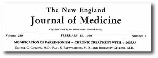
Title: Modification of Parkinsonism – chronic treatment with L-dopa.
Authors: Cotzias GC, Papavasiliou PS, Gellene R.
Journal: New England Journal of Medicine. 1969 Feb 13;280(7):337-45.
PMID: 4178641

George Cotzias was one of the first physicians to give L-dopa to people with Parkinson’s disease.

50396550-1200x800

Dr George Cotzias. Source: NewScientist

Cotzias and colleagues administered L-dopa to 28 people with Parkinson’s disease (17 males and 11 females) and observed modest to moderate response in 8 of them, a marked response in 10, and dramatic responses in the other 10 people. During their two year observation period, they also reported seeing involuntary movements (dyskinesias) in half of the subjects in the study (14/28). They ranged from rare and fleeting (eg. grimacing or gnawing and wave-like motions of the head) to severe (eg. full body/limb movements). They noted that the dyskinesias were most severe in the people with the longest duration of the disease.

It should be noted that the speed with which some of the patients (that Cotzias was treating) developed their dyskinesias – less than 2 years – was a reflection on the late stage of the condition at which the treatment was begun. When the administration of L-dopa is started at an earlier stage, the window of effective treatment is generally longer (5-10 years, depending on individual cases).

So what causes the dyskinesias?

Oh boy.

This question is the source of much debate.

Volumes of text have been bashed out and sides have been taken. We are going to have to tread very carefully here for fear of upsetting folks is the world of Parkinson’s research.

There is some agreement, however, that the factors associated with the development of L-dopa-induced dyskinesias include:

  • the duration of the disease
  • the severity of the disease
  • the dose of L-dopa (cue the debating)
  • young age onset

There are also some genetic forms of Parkinson’s disease that can have an impact on the chances of developing dyskinesias.

Duration/severity of the disease – Experimental studies in animal models of Parkinson’s disease indicate that, if L-dopa is given to the animals, involuntary movements will only develop when the loss of dopamine in the brain exceeds 80–85% of normal. Clinical observations, however, indicate that the severe loss of dopamine in the brain is not sufficient for patients to develop dyskinesias.

This has lead to theories regarding intact part of the brain, suggesting that there are changes in the neurons that the dopamine is acting on. And indeed postmortem analysis of brains from people with & without dyskinesias suggests that there are differences in the neurons that dopamine act on (Click here and here for more on this).

The dose of L-dopa – in a large clinical study, the researchers found that an average daily L-dopa dose of 338 mg was not associated with dyskinesias, while an average daily dose of 387 mg was (Click here and here to read more on this).

Young age onset – Given the length of time that people with early-onset Parkinson’s disease will be on L-dopa, there is a strong association between early-onset and dyskinesias.


EDITORIAL NOTE: We are now about to discuss what can be done to alleviate dyskinesias. Before doing so, we here at the Science of Parkinson’s disease would just like to repeat our standard warning that the contents provided on this website is of an informative nature, and no actions should be taken based on what you have read without first consulting your doctor. Please seek medical advice before changing or experimenting with your treatment regime.


And what can be done to alleviate dyskinesias?

There are several methods of reducing dyskinesias:

Reducing L-dopa dose

Obviously, one can lower the dose of L-dopa. This almost always results in a reduction of dyskinesias. BUT, this almost always results in a worsening of Parkinson’s disease motor features, so it can’t really be considered a solution.

Dopamine receptor agonists

Rather than giving the brain L-dopa or dopamine, chemicals that behave exactly like dopamine can be administered. Dopamine receptor agonists are drugs that act on the receptors of dopamine that are present on the cells that dopamine acts on. These drugs have a longer half‐life than levodopa, meaning that they hang around in the brain for longer (eg. they are not broken down or used up as quickly as L-dopa).

In a large double‐blind study that compared the safety and efficacy of a dopamine receptor agonist – ‘Ropinirole’ – with that of levodopa over a period of five years, researchers found that the incidence of dyskinesia (regardless of levodopa supplementation) was 20% in the ropinirole group and 45% in the levodopa-only group (Click here for more on that study, and click here for a similar study with the dopamine agonist pramipexole).

One cautionary note – Dopamine agonists have been associated with the development of compulsive and impulsive behaviours (Click here for more on this).

Drugs acting on NMDA receptors

N-methyl-D-aspartate receptors (NMDA receptors) are receptors of the chemical glutamate. They are widely found in the brain, but during dyskinesias they appear to become more abundant. As a result, researchers have used drugs that block NMDA receptors (called NMDA receptor antagonists) as potential treatment for dyskinesias. And they appear to help in many cases.

In a double‐blind, placebo‐controlled study of 18 people with Parkinson’s disease, researchers found that the NMDA receptor antagonist ‘Amantidine’ reduced the duration of L-dopa-induced dyskinesias by 60% (Click here for more on this).

Drugs acting on serotonergic systems

Recently there has been a lot of attention focused on the role in dyskinesias of another chemical in the brain: serotonin. There is significant loss of serotonergic cells and fibres in the brain of people with Parkinson’s disease, though not to the same scale as dopamine.

A recent clinical study investigating the use of drugs that prolong the serotonin floating around in the brain (called selective serotonin reuptake inhibitors or SSRIs), found that they did not protect people with Parkinson’s disease from dyskinesias, but may delay their onset (Click here for more on this). There are also clinical trials investigating the use of serotonin receptor agonists in Parkinson’s disease with dyskinesias, based on positive results from preclinical studies (Click here for more on this).

More recently researchers have been investigating the role of serotonin cells in the production of dopamine from L-dopa. Serotonin cells are known to absorb L-dopa and to convert it into dopamine, but they do not have any means of storing it and they release it in an uncontrolled fashion. Recent studies in rodent models of L-dopa-induced dyskinesias have reported reductions in dyskinetic behaviour as a result of lesioning the serotonin cells or blocking specific serotonin receptors. The clinical relevance of these finding is yet to be tested, however.

Neurosurgery

The use of ‘pacemaker’ surgeries (such as deep brain stimulation targeting regions such as the globus pallidum or subthalamic nucleus) have been shown to be effective in treating advanced Parkinson’s disease. The resulting motor improvements are also associated with a reduction in dyskinesias.

A blinded pilot study comparing the safety and efficacy of deep brain stimulation in people with advanced Parkinson’s disease reported a 60-90% reduction in dyskinesias, depending on the region of the brain that was targeted (Click here for more on this).

Surgical lesions targeting the thalamus, globus pallidum or subthalamic nucleus have also been used in the treatment of advanced Parkinson’s disease, with reductions in dyskinesias also being observed. It is effective in both young as well as elderly subjects, with benefit persisting for up to 5 years. These surgical lesion procedures, however, are irreversible.

Recent advances in our understanding

We always like to bring you new research here at the Science of Parkinson’s disease and recently there have been some interesting results published. For example, this one:

Roussakis_title

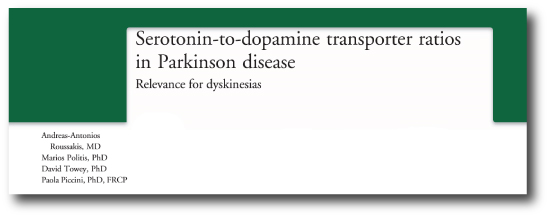
Title: Serotonin-to-dopamine transporter ratios in Parkinson disease: Relevance for dyskinesias.
Authors: Roussakis AA, Politis M, Towey D, Piccini P.
Journal: Neurology. 2016 Published Feb 26.
PMID: 26920358

The researchers in this study conducted brain imaging on people with Parkinson’s disease who did have dyskinesias (17 people) and did not have dyskinesias (11 people). Specifically they were looking to see the difference in the density of dopamine and serotonin fibres in particular areas of the brain affected by dyskinesias. They found that people with Parkinson’s disease AND dyskinesias had a higher ratio of serotonin fibres to dopamine fibres than people with Parkinson’s disease but no dyskinesias. This result adds further support to the role that serotonin cells are playing in the development of L-dopa-induced dyskinesias.


Phew, long post.

If you have got this far and you are still reading – thanks! We hope it was informative.

In (shorter) future posts, we will be assessing new research dealing the mechanisms of and novel ways to treat dyskinesias. This post was meant to be an introduction that we will refer back to from time to time.

Stay tuned!

A sense of (and the science of) smell

Losing the sense of smell is a common feature associated with Parkinson’s disease. But this feature of the condition may help us to better understand the condition. Some autopsy studies have suggested that the olfactory system is one of the first structures in the brain to be affected by the disease.

bad-smell-001

Source: Guardian

How do we smell?

Bad.

That’s both a pathetic attempt at humour and a serious answer. Compared with fellow members of the mammalian family, human beings have a pretty poor sense of smell.

The process of smelling stuff is conducted through structures called the olfactory bulbs. The human olfactory bulbs lie on the base of our brains, protruding forward towards our nose (and nasal cavity).

olfactory_bulb_(inferior_view)1321478082612

A view of the human brain from below (olfactory bulbs are in yellow). Source: StudyBlue

olfactorybulbq1318387403476

A view of the human brain from in front (olfactory bulbs are in yellow). Source: StudyBlue

Inside your nose there is an area of smell sensitive cells that lies on the roof of the nasal cavity (about 7 cm behind your nostrils). That area is called the olfactory epithelium, and it plays a critical role in our sense of smell.

The size of the human olfactory epithelium is rather small and reflects our poor sense of smell, especially when compared, for example, to a dog  (humans have about 10 cm2 (1.6 sq in) of olfactory epithelium, while some dogs have 170 cm2 (26 sq in)).

odorant_eng

The human olfactory system. Source: Biology junction.com

When you inhale an odor (or odorant molecules) through your nose, there are tiny receptors (called olfactory receptors) on the  olfactory epithelium that are the first step in detecting the smell. Every single olfactory receptor cell presents just one (and only one) type type of odorant receptor. When they detect that odor, the olfactory receptor cell reacts by sending an electrical signal along its branch (called an axon) to the olfactory bulbs in the brain.

As the axon of olfactory receptor cell enters the olfactory bulb it forms clusters with other olfactory receptor cell axons, and these clusters are called glomeruli. Inside the glomerulus (singular), the axons make contact the branches of a type of brain cell called a mitral cell. Mitral cells send their axons to many different areas of the brain, including the anterior olfactory nucleus, piriform cortex, the amygdaloid complex, the entorhinal cortex, and the olfactory tubercle.

fulllengths-olfaction-2

Source: YaleScientific

From here our understanding of olfactory processing is less well understood. The piriform cortex is considered the area most likely associated with identifying particular odor. The amygdala is involved in emotional and social functions (eg. mating and recognition), while the entorhinal cortex (and connected hippocampus) is associated with memory – this area is probably activated when a particular smell reminds us of something in our childhood.

What is known about our sense of smell in Parkinson’s disease?

In 1975, two researchers in Minnesota noticed that many of their people with Parkinson’s disease that they were assessing had reduced olfactory abilities. They decided to test this observation:

olfactory-title

Title: Olfactory function in patients with Parkinson’s disease.
Authors: Ansari KA, Johnson A.
Journal: J Chronic Dis. 1975 Oct;28(9):493-7.
PMID: 1176578

The researchers took 22 people with Parkinson’s disease and 37 age/sex-matched controls and repeatedly tested them in a double blind study to determine their olfactory acuity. In each test, the subjects were given five test tubes. Two of the tubes in each set contained 0.5 ml of diluted amyl acetate (which has a distinct smell). The other three tubes contained just water. The subjects were asked to inhale through their nose and then identify which two tubes in each set contained the amyl acetate. The highest dilution (the weakest smelling solution) at which the subject could correctly identify the two amyl acetate containing tubes was designated as their olfactory threshold.

The researchers found that people with Parkinson’s disease had a significantly reduced olfactory acuity (a lower olfactory threshold than compared to control subjects). They also noted that subjects with more progressive forms of the disease exhibited a worse performance on the test. Numerous studies have now replicated this overall result, including a recent study that indicated that smoking may have a protective role on the olfactory ability (Click here and here for more on this).


EDITORIAL NOTE: Please understand that the loss of smell in Parkinson’s disease does not immediately mean that you will have a more progressive form of the condition. There is simply a trend in the data that suggests the loss of smell is a risk factor for having a more progressive version of the condition. 

We would also like to discourage any thoughts of taking up smoking in order to protect your sense of smell.


So what is actually happening in the Parkinson’s disease brain?

This is Prof Heiko Braak:

heiko-braak-01

He’s a dude. We’ve mentioned him before in a previous post.

Many years ago, he and his colleagues were intrigued with the hyposmia (reduction in olfactory ability) in Parkinson’s disease. They conducted a series of autopsy studies, looking at 413 brains! Specifically, they were looking for deposits of the Parkinson’s disease-related protein, alpha synuclein, in the brains and where the protein was accumulating. The accumulation of alpha synuclein is believed to be associated with the loss of cells in the brain.

In total they found 30 brains that exhibited accumulation of alpha synuclein. Of interest, they found that 16 of those brains had accumulation of alpha synuclein in the olfactory bulb. And in one particular case, the olfactory bulb was the only affected part of the brain, except for a tiny region of the brain stem.

The researchers were curious about the possibility that the olfactory system could be a potential starting point for Parkinson’s disease, but they were quick to point out that only half the cases they analysed (16/30) had accumulation of alpha synuclein in the olfactory bulb. Thus, while the olfactory system may be involved, it seems unlikely that the nose is the sole induction site of Parkinson’s disease.

After this study was published, however, Braak and his colleagues went on to analyse the accumulation of alpha synuclein in the lining of the gut and their results suggested this as another possible site of induction (we have written about this in a previous post). They have subsequently proposed a model of disease spread based on entry to the brain via the nose and gut:

nrneurol.2012.80-f1

The Braak stages of Parkinson’s disease. Source: Nature Reviews Neurology.

It is interesting to observe that studies by other scientists have indicated that the nasal epithelium of people with Parkinson’s disease (both with and without the loss of olfactory abilities) is not damaged or presenting an accumulation of alpha synuclein (Click here for more on this).

So what happens to the olfactory bulbs in Parkinson’s disease?

A recent review of the previous studies investigating olfactory bulb volume in people with Parkinson’s disease was published in the Open Access journal PlosOne:

Olfactory_title

Title: Changes in Olfactory Bulb Volume in Parkinson’s Disease: A Systematic Review and Meta-Analysis.
Authors: Li J, Gu CZ, Su JB, Zhu LH, Zhou Y, Huang HY, Liu CF.
Journal: PLoS One. 2016 Feb 22;11(2):e0149286.
PMID: 26900958  (this report is OPEN ACCESS if you would like to read it)

The authors of the study conducted a systematic review (or meta-analysis) of all of the previous studies (six in total) that have measured the size of the olfactory bulb in the brains of people with Parkinson’s disease (using brain imaging techniques). They found that in all of the 6 studies (collectively 216 PD patients and 175 healthy controls) there was a significant reduction in the size of the olfactory bulbs of people with Parkinson’s disease. Strangely, they authors also found the right olfactory bulb was larger than the left in subjects with Parkinson’s disease across all of the studies, and this effect was not found in the healthy controls.

The motor features of Parkinson’s disease usually begin asymmetrically – by this we mean that the left arm is affected before the right, or the right leg has tremor before the left. This is different for each person, as the disease has no particular preference for either side of the body. So why on earth is the right olfactory bulb more affected than the left?

There is your homework question for tonight!

I’ll expect your answers tomorrow.


Diagnosed 2500 years ago? No problem.

Something different for you today – a history lesson…with some science.

The history of Parkinson’s disease dates back well before Dr James Parkinson made his observations about 6 patients 199 years ago (oh, big anniversary coming up! Who knew)

But it may surprise you to know that the history of Parkinson’s disease dates back before even Jesus turned up.

You actually have to go back a long back in order to get to the beginning…


If you were demonstrating the early features of Parkinson’s disease in the year 500 BCE, there was really only one place in the world that you wanted to be:

india-2do290z

India. Source: blogs.umb.edu

Not only did India have a extremely sophisticated system of diagnosis for what we call Parkinson’s disease, but they also have a VERY effective treatment!

Don’t believe me? Read on.

The diagnosis

Around 5000 BCE, the wise and farsighted members of the Indian medical establishment began pooling their collective knowledge – firstly in an oral form, but then eventually in a written format. That written material became the text known as the Ayurveda (/aɪ.ərˈveɪdə/; Sanskrit for “the science of life” or “Life-knowledge”).

It can not be understated how sophisticated the Ayurveda was for its time. This was a period bridging the ‘new stone age’ and the ‘Bronze age’. People’s understanding of medical afflictions was basically limited to what the Gods and evil spirits were doing to them.

The earliest account of Parkinson’s disease features in the Ayurveda  was compiled by Susruta (the 600 BC author of “Susruta Samhita”). He described slowness (cestasanga in Sanskrit) and akinesia (cestahani) in certain individuals, and also (curiously) reported that certain poisons could cause rigidity and tremor.

To demonstrate to you just how sophisticated the Ayurveda was, consider this: when faced with a person exhibiting tremor a practitioner using the Ayurveda could chose between six different types of tremor:

  1. Vepathu (a generalised tremor)
  2. Prevepana (excessive shaking)
  3. Kampa vata (tremors due to vata)
  4. Sirakampa (head tremor)
  5. Spandin (quivering)
  6. Kampana (tremors)

Number 3 (Kampa vata) on that list is what we now refer to as Parkinson’s disease. Kampa basically means ‘tremor’, while Vata is more difficult to define – it is essentially the property/force that governs all movement in the mind and body (blood flow, breathing, etc – even the movement of thoughts).

The treatment

Since the 3rd century BCE, practitioner of the Ayurveda have been using the seeds of Mucuna pruriens in treating conditions of tremor. 

Mucuna-fruit

Mucuna Prurien seeds. Source: Kisalaya

Commonly known as the cowhage plant, Mucuna pruriens are a tropical legume. They are called atmagupta in Sanskrit. Powdered seeds of atmagupta mixed in milk was generally given to treat Kampa vata. And it worked very effectively!

How did it work?

In 1937, a pair of chemist discovered the secret ingredient that allowed Mucuna pruriens seeds to work their magic. 

Mucuna_title
Title: Isolation of l-3:4-dihydroxyphenylalanine from the seeds of Mucuna pruriens.
Authors: Damodaran M, Ramaswamy R.
Journal: Biochem J. 1937 Dec;31(12):2149-52. No abstract available.
PMID: 16746556   (this article is OPEN ACCESS and available to read if you would like)

They found that the seeds contained very high concentrations of a chemical that you and I are familiar with: L-dopa.
Remarkably, Mucuna Pruriens are approximately 4-6% L-dopa, making them mother nature’s natural treatment for Parkinson’s disease. And remember that for over 2000 years, this treatment (atmagupta) has been utilised in the treatment of Kampa vata  in India!

EDITORIAL NOTE: This will probably get me in trouble with the major drug companies, but it would be a worthwhile enterprise for an NGO to set up some Mucuna pruriens plantations in strategically located positions around the world, in order to supply the growing number of people with Parkinson’s disease in the 3rd world. Just a thought. 

How does atmagupta compare with modern L-dopa?

Interesting question.

One that has already been tested:

Katzenschlager_title

Title: Mucuna pruriens in Parkinson’s disease: a double blind clinical and pharmacological study.
Authors: Katzenschlager R, Evans A, Manson A, Patsalos PN, Ratnaraj N, Watt H, Timmermann L, Van der Giessen R, Lees AJ.
Journal: J Neurol Neurosurg Psychiatry. 2004 Dec;75(12):1672-7.
PMID: 15548480   (this report is OPEN ACCESS if you would like to read it)

In this double blind clinical study, the researchers gave 8 people with Parkinson’s disease with a short duration L-dopa response and dyskinesias single doses of 200/50 mg L-dopa or 15-30 g of mucuna preparation. They gave these treatments in a randomised fashion at weekly intervals. They found that mucuna seed powder formulation had a more rapid onset of action and a longer period without dyskinesias. The researchers concluded that ‘this natural source of L-dopa might possess advantages over conventional L-dopa preparations in the long term management of PD’. A grand conclusion, but they also note that a more long term assessment is required.

And that concludes your history lesson for today – hope you liked it!

Cleaning up with Ambroxol

Exciting news recently with the announcement of the Ambroxol study starting.

Exciting for two reasons:

  1. Ambroxol has the potential to make a major impact in the lives of some people with Parkinson’s disease.
  2. It illustrates how FAST things are moving in the world of Parkinson’s disease!


Inside each and every cell, there are millions of tiny actions taking place. Minute processes all working in a collective manner allowing the cell to function normally. There are lots of proteins helping to make other proteins, lots of proteins helping other proteins to get to where they need to be, and lots of proteins helping to break down other proteins after they have done their job.

All this activity generates a lot of waste. And a fundamental part of the activity in any cell is waste disposal. If that does not function properly, the cell is in serious trouble.

One of the most common genetic mutations associated with Parkinson’s disease – called GBA – results in cells having trouble getting rid of waste.

GBA-cartoon

Adapted from a cartoon by Dr Jing Pu. Source: The Nichd connection

What is GBA?

Glucocerebrosidase (or GBA) is an enzyme that helps with the recycling of waste. It is active in inside ‘lysosomes‘.

What are Lysosomes?

Lysosomes are small structures inside cells that act like recycling centers. Waste gets put inside the lysosome where enzymes like GBA help to break it down into useful parts. Mutations in the GBA gene can result in an abnormally short, non-functioning version of the enzyme. And in those cases the breaking down of waste inside the lysosome because inhibited.

What is the connection between GBA and Parkinson’s disease?

GBA mutations are the most common genetic anomaly associated with Parkinson’s disease. People with a mutation in their GBA gene are at higher risk of developing Parkinson’s disease than the general population. And people with Parkinson’s are approximately five times more likely to carry a GBA mutation than healthy control subjects.

So what is Ambroxol?

Ambroxol is a commonly used treatment for respiratory diseases. It promotes mucus clearance and eases coughing. Ambroxol is also anti-inflammatory, reducing redness in a sore throat.

Ok, but why the excitement for Parkinson’s disease?

In May of 2014 – less than 2 years ago – this study was published:

McNeil1

Title: Ambroxol improves lysosomal biochemistry in glucocerebrosidase mutation-linked Parkinson disease cells.
Authors: McNeill A, Magalhaes J, Shen C, Chau KY, Hughes D, Mehta A, Foltynie T, Cooper JM, Abramov AY, Gegg M, Schapira AH.
Journal: Brain. 2014 May;137(Pt 5):1481-95.
PMID: 24574503    (This report is OPEN ACCESS if you want to read it)

It was the first time that Ambroxol – a commercially available drug – had been tested in a Parkinson’s disease related context.

In this study the researchers collected skin cells (called fibroblasts) from eleven people with GBA mutations (some had been diagnosed with Parkinson’s disease). They measured the amount of glucocerebrosidase protein and enzyme activity in these cells, and they found that glucocerebrosidase enzyme activity was significantly reduced in fibroblasts from GBA mutations (on average just the enzyme was acting at just 5% of normal levels). They found that ambroxol increased glucosylceramidase activity in fibroblasts from people with GBA mutations AND in fibroblasts from healthy controls. Ambroxol treatment also reduced markers of oxidative stress in GBA mutant cells.

Given the increase in glucocerebrosidase activity after ambroxol treatment, the researchers wondered whether the drug would reduce alpha-synuclein levels in cells that were over-expressing this protein. Amazingly, after 5 days of ambroxol treatment, levels of alpha-synuclein had decreased significantly (15% on average 15%).

You can understand why the researchers were a little bit excited by these results. Here was a drug that re-activated the recycling unit in the cell and reduced levels of one of the main proteins associated with Parkinson’s disease. If the drug can reduce the levels of alpha synuclein in the brains of people with Parkinson’s disease, maybe the researchers will be able to slow down (or even halt) the disease!

Additional studies have now been reported which have confirmed the initial results.

And now the clinical trial?

Funded by the Cure Parkinson’s Trust and the Van Andel Research Institute (USA), it was announced this week that they had started recruiting subjects to be involved in a clinical trial at the Royal Free Hospital in London. The trial is a phase 1 study which will test the safety of Ambroxol in Parkinson’s disease. The researchers will also look to see if Ambroxol can increase levels of glucocerebrosidase and whether this has any beneficial effects in the subjects. The study will be conducted on 20 people with Parkinson’s disease who also have GBA mutations. They will be given the drug and followed over the next 24 months.

These are exciting times for the world of Parkinson’s disease as these drugs are no longer simply reducing the motor features of the condition, but actually attempting to slow/halt the disease.

And as we suggested at the start of the post the pace of these developments is becoming hard to keep up with.

Diabetes, Monster saliva, and Parkinson’s disease


If I were to tell you that there exists a miraculous elixir derived from the saliva of a monster and it may aid us in the treatment of Parkinson’s disease, would you think me mad?


In 1974, a small study was published in the Journal of Chronic Diseases that presented a rather startling set of results:

Lipman-title

In the study, Lipman and colleagues conducted some routine glucose tolerance tests on a group of 56 people with Parkinson’s Disease (7 additional subjects with Parkinson’s were excluded because they had been previously diagnosed with diabetes).

After being asked to fast overnight, the  subjects were then given 100g of glucose and blood samples were collected from them every hour for 3 hours. When the glucose levels in the blood were measured and compared with the results of 5 previous studies conducted on normal healthy adults of the same age (one of those studies involved 7000 participants), it was found that the people with Parkinson’s disease in the Lipman study had a much higher average level of glucose in their blood than all of the other 5 studies looking at healthy individuals.

Shockingly, almost half (46.4%) of the participants in the Lipman study actually fulfilled the criteria for a diagnosis of diabetes.

More recent survey data has revealed that diabetes is established in between 8–30% of people with Parkinson’s disease (click here for more on this) – obviously this is in excess of the approximately 6% prevalence rate in the general public (Source: DiabetesUK).

What is diabetes?

‘Diabetes mellitus’ is what we commonly refer to as diabetes. It is basically a group of metabolic diseases that share a common feature: high blood sugar (glucose) levels for a prolonged period.

Diabetic patient doing glucose level blood test
Diabetic patient doing glucose level blood test using ultra mini glucometer and small drop of blood from finger and test strips isolated on a white background. Device shows 115 mg/dL which is normal

Source: Gigaom

There are three types of diabetes:

  • Type 1, which involves the pancreas being unable to generate enough insulin. This is usually an early onset condition (during childhood) and is controlled with daily injections of insulin.
  • Type 2, which begins with cells failing to respond to insulin. This is a late/adult onset version of diabetes that is caused by excess weight and lack of exercise.
  • Type 3, occurs during 2-10% of all pregnancies, and is transient except in 5-10% of cases.

What is this stuff called insulin?

Insulin is a hormone – that our body makes – which allows us to use sugar (glucose) from the food that you eat. Glucose is a great source of energy. After eating, our body is releases insulin which then attaches to cells and signals to those cells to absorb the sugar from our bloodstream. Without insulin, our cells have a hard time absorbing glucose. Think of insulin as a “key” which unlocks cells to allow sugar to enter the cell.

Ok, so how is it all connected to Parkinson’s disease?

The short answer is ‘we currently don’t know’.

There have, however, been numerous studies now that suggest an association between diabetes and Parkinson’s disease. The first of these studies was:

Driver_title
Title: Prospective cohort study of type 2 diabetes and the risk of Parkinson’s disease.
Authors: Driver JA, Smith A, Buring JE, Gaziano JM, Kurth T, Logroscino G.
Journal: Diabetes Care. 2008 Oct;31(10):2003-5.
PMID: 18599528

In this study, 21,841 male doctors (participants in the Physicians’ Health Study) were followed over 23 years. The researchers found that people with diabetes had an increased risk of developing Parkinson’s disease risk. Interestingly they reported that the highest Parkinson’s disease risk was seen in individuals with short-duration, older-onset diabetes.

In another study:

Xu_title
Title: Diabetes and risk of Parkinson’s disease.
Authors: Xu Q, Park Y, Huang X, Hollenbeck A, Blair A, Schatzkin A, Chen H.
Journal: Diabetes Care. 2011 Apr;34(4):910-5. doi: 10.2337/dc10-1922. Epub 2011 Mar 4.
PMID: 21378214

This study came from another long term study, which was following 288,662 participants of the National Institutes of Health-AARP Diet and Health Study. The researchers found that the risk of Parkinson’s disease was approximately 40% higher among diabetic patients than among participants without diabetes. In this study, however, the analysis showed that the risk was largely limited to individuals who had diabetes for more than 10 years.

A third study:

Schernhammer_title

Title: Diabetes and the risk of developing Parkinson’s disease in Denmark.
Authors: Schernhammer E, Hansen J, Rugbjerg K, Wermuth L, Ritz B.
Title: Diabetes Care. 2011 May;34(5):1102-8.
PMID: 21411503

Using data from the nationwide Danish Hospital Register hospital records, the researchers found that having diabetes was associated with a 36% increased risk of developing Parkinson’s disease. Interestingly, they reported that the risk was stronger in women and patients with early-onset Parkinson’s disease (eg. diagnosed before the age of 60 years).


EDITORIAL NOTE HERE: It is important to understand that these studies do not suggest that having diabetes will naturally lead to Parkinson’s disease. They are simply pointing out that diabetics have an increased risk of developing the condition. We present this data here for informative purposes and to make people aware.

It is of interest to note that there is also an association between diabetes and Alzheimer’s disease (click here and here for more on this). Thus Parkinson’s disease is not the only neurodegenerative condition associated with diabetes. 


Is the association between Parkinson’s disease and diabetes genetic?

At present, the answer is no.

The connection between diabetes and Parkinson’s disease does not appear to be genetic, as genome wide sequencing studies have found no common mutations or associations between the two conditions (click here for more on this).

So what are we doing with this knowledge?

Dragons

This is the Gila monster (Source: Californiaherps).

Cute huh?

Named after the Gila River Basin of New Mexico and Arizona, where these lizards are found found, the saliva of the Gila monster was found to have some rather amazing properties with regards to the management of type 2 diabetes. This was due largely to a protein extracted from the saliva, called exendin-4. Scientists have made a synthetic version of exendin-4 which they have called Exenatide.

When tested in a three year clinical trial, Exenatide was found to return people with type 2 diabetes to healthy sustained glucose levels and progressive weight loss.

Exenatide is a glucagon-like peptide-1 (GLP-1) agonist. These types of drugs work by mimicking the functions of the natural hormones in your body that help to lower blood sugar levels after meals. They do this by aiding the release of insulin from the pancreas, blocking a hormone that causes the liver to release its stored sugar into the bloodstream, and slowing glucose absorption into the bloodstream.

Great, but what has this got to do with Parkinson’s disease?!?

Exenatide has also been found to have impressive results in both animal models of Parkinson’s disease and in an open-label clinical trial.

The first study to report a positive effect of Exenatide in a Parkinson’s disease model was:

Bertilsson-title

Title: Peptide hormone exendin-4 stimulates subventricular zone neurogenesis in the adult rodent brain and induces recovery in an animal model of Parkinson’s disease.
Authors: Bertilsson G, Patrone C, Zachrisson O, Andersson A, Dannaeus K, Heidrich J, Kortesmaa J, Mercer A, Nielsen E, Rönnholm H, Wikström L.
Journal: J Neurosci Res. 2008 Feb 1;86(2):326-38.
PMID: 17803225

In this study, the scientists found that exendin-4 (aka Exenatide) improved both behavioural motor ability and protected dopamine neurons in a rodent model of Parkinson’s disease (and the drug was given 5 weeks after the animals developed the motor features). How these results were achieved – the biology behind the results – is unclear, but the effect was interesting enough to encourage other groups to also test Exenatide and they found similar results:

Harkavyi-title

Title: Glucagon-like peptide 1 receptor stimulation reverses key deficits in distinct rodent models of Parkinson’s disease.
Authors: Harkavyi A, Abuirmeileh A, Lever R, Kingsbury AE, Biggs CS, Whitton PS.
Journal: J Neuroinflammation. 2008 May 21;5:19. doi: 10.1186/1742-2094-5-19.
PMID: 18492290    (This study is OPEN ACCESS if you would like to read it)

The scientists in this study tested exendin-4 (aka Exenatide) on two different rodent models of Parkinson’s disease and they found similar results to the previous study. The drug was given 1 week after the animals developed the motor features, but still positive effects were observed.

These (and other) initial results led to the initiation of a clinical trial. Given that Exenatide is already approved for use with diabetes, testing the drug in Parkinson’s disease was a relatively straightforward process (funded by the Cure Parkinson’s Trust).

Olmos-title

Title: Exenatide and the treatment of patients with Parkinson’s disease.
Authors: Aviles-Olmos I, Dickson J, Kefalopoulou Z, Djamshidian A, Ell P, Soderlund T, Whitton P, Wyse R, Isaacs T, Lees A, Limousin P, Foltynie T.
Journal: J Clin Invest. 2013 Jun;123(6):2730-6.
PMID: 23728174     (This study is OPEN ACCESS if you would like to read it)

The researchers running the clinical study gave Exenatide to a group of 21 patients with moderate Parkinson’s disease and evaluated their progress over a 14 month period (comparing them to 24 control subjects with Parkinson’s disease). Exenatide was well tolerated by the participants, although there was some weight loss reported amongst many of the subjects (one subject could not complete the study due to weight loss). Importantly, Exenatide-treated patients demonstrated improvements in their clinician assessed PD ratings, while the control patients continued to decline.

Importantly, in a two year follow up study – this was 12 months after the patients stopped receiving  Exenatide – the researchers found that patients previously exposed to Exenatide demonstrated a significant improvement (based on a blind assessment) in their motor features when compared to the control subjects involved in the study.

The results of this initial clinical study are intriguing and exciting, but it is important to remember that the study was open-label: the subjects knew that they were receiving the drug. This means that we can not discount the placebo effect causing some of the beneficial effects reported.

There is, however, a follow up double blind clinical study currently underway – funded by the Michael J Fox Foundation – which will be completed in June of this year (2016).

We look forward to reading the results of that trial.

And Exenatide is not the only diabetes drug being tested

Pioglitazone is another licensed diabetes drug that is now being tested in Parkinson’s disease. It reduces insulin resistance by increasing the sensitivity of cells to insulin. Pioglitazone has been shown to offer protection in animal models of Parkinson’s disease (click here and here for more on this). And the drug is currently being tested in a clinical trial.

We look forward to reading these results as well.

Summing up

As with melanoma and red hair, there appears to be a curious connection between diabetes and Parkinson’s disease. Large longitudinal studies point to people with diabetes as having a higher risk of Parkinson’s disease than non-diabetic individuals. Why this is remains unclear, but some of the drugs used for treating diabetes may provide novel therapeutic options in the treatment of Parkinson’s disease. We will continue to follow this research and report results as they come to hand.

And you didn’t believe me about the monster saliva!