New criteria for Parkinson’s disease

eligibility_criteria16236bd6e

Diagnosing Parkinson’s disease is actually hard work, and mistakes can be made (click here for more on this). A new criteria has been proposed by a group of experts. In today’s post we will have a look at what is included (and excluded) from this new criteria for Parkinson’s disease.


gr2_lrg

Brain imaging of a normal brain (left) and two Parkinsonian brains. Source: the Lancet

In the United Kingdom, the most commonly used criteria for Parkinson’s disease is the UK Brain bank criteria. It is a three step criteria that clinicians can use in their assessments of individuals suspected of having Parkinson’s disease.

a04tab01

The UK Brain bank criteria. Source: Scielo

In the USA, many physicians use the United Parkinson’s Disease Rating Scale (UPDRS) for diagnosing Parkinson’s disease. UPDRS is a rating scale of Parkinson’s features. There is also a growing trend towards the use of a brain imaging technique called a DAT-Scan, which is an FDA-approved approach for differentiating Parkinson’s disease from essential tremor (but it cannot distinguish between PD and parkinsonian subtypes).

DaTscan-Image-Interpretation_v2

DAT-Scan. Source: GEHealthcare

Ok, so why do we need a new criteria from Parkinson’s disease?

There have been major advances made since Dr James Parkinson first described Parkinson’s disease in 1817 (200 year anniversary coming up!!!). All that progress is changing in the way we look at the condition, for example only in the last two decades has our understanding of the genetics underlying Parkinson’s really started to blossom.

Scientific advances have also complicated our view of Parkinson’s disease. To date, a definitive diagnosis of Parkinson’s disease can only be made at the postmortem stage, with an analysis of the brain itself. That examination involves looking for clusters of proteins in the brain, called Lewy bodies. Recently, however, it has been observed that many people with Parkinson’s disease that have a mutation in the Lrrk2 gene do not have Lewy bodies. Why this is? We do not know. It is one of many complicating factors in the diagnosis of Parkinson’s disease.

Given this state of affairs, it was decided that an updated definition/criteria for Parkinson’s disease was required.

Who decides what is Parkinson’s disease?

In 2014, the International Parkinson and Movement Disorder Society (MDS) organised a task force with the goal of providing an updated definition/criteria for Parkinson’s disease.

That group of experts held two ‘brainstorming’ teleconferences and then a physical meeting that all attended. From those meetings a first draft document was produced. Over the next 6 months a revision process was undertaken. The final version of the new criteria was ratified in San Diego (California) in June 2015.

What is the new criteria?

If you would like to read the new criteria in full – you can find it by clicking here.

Below we present a layman summary of the criteria. Central to the new criteria is firstly establishing that an individual has Parkinsonism, and then determining if Parkinson’s disease is the cause of that Parkinsonism.

Now that sounds a bit weird, but it does make sense. Here is how it works: Parkinsonism embodies a set of conditions that are characterized by tremor, bradykinesia, rigidity, and postural instability. Parkinson’s disease is the most common type of parkinsonism. Another form of Parkinsonism is vascular parkinsonism, in which blood vessel issues cause the tremor, bradykinesia, and rigidity features. Approximately 7% of people who are diagnosed with parkinsonism have developed their features after using (or treatment with) particular medications (such as neuroleptic antipsychotics). Thus, it is important to determine that a person’s parkinsonism is caused by Parkinson’s disease itself.

How do you establish Parkinsonism?

Ever since Dr James Parkinson’s first description of Parkinson’s disease, the clinical criteria for the parkinsonism have centred around the motor features. The new criteria continues this tradition, defining of Parkinsonism being based on the three cardinal motor features. These are:

Bradykinesia, which is defined as slowness of movement AND decrement in amplitude or speed as movements are continued (eg. progressive hesitations/halts). Bradykinesia can be evaluated by using finger tapping, hand movements, pronation-supination movements (for example, twisting the forearm so that the palm is facing up and then down), toe tapping, and foot tapping.

5404237

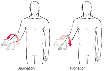
Pronation-supination movements. Source: YogiDoc

Importantly: Although bradykinesia can also occur in the voice, the face, and axial or gait domains, limb bradykinesia must be documented to establish a diagnosis of Parkinsonism.

Rigidity – Rigidity is determined on the “slow passive movement of major joints with the patient in a relaxed position and the examiner manipulating the limbs and neck.”
Rigidity deals with resistance and is referred to as ‘Lead-pipe rigidity’. This occurs when an increase in muscle tone causes a sustained resistance to passive movement (without fluctuations) through an entire range of motion.

Cogwheel rigidity is a combination of lead-pipe rigidity and tremor, presenting as a jerky resistance to passive movement – caused by muscles tensing and relaxing. Cogwheel rigidity is often present in Parkinson’s disease, but without lead-pipe rigidity Cogwheeling does not fulfill minimum requirements for rigidity.

Resting Tremor – this involves the shaking of 4 to 6-Hz in a fully resting limb. Importantly, for diagnosis of Parkinson’s disease, the tremor must be suppressed during movement initiation. The assessment of resting tremor can be made during the entire period of examination. And although postural instability is a feature of Parkinson’s disease, alone it does not qualify for a diagnosis of the condition.


Once it has been determined that the person has parkinsonism, the examiner will then determine whether the patient meets criteria for Parkinson’s disease as the cause of this parkinsonism. This determination is based on three requirements:

  1. Absence of absolute exclusion criteria
  2. At least two supportive criteria
  3. No red flags

1.  Absolute Exclusion Criteria
The exclusion criteria is a list of clinical aspects that indicate alternative possible causes of Parkinsonism. The presence of any of the following features will result in Parkinson’s disease being ruled out as the cause of the Parkinsonism:

– Indications of cerebellar abnormalities, such as cerebellar gait, limb ataxia, or cerebellar oculomotor abnormalities.
– Downward vertical supranuclear gaze palsy (difficulty looking down), or selective slowing of downward vertical eye movements
– Diagnosis of probable behavioral variant frontotemporal dementia (BvFTD) or primary progressive aphasia (a rare neurological syndrome in which language capabilities become slowly and progressively impaired, while other mental functions remain preserved)
– The Parkinsonian motor features restricted to only the lower limbs for more than 3 years
– Treatment with any dopamine receptor blockers or dopamine-depleting agents in doses and a time-course consistent with drug-induced parkinsonism
– The absence of any observable response to a high-dose of levodopa
– Unequivocal cortical sensory loss (eg., graphesthesia or the ability to recognize writing on the skin purely by the sensation of touch), clear limb ideomotor apraxia, or progressive aphasia
– Normal functional neuroimaging of the presynaptic dopaminergic system (this could be the DATScan mentioned above)
– Documentation of an alternative condition known to produce parkinsonism and plausibly connected to the patient’s symptoms

2. Supportive Criteria

The Supportive criteria is a list of clinical findings that support the indication that the Parkinsonism is caused by Parkinson’s disease. These include:

  1. An obvious beneficial response (return to normal or near-normal level of functioning) in response to dopaminergic therapy (L-dopa treatment)
  2. The presence of levodopa-induced dyskinesias
  3. Resting tremor of a limb, documented on clinical examination
  4. Positive results from at least one ancillary diagnostic test. Currently available tests that meet this criterion include:

3. Red Flags
Red flags are indications of an alternative explanation for the Parkinsonism. While the presence of red flags can be counterbalanced by supportive criteria items, if there are more than two red flags, clinically probable PD cannot be diagnosed. The red fags include:

  1. Rapid progression of gait impairment requiring regular use of wheelchair within 5 years of onset of features
  2. A complete absence of progression of motor symptoms or signs over 5 or more years unless stability is related to treatment
  3. Early bulbar dysfunction, defined as one of severe dysphonia, dysarthria (speech unintelligible most of the time), or severe dysphagia (requiring soft food, NG tube, or gastrostomy feeding) within the first 5 years of disease.
  4. Inspiratory respiratory dysfunction defined as either diurnal or nocturnal inspiratory stridor or frequent inspiratory sighs
  5. Severe autonomic failure in the first 5 y of disease. This can include Orthostatic hypotension or severe urinary incontinence or urinary retention in the first 5 years of disease.
  6. Recurrent (>1/y) falls because of impaired balance within 3 years of onset.
  7. The presence of disproportionate anterocollis (dystonic in nature) or contractures of hand or feet within the first 10 years.
  8. Absence of any of the common nonmotor features of disease despite 5 years disease duration. These include sleep dysfunction, constipation, daytime urinary urgency, Hyposmia, Psychiatric dysfunction (depression, anxiety, or hallucinations)
  9. Otherwise unexplained pyramidal tract signs, defined as pyramidal weakness or clear pathologic hyperreflexia (excluding mild reflex asymmetry in the more affected limb, and isolated extensor plantar response).
  10. Bilateral symmetric parkinsonism throughout the disease course. The patient or caregiver reports bilateral symptom onset with no side predominance, and no side predominance is observed on objective examination.

This new criteria for Parkinson’s disease will now be through a period of clinical evaluation and may be adjusted based on that assessment process.

It is interesting to see the condition becoming more defined and specified.


The banner for today’s post was sourced from Help to buy SES

Your appendix and Parkinson’s disease

appendix-and-surrounding-anatomy-enlarged

The appendix was long considered an odd little organ in the body. It was a potentially troublesome, rather redundant appendage to the lower colon of the intestinal tract, and biologists were baffled as to its true function. Recently there were suspicions that it may be playing a role in Parkinson’s disease. This week, however, new research suggests that this may not be the case.


We have previously discussed the idea that Parkinson’s may possibly start in the gut (click here to read more on this). Some in the research community suspect that there is a particular part of the gut where it may start: the Appendix.

What is the Appendix?

The human appendix is a small (averaging 9 cm in length) tube attached to the beginning of the large intestine. Most of us only ever think of the appendix when we are affected by it in the case of Appendicitis.


1300x865x14.jpg.pagespeed.ic.FUdUihmV5G

Source: Healthversed

The Appendix was long considered functionless, an oddity, and by some an mistake or accident of evolution. More recently, however, a new image has started to appear with regards to the appendix. And it has to do with the bacteria of the gut.

We have previously written about Helicobacter pylori and the possible associations with Parkinson’s disease, and in that post we discussed the wide variety of bacteria in the gut. These populations of bacteria are constantly changing, based on our interactions with the world around us (eg. what we are eating, geographically where we are, etc). The developing image of the appendix is that this small organ represents a safe house for bacteria, that is to say: ‘the appendix serves as a haven for useful bacteria when illness flushes those bacteria from the rest of the intestines’ (Wikipedia).

So what would this have to do with Parkinson’s disease?

We have previously discussed the idea that the gut may be one of the starting points for Parkinson’s disease. Many researchers believe that some unknown agent or causal factor is accessing the brain via the nerve fibers surrounding the gut. This theory is supported by reports that sectioning those nerves (to treat ulcers) can reduce your chance of Parkinson’s disease  (click here for more on this).

When looking at the nerve fibres surrounding the intestinal system, one can not help but notice that the appendix is densely innervated. And this is why some researchers suspect that the appendix may be playing a role in Parkinson’s disease.

Gray536

 Blood vessels of the Appendix. Source: Wikipedia

What evidence exists for a connection between the Appendix and Parkinson’s disease?

In 2014, a group of research looked at tissue of the appendix from normal people and they found something interesting.

Appendix1

Title: Alpha-synuclein in the appendiceal mucosa of neurologically intact subjects.
Authors: Gray MT, Munoz DG, Gray DA, Schlossmacher MG, Woulfe JM.
Title: Mov Disord. 2014 Jul;29(8):991-8. doi: 10.1002/mds.25779. Epub 2013 Dec 18.
PMID: 24352892

The researchers looked at biopsies of the appendix from 20 normal people (no history of Parkinson’s disease). In all cases they found high levels of the Parkinson’s disease associated protein, Alpha synuclein (Click here to read more on this), in the nerve fibres surrounding the Appendix. When they looked at other areas of the intestinal system, they found little or no alpha synuclein.

This result got a lot of attention.

A group of researchers then took a  large cohort of people  with Parkinson’s disease and asked which of them had ever had an appendectomy (removal of the Appendix).

Appendix2

Title: Appendectomy may delay Parkinson’s disease Onset
Authors: Mendes A, Gonçalves A, Vila-Chã N, Moreira I, Fernandes J, Damásio J, Teixeira-Pinto A, Taipa R, Lima AB, Cavaco S.
Journal: Mov Disord. 2015 Sep;30(10):1404-7. doi: 10.1002/mds.26311. Epub 2015 Jul 30.
PMID: 26228745

Of the 295 people with Parkinson’s disease involved in the study, 34 were found to have had an appendectomy. There was no significant difference in age of onset across the entire group of people involved in the study, but in people with late onset Parkinson’s (after the age of 55 years) the authors found that found evidence that an appendectomy significantly delayed the onset of Parkinson’s symptoms.

This result led some researchers to conclude that the appendix may have some role in Parkinson’s disease.

What was found in the study this week?

Before you rush out and order yourself an appendectomy, please read the following – This week, any role of the Appendix in Parkinson’s disease has been called into question with the publication of this study:

Appendix

Title: Appendectomy in mid and later life and risk of Parkinson’s disease: A population-based study.
Authors: Marras C, Lang AE, Austin PC, Lau C, Urbach DR.
Journal: Mov Disord. 2016 May 31. doi: 10.1002/mds.26670. [Epub ahead of print]
PMID: 27241338

The researchers involved in this study looked at the medical records of the 14 million residents of Ontario (Canada) who have health care insurance. They found 42,999 had undergone an appendectomy. When the researchers compared people with appendectomies with people without an appendectomy (the control group) and people who had a cholecystectomy (removal of the gallbladder – a surgical control group), they found no difference in the risk of Parkinson’s disease. The researchers concluded that their data did not support an association between mid to late life appendectomy and Parkinson’s disease.

These results are based on large numbers of people and it will be interesting to see how the research community reacts to them. We’ll keep you posted.


UPDATE (23/09/16): A new study came out last week from a group in Denmark that suggests Appendectomies ARE associated with a small increase in risk of developing Parkinson’s disease, but importantly this is only at 10 or more years post surgery.

Title: Appendectomy and risk of Parkinson’s disease: A nationwide cohort study with more than 10 years of follow-up.
Authors: Svensson E, Horváth-Puhó E, Stokholm MG, Sørensen HT, Henderson VW, Borghammer P.
Journal: Mov Disord. 2016 Sep 13.
PMID: 27621223


Today’s banner, illustrating the location of the Appendix was sourced from UCDenver

Muhammad Ali (1942-2016)

wOIYJTo

The world today is mourning the passing of the boxing great, Cassius Clay jr (aka Muhammad Ali). He was many different things to many different people – a boxer, an entertainer, a civil rights activist, an anti-war protestor, a philanthropist, a legend – but he was definitely one of the defining figures of the late 20th century.

During the last third of his life, however, he lived with Parkinson’s disease. You will find a great deal written about Ali and his sporting achievements elsewhere on the web, but today’s post here at SoPD will explore his battle with Parkinson’s.


Many famous figures throughout history have been affected by Parkinson’s disease ( Pope John Paul II, Adolf Hitler, Mao Zedong,…), but very few of them have dealt with their condition in the public eye as much as Muhammad Ali.

Ali was first diagnosed with Parkinson’s disease in 1984.

It was in September of that year – just three years into retirement from boxing – that Ali became concerned about tremors, slowness of movement, slurred speech and unexplained fatigue. He travelled with his entourage to New York, and he was evaluated for a week by Dr Stanley Fahn, M.D., a neurologist at Columbia-Presbyterian Medical Center (New York), before Fahn finally gave Ali his diagnosis.

maxresdefault

Dr Stanley Fahn. Source: Youtube

Given his long boxing career, Dr. Fahn suspected that the head trauma inflicted on Ali could be the cause of his condition. In fact, one of the early complaints from Ali was of numbness in his lips and face, which Dr Fahn assumed meant damage to the brain stem – most likely resulting from the boxing.

Neurodegeneration is a serious issue for boxing. Many retired boxers suffer from what is called Dementia pugilistica – a neurodegenerative condition with Alzheimer’s-like dementia. Some estimates suggest that 15-20% of boxers may be affected, with symptoms usually starting 12-16 years after the start of a career in boxing. Some very famous boxers have been diagnosed with this condition, including world champions Floyd Patterson, Joe Louis, Sugar Ray Robinson and boxer/coach Freddie Roach.

In the case of Ali, however, subsequent follow up assessments over many years highlighted the steady progression of his condition, a disease course more indicative of classic Parkinson’s disease. Dr. Fahn admits, however, that – as with all cases of Parkinson’s disease – “the proof is only going to come at his autopsy”.

image-3-for-muhammad-ali-70-pictures-at-70-gallery-443788110

Ali and a young fan. Source: Pinterest

Being diagnosed at 42 years of age basically placed Ali in the ‘young onset’ group of people with Parkinson’s disease. The average age of diagnosis for Parkinson’s disease is 65 years, but 5-10% of the Parkinson’s community is diagnosed at or below the age of 40. And there are many anecdotal bits of evidence to suggest that Ali was possibly affected by the disease before the age of 40. Ali’s trainer, Angelo Dundee, suspected that Ali’s condition was present during the last few years of his boxing. He remembers Ali gradually slowing down and the newspaper reporters having to lean in to hear what Ali was saying during some of the later interviews. Sports Illustrated senior writer William Nack also noted that “You could see back then that he was just not right”. So although Ali was diagnosed at 42 years of age, the condition may have been affecting him much earlier.

Following the diagnosis, Ali stepped away from the public eye. Parkinson’s affected both of Ali’s most defining characteristics: his moves and his voice. It would have been very understandable for a man as proud as Ali to decide to disappear completely while dealing with his condition. A decade later, however, Ali lit the Olympic caldron at the opening ceremonies of the Atlanta Games (1996), and he was rarely out of the public eye. Attending regular events not only in support of Parkinson’s disease, but also in his role of globetrotting ambassador for peace. Within the Parkinson’s community, Ali lent his name to the ‘Muhammad Ali Parkinson Research Center‘ (Phoenix) and also served as an ambassador for Parkinson’s causes.


In writing this post I have learned a great deal about Ali that I did not know. I have also enjoyed watching and re-watching many of the video interviews of Ali on the internet (Michael Parkinson’s ones are particularly good). Beyond everything the man did and the disease that later came to define him, Ali was an amazing character. It is difficult to think of his equal in the modern world of sports (or beyond).

Truly a sad day.

scKebXi

Inspirational words from the man. Source: Wallpapercave


Today’s banner was sourced from Pinterest. And much of the information for this post was sourced from an article written about Ali by the American Academy of Neurology.