I was recently presenting a talk at a Parkinson’s support group meeting. Afterwards I sat with some of the attendees and we chatted over tea and cookies. At one point the lady sitting beside me tapped me on the arm and said:
“The other day we were discussing some of the commonalities that we [people with Parkinson’s] share. I wonder if they would be of interest to you?”
“Absolutely”‘ I replied, “Let’s hear them”
“Well, firstly, most of us have little or any sense of smell” she said
And I nodded, “this is a common feature amongst people with Parkinson’s disease” (Click here for more on this)
“Ok. Number two, we all have trouble doing ‘number twos'”
I nodded again, and explained that constipation and gastrointestinal problems are also common features of Parkinson’s disease. (Click here for more on this)
“Interesting”, she said, before aiding: “Thirdly, none of us have ever had chickenpox”
I confess I looked at her a long time.
I was speechless.
I had never heard of anything like that.
In science, we are always looking for the presence of possible causal agents – not their absence. I was so intrigued that I took her contact details and told her that I would go away and do some homework on the matter.
I’d like to share my findings here as part of a larger discussion on viruses and Parkinson’s disease.
Given the random and indiscriminate way in which Parkinson’s disease attacks people, scientists have looked for a virus that may be causing the condition.

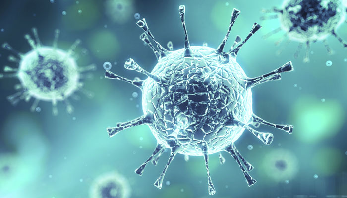
Throughout our lives, our immune system is constantly under attack from viruses. They are small infectious agent that thrive by replicating themselves inside the living cells of other organisms. Technically speaking they are not alive as they lack most of the machinery which characterizes ‘life’ (most importantly the components that is necessary for reproduction). We currently know of approx. 3000 viruses, but we can only guess at the total number of viruses (it may be in the millions!).
There are several different ways that Parkinson’s disease could theoretically be caused in some way by a virus:
1. There may be a specific virus that we are unaware of that infects the body at some point in one’s life causing the slow progressive disease. This could be consider the ‘lightning bolt’ theory – a single unlucky event with terrible consequences. Such a theory has weight as it would explain why some clusters of Parkinson’s disease is sometimes observed. People often use the example of Michael J Fox and his TV work colleagues in this theory.

Actor Michael J Fox and three other people who worked on the Canadian TV show ‘Leo & me’ went on to develop Parkinson’s disease. Image source: Michael J Fox Foundation
2. A virus attacking the body coincides with a secondary event (e.g. a bacterial infection) that may result in the slow progressive events that result in Parkinson’s disease. The secondary event may be a genetic mutation or exposure to an environmental toxin. The virus attack in itself may not be enough in itself.
The two theories outlined above are just theories. We do not know if Parkinson’s disease is caused by a viral infection.
There is, however, some lines of evidence supporting the idea:
Influenza and Parkinson’s disease
Between January 1918 and December 1920 there were two outbreaks of an influenza virus during an event that became known as the 1918 flu pandemic. Approximately 500 million people across the globe were infected, and this resulted in 50 to 100 million deaths (basically 3-5% of the world’s population). Given that is occurred during World War 1, censors limited the media coverage of the pandemic in many countries in order to maintain morale. The Spanish media were not censored, however, and this is why the 1918 pandemic is often referred to as the ‘Spanish flu’.
Influenza is the virus that causes ‘the flu’. Most commonly in a mild form (runny nose, sore throat, coughing, and fatigue), the symptom will arise two days after exposure and last for about a week. There are three types of influenza viruses, called Type A, Type B, and Type C. Type A are the most virulent in humans. The influenza virus behind both of the outbreaks in the 1918 pandemic was a Type A. It was called H1N1.
NOTE: The “H” (hemagglutinin) and the “N” (neuraminidases) are both proteins that are found on the outer surface of the virus. Different viruses have different hemagglutinin and neuraminidase proteins, hence the numbering.
At the same time that H1N1 was causing havoc, a Romanian born neurologist named Constantin von Economo reported a number of unusual symptoms which were referred to as encephalitis lethargica (EL). This disease left many of the victims in a statue-like condition, both motionless and speechless. You may be familiar with the Oliver Sacks book ‘Awakenings’ which was turned into a film starring Robin Williams and Robert De Niro – the patients in that book were victims of EL.

Robin Williams and Robert De Niro in Awakenings
Historically, it was believed that EL was caused by the influenza virus from the 1918 Spanish influenza pandemic. This was largely due to a temporal association and the finding of influenza antigens in some of the suffers of EL. More recent evidence rejects this hypothesis (e.g. an absence of viral RNA recovered from the brains of postencephalitic PD patients – click here for more on this). We genuinely don’t know what caused EL.
But there has recently been some evidence suggesting a link between Parkinson’s disease and influenza:

Title: Highly pathogenic H5N1 influenza virus can enter the central nervous system and induce neuroinflammation and neurodegeneration.
Author: Jang H, Boltz D, Sturm-Ramirez K, Shepherd KR, Jiao Y, Webster R, Smeyne RJ.
Journal: Proc Natl Acad Sci U S A. 2009 Aug 18;106(33):14063-8.
PMID: 19667183
The researchers in this study found that when they injected the highly infectious H5N1 influenza virus into mice, the virus progressed from the periphery into the brain, where it induced Parkinson’s disease-like symptoms. The virus also caused a significant increase in the aggregation of the protein Alpha Synuclein. Importantly, they witnessed the loss of dopamine neurons in the midbrain of the mice 60 days after resolution of the infection.
This study supports the theory we discussed above (theory 1.) of a virus possibly causing Parkinson’s disease. These same researchers have also looked at other influenza viruses and found additional results:

Title: Induction of microglia activation after infection with the non-neurotropic A/CA/04/2009 H1N1 influenza virus.
Author: Sadasivan S, Zanin M, O’Brien K, Schultz-Cherry S, Smeyne RJ.
Journal: PLoS One. 2015 Apr 10;10(4):e0124047.
PMID: 25861024
In this second study, however, the different type of influenza (H1N1) did not infect the brain, but did cause the immune system to flare up. This is an interesting example of the second theory we discussed above (theory 2.), the double hit theory of Parkinson’s disease, in which the virus doesn’t necessarily cause Parkinson’s disease but plays a supportive role to some other toxic agent in attacking the body.
In a follow up study to their 2009 report on H5N1, these same researchers found that the Parkinson’s disease-like symptoms that they observed were actually only temporary:

Title: Inflammatory effects of highly pathogenic H5N1 influenza virus infection in the CNS of mice.
Authors: Jang H, Boltz D, McClaren J, Pani AK, Smeyne M, Korff A, Webster R, Smeyne RJ.
Journal: Journal for Neuroscience, 2012 Feb 1;32(5):1545-59.
PMID: 22302798
This third study may give further support to the double hit theory (theory 2.), but also indicates how complicated a viral component to Parkinson’s disease can be.
And influenza is not the only virus to be associated with Parkinson’s disease.
Hepatitis C and Parkinson’s disease
Hepatitis C is a contagious liver disease, which is caused by the hepatitis C virus (HCV). The virus has been found in the brains of infected people, and it has also been shown to kill dopamine neurons in cell culture. Only in the last few months, however, has a more direct association with Parkinson’s disease been proposed:
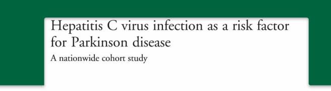
Title: Hepatitis C virus infection as a risk factor for Parkinson disease: A nationwide cohort study.
Authors: Tsai HH, Liou HH, Muo CH, Lee CZ, Yen RF, Kao CH.
Journal: Neurology, 2015 Dec 23. Published early online.
PMID: 26701382
The researchers in this study wanted to investigate whether hepatitis C could be a risk factor for Parkinson’s disease. They did this by analyzing data from 2000-2010 drawn from the Taiwan National Health Insurance Research Database.
The database included 49,967 people with either hepatitis B, hepatitis C or both, in addition to 199,868 people without hepatitis. During the 12 year period, 270 participants who had a history of hepatitis developed Parkinson’s disease (120 still had hepatitis C). This compared with 1,060 participants who were free of hepatitis, but went on to develop Parkinson’s disease.
When the researchers controlled for potentially confounding factors (such as age, sex, etc), the researchers found participants with hepatitis C had a 30% greater risk of developing Parkinson’s disease than the controls.
Summary
It is tempting to consider a viral theory for Parkinson’s disease, especially as the condition seems to strike indiscriminately from out of the blue. Maybe a virus works in cahoots with another factor (unable to do the job alone). The evidence of this, however, has not been apparent to allow for a definitive conclusion.
Finally, regarding my homework…
Never having Chickenpox could mean two different things – never being exposed to it OR being exposed to it and not getting infected (missing a particular protein required for infection). The first would suggest that exposure to the virus would given some kind of resistance to Parkinson’s disease. The latter would suggest that a protein which makes you vulnerable to Chickenpox gives you resistance to Parkinson’s disease.
As to the scientific literature, there have been two studies published regarding Chickenpox and Parkinson’s disease. The first:

Title: Parkinson’s disease: a test of the multifactorial etiologic hypothesis.
Authors: Semchuk KM, Love EJ, Lee RG.
Journal: Neurology, 1993 Jun;43(6):1173-80.
PMID: 8170564
In this study, the researchers collected life-time information (family history, occupational and medical records, etc) from 130 people with Parkinson’s disease. When the looked at all of the variables, they noted that a family history of Parkinson’s had the strongest association with Parkinson’s disease. This was followed by head trauma and occupational herbicide use. The subjects with Parkinson’s disease did not differ from control subjects with regards to:
- exposure to smoking or ionizing radiation
- family history of essential tremor
- work-related contact with aluminum, carbon monoxide, cyanide, manganese, mercury, or mineral oils
- history of arteriosclerosis, chicken pox, encephalitis, hypertension, hypotension, measles, mumps, rubella, or Spanish flu.
They proposed that the results supported the idea of a multifaceted cause of Parkinson’s disease, “probably involving genetic, environmental, trauma, and possibly other factors”.
And the second published study was:
Title: Infections as a risk factor for Parkinson’s disease: a case-control study.
Authors: Vlajinac H, Dzoljic E, Maksimovic J, Marinkovic J, Sipetic S, Kostic V.
Journal: Int J Neurosci. 2013 May;123(5):329-32. doi: 10.3109/00207454.2012.760560. Epub 2013 Feb 4.
PMID: 23270425
In this study the researchers found that Parkinson’s Disease was significantly associated to mumps, scarlet fever, influenza, whooping cough and herpes simplex infections. But they found no association between Parkinson’s disease and Tuberculosis, measles or chickenpox.
So it would appear that chickenpox is not associated with Parkinson’s disease. And at a subsequent Parkinson’s support group meeting I asked the audience for a raise of hands as to who has had chickenpox and there was a sea of hands.
Back to the drawing board I guess.