G.P.N.M.B

# # # #

There is a critical need for good biomarkers in neurodegenerative research.

A biomarker is an objective indicator of a medical state that can be assessed from outside a patient, and can be measured accurately and reproducibly. It could come in the form of a medical imaging application or a biological sample (such as a blood test).

Recent research points towards a particular protein (referred to as GPNMB) that could be a potential biomarker for a specific subtype of Parkinson’s.

In today’s post, we will review some of the research on this topic and consider how a biomarker could potentially be used in Parkinson’s research.

# # # #


Source: TED

Back in 2016, some researchers reported something interesting.

They had been investigating cells collected from people with a condition called Gaucher disease (Pronounced: ‘Go-Shay’; don’t ask – we’ll discuss what it is in a moment, just let me get the intro out of the way). Specifically, the scientists were seeking potential biomarkers for Gaucher disease… and they might have found one.

Here is their report:

Title: Elevation of glycoprotein nonmetastatic melanoma protein B in type 1 Gaucher disease patients and mouse models.
Authors: Kramer G, Wegdam W, Donker-Koopman W, Ottenhoff R, Gaspar P, Verhoek M, Nelson J, Gabriel T, Kallemeijn W, Boot RG, Laman JD, Vissers JP, Cox T, Pavlova E, Moran MT, Aerts JM, van Eijk M.
Journal: FEBS Open Bio. 2016 Jul 30;6(9):902-13.
PMID: 27642553                 (This report is OPEN ACCESS if you would like to read it)

In the study, the researchers collected cells from the spleen of an individual with Gaucher disease and looked for proteins in the cells that were higher than normal.

They found glycoprotein non-metastatic melanoma protein B (GPNMB, also known as osteoactivin) was ridiculously high. Off the charts high. They then compared GPNMB levels in blood samples collected from 59 people with Gaucher disease and 20 healthy controls. As you can see in the graph below, GPNMB levels were on average 25‐fold higher in all of the 59 people with Gaucher disease (Note: the Y axis is logarithmic):

Source: PMC

Interestingly, when the individuals with Gaucher disease started the standard treatment for the condition, the levels of GPNMB collectively dropped:

Source: PMC

And this result has been independently validated (Click here to read that report). The second study used a larger cohort of individuals with Gaucher disease (155 patients) and they found a >15-fold elevation of GPNMB in the blood of this group (compared to controls). And again these high levels were reduced when the Gaucher group started treatment:

Source: PMC

A third study found that GPNMB levels in the brains of a mouse model of Gaucher disease correlated with disease severity in the mice, and also reported elevated GPNMB levels in brain samples from patients with Gaucher disease.

All of the research groups concluded that their data supports the potential utility of GPNMB as a biomarker of Gaucher disease.

Great! But what is Gaucher disease and why is this on a website for Parkinson’s research?

Continue reading “G.P.N.M.B”

The Melanoma drug from MODAG

Melanoma

A build up of a protein called alpha synuclein inside neurons is one of the characteristic feature of the Parkinsonian brain. This protein is believed to be partly responsible for the loss of dopamine neurons in this condition.

A similar build up of alpha synuclein is also seen in the deadly skin cancer, Melanoma… but those cells don’t die (?!?)… in fact, they just keep on dividing.

Why is there this critical difference?

In today’s post we look at an interesting new study that may have solved this mystery.


o-melanoma-facebook

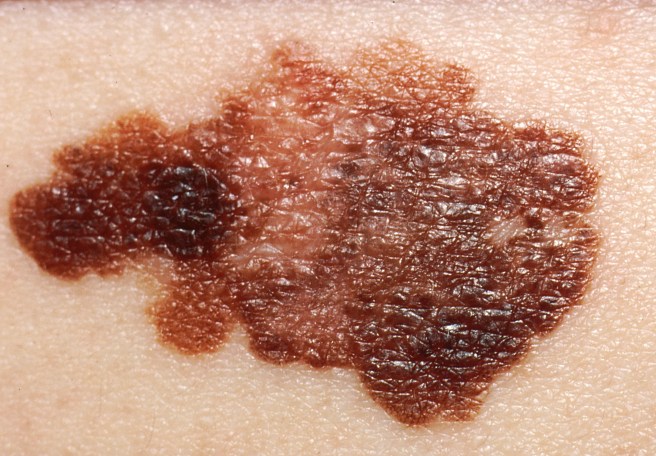
A melanoma. Source: Huffington Post

Parkinson’s disease has a very strange relationship with the skin cancer melanoma.

As we have stated in previous posts (Click here, herehere and here to read those posts) people with Parkinson’s disease are 2-8 times more likely to develop melanoma than people without Parkinson’s (And this finding has been replicated a few times: Olsen et al, 2005; Olsen et al, 2006; Driver et al 2007; Gao et al 2009; Lo et al 2010; Bertoni et al 2010;Schwid et al 2010; Ferreira et al, 2010Inzelberg et al, 2011; Liu et al 2011; Kareus et al 2012; Wirdefeldt et al 2014; Catalá-López et al 2014; Constantinescu et al 2014; Ong et al 2014).

The truly baffling detail in this story, however, is that this relationship is reciprocal – if you have melanoma you are almost 3 times more likely to develop Parkinson’s disease than someone without melanoma (Source: Baade et al 2007; Gao et al 2009).

What is melanoma exactly?

Melanoma is a type of skin cancer.

It develops from the pigment-containing cells known as melanocytes. Melanocytes are melanin-producing cells located in the bottom layer (the stratum basale) of the skin’s outer layer (or epidermis).

Blausen_0632_Melanocyte

The location of melanocytes in the skin. Source: Wikipedia

Melanocytes produce melanin, which is a pigment found in the skin, eyes, and hair. It is also found in the brain in certain types of cells, such as dopamine neurons (where it is referred to as neuromelanin).

subnigmicro

Neuromelanin (brown) in dopamine neurons. Source: Schatz

Melanomas are usually caused by DNA damage resulting from exposure to ultraviolet radiation. Ultraviolet radiation from tanning beds increases the risk of melanoma (Source), as does excessive air travel (Source), or simply spending to much time sun bathing.

Approximately 2.2% of men and women will be diagnosed with melanoma at some point during their lives (Source). In women, melanomas most commonly occur on the legs, while in men they are most common on the back. Melanoma makes up 5% of all cancers (Source).

Generally, melanomas is one of the safer cancers, as it can usually be detected early by visual inspection. This cancer is made dangerous, however, by its ability to metastasise (or spread to other organs in the body).

melanoma-progression

The stages of melanoma. Source: Pathophys

Are there any genetic associations between Parkinson’s disease and melanoma?

No.

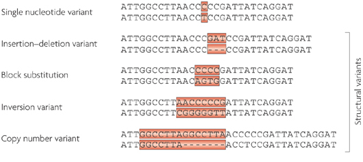
When the common genetics mutations that increase the risk of both conditions were previously analysed, it was apparent that none of the known Parkinson’s mutations make someone more susceptible to melanoma, and likewise none of the melanoma-associated genetic mutations make a person vulnerable to Parkinson’s disease (Meng et al 2012;Dong et al 2014; Elincx-Benizri et al 2014).

In fact, researchers have only found very weak genetic connections between two conditions (Click here to read our previous post on this). It’s a real mystery.

Are there any other connections between Parkinson’s disease and melanoma?

Yes.

Another shared feature of both Parkinson’s disease and melanoma is the build up of a protein called alpha synuclein. Alpha synuclein is believed to be one of the villains in Parkinson’s disease – building up inside a cell, becoming toxic, and eventually killing that cell.

But recently researchers noticed that melanoma also has a build up of alpha synuclein, but those cells don’t die:

Melan2

Title: Parkinson’s disease-related protein, alpha-synuclein, in malignant melanoma
Authors: Matsuo Y, Kamitani T.
Journal: PLoS One. 2010 May 5;5(5):e10481.
PMID: 20463956               (This article is OPEN ACCESS if you would like to read it)

In this study, researchers from Japan found that alpha synuclein was detected in 86% of the primary and 85% of the metastatic melanoma. Understand that the protein is not detectable in the non-melanoma cancer cells.

So what is it doing in melanoma cells?

Recently, researchers from Germany believe that they have found the answer to this question:

Melanoma

Title: Treatment with diphenyl-pyrazole compound anle138b/c reveals that α-synuclein protects melanoma cells from autophagic cell death
Authors: Turriani E, Lázaro DF, Ryazanov S, Leonov A, Giese A, Schön M, Schön MP, Griesinger C, Outeiro TF, Arndt-Jovin DJ, Becker D
Journal: Proc Natl Acad Sci U S A. 2017 Jun 5. pii: 201700200. doi: 10.1073/pnas.1700200114
PMID: 28584093

In their study, the German researchers looked at levels of alpha synuclein in melanoma cells. They took the melanoma cells that produced the most alpha synuclein and treated those cells with a chemical that inhibits the toxic form of alpha synuclein (which results from the accumulation of the protein).

What they observed next was fascinating: the cell morphology (or physically) changed, leading to massive melanoma cell death. The investigators found that this cell death was caused by instability of mitochondria and a major dysfunction in the autophagy process.

Mitochondria, you may recall, are the power house of each cell. They keep the lights on. Without them, the lights go out and the cell dies.

Mitochondria

Mitochondria and their location in the cell. Source: NCBI

Autophagy is the garbage disposal/recycling process within each cell, which is an absolutely essential function. Without autophagy, old proteins and mitochondria will pile up making the cell sick and eventually it dies. Through the process of autophagy, the cell can break down the old protein, clearing the way for fresh new proteins to do their job.

Print

The process of autophagy. Source: Wormbook

Waste material inside a cell is collected in membranes that form sacs (called vesicles). These vesicles then bind to another sac (called a lysosome) which contains enzymes that will breakdown and degrade the waste material. The degraded waste material can then be recycled or disposed of by spitting it out of the cell.

What the German research have found is that the high levels of alpha synuclein keep the mitochondria stable and the autophagy process working at a level that helps to keeps the cancer cell alive.

Next, they replicated this cell culture research in mice with melanoma tumors. When the mice were treated with the chemical that inhibits the toxic form of alpha synuclein, the cancer cancer became malformed and the autophagy process was blocked.

The researchers concluded that “alpha synuclein, which in PD exerts severe toxic functions, promotes and thereby is highly beneficial to the survival of melanoma in its advanced stages”.

So what does all of this mean for Parkinson’s disease?

Well, this is where the story gets really interesting.

You may be pleased to know that the chemical (called Anle138b) which was used to inhibit the toxic form of alpha synuclein in the melanoma cells, also works in models of Parkinson’s disease:

Wagner

Title: Anle138b: a novel oligomer modulator for disease-modifying therapy of neurodegenerative diseases such as prion and Parkinson’s disease.
Authors: Wagner J, Ryazanov S, Leonov A, Levin J, Shi S, Schmidt F, Prix C, Pan-Montojo F, Bertsch U, Mitteregger-Kretzschmar G, Geissen M, Eiden M, Leidel F, Hirschberger T, Deeg AA, Krauth JJ, Zinth W, Tavan P, Pilger J, Zweckstetter M, Frank T, Bähr M, Weishaupt JH, Uhr M, Urlaub H, Teichmann U, Samwer M, Bötzel K, Groschup M, Kretzschmar H, Griesinger C, Giese A.
Journal: Acta Neuropathol. 2013 Jun;125(6):795-813
PMID: 23604588              (This article is OPEN ACCESS if you would like to read it)

In this first study the researchers discovered Anle138b by conducted a large screening study to identify for molecules that could inhibit the toxic form of alpha synuclein.

They next tested Anle138b in both cell culture and rodent models of Parkinson’s disease and found it to be neuroprotective and very good at inhibiting the toxic form of alpha synuclein. And the treatment looks to be very effective. In the image below you can see dark staining of toxic alpha synuclein in the left panel from the brain of an untreated mouse, but very little staining in the right panel from an Anle138b treated mouse.

NL_2014_01_modag


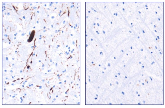
Toxic form of alpha synuclein (dark staining). Source: Max-Planck

Importantly, Anle138b does not interfere with normal behaviour of alpha synuclein in the mice (such as production of the protein, correct functioning, and eventual degradation/disposal of the protein), but it does act as an inhibitor of alpha synuclein clustering or aggregation (the toxic form of the protein). In addition, the investigators found no toxic effects of Anle138b in any of their experiments even after long-term high-dose treatment (more than one year).

And in a follow up study, the drug was effective even if it was given after the disease model had started:

Olig2

Title: The oligomer modulator anle138b inhibits disease progression in a Parkinson mouse model even with treatment started after disease onset
Authors: Levin J, Schmidt F, Boehm C, Prix C, Bötzel K, Ryazanov S, Leonov A, Griesinger C, Giese A.
Journal: Acta Neuropathol. 2014 May;127(5):779-80.
PMID: 24615514                (This article is OPEN ACCESS if you would like to read it)

During the first study, the researchers had started Anle138b treatment in the mouse model of Parkinson’s disease at a very young age. In this study, however, the investigators began treatment only as the symptoms were starting to show, and Anle138b was found to significantly improve the overall survival of the mice.

One particularly interesting aspect of Anle138b function in the brain is that it does not appear to change the level of the autophagy suggesting that the biological effects of treatment with Anle138b is cell-type–specific (Click here to read more about this). In cancer cells, it is having a different effect to that in brain cells. These differences in effect may also relate to disease conditions though, as Anle138b was not neuroprotective in a mouse model of Multiple System Atrophy (MSA; Click here to read more about this).

Is Anle138b being tested in the clinic?

Not yet.

Ludwig-Maximilians-Universität München and the Max Planck Institute for Biophysical Chemistry (Göttingen) have spun off a company called MODAG GmbH that is looking to advance Anle138b to the clinic (Click here for the press release). The Michael J Fox Foundation are helping to fund more preclinical development of this treatment (Click here to read more about this).

We will be watching their progress with interest.

What does it all mean?

Summing up: There are many mysteries surrounding Parkinson’s disease, but some researchers from Germany may have just solved one of them and at the same time developed a potentially useful new treatment.

They have discovered that the Parkinson’s associated protein, alpha synuclein, which is produced in large amounts in the skin cancer melanoma, is actually playing an important role in keeping those cancer cells alive. By finding a molecule that can block the build up of alpha synuclein, they have not only found a treatment for melanoma, but also potentially one for Parkinson’s disease.

And given that both diseases are closely associated, this could be seen as a great step forward. Two birds with one stone as the saying goes.


The banner for today’s post was sourced from Wikipedia

PARK2 and the big C

cancer

Recently it has been announced that the Parkinson’s disease-associated gene PARK2 was found to be mutated in 1/3 of all types of tumours analysed in a particular study.

For people with PARK2 associated Parkinson’s disease this news has come as a disturbing shock and we have been contacted by several frightened readers asking for clarification.

In today’s post, we put the new research finding into context and discuss what it means for the people with PARK2-associated Parkinson’s disease.


4bd6f881-a898-4829-8318-65e1b827d1ac

The As, the Gs, the Ts, and the Cs. Source: Cavitt


The DNA in almost every cell of your body provides the template for making a human being.

All the necessary information is encoded in that amazing molecule. The basic foundations of that blueprint are the ‘nucleotides’ – which include the familiar A, C, T & Gs – that form pairs (called ‘base pairs’) and which then join together in long strings of DNA that we call ‘chromosomes’.

Chemical-structure-of-DNA

The basics of genetics. Source: CompoundChem

If DNA provides the template for making a human being, however, it is the small variations (or ‘mutations’) in our individual DNA that ultimately makes each of us unique. And these variations come in different flavours: some can simply be a single mismatched base pair (also called a point-mutation or single nucleotide variant), while others are more complicated such as repeating copies of multiple base pairs.

nrg2554-f1

Lots of different types of genetic variations. Source: Nature

Most of the genetic variants that define who we are, we have had since conception, passed down to us from our parents. These are called ‘germ line’ mutations. Other mutations, which we pick up during life and are usually specific to a particular tissue or organ in the body (such as the liver or blood), are called ‘somatic’ mutations.

germlinesomatic1

Somatic vs germ line mutations. Source: AutismScienceFoundation

In the case of germ line mutations, there are several sorts. A variant that has to be provided by both the parents for a condition to develop, is called an ‘autosomal recessive‘ variant; while in other cases only one copy of the variant – provided by just one of the parents – is needed for a condition to appear. This is called an ‘autosomal dominant’ condition.

Auto

Autosomal dominant vs recessive. Source: Wikipedia

Many of these tiny genetic changes infer benefits, while other variants can result in changes that are of a more serious nature.

What does genetics have to do with Parkinson’s disease?

Approximately 15% of people with Parkinson disease have a family history of the condition – a grandfather, an aunt or cousin. For a long time researchers have noted this familial trend and suspected that genetics may play a role in the condition.

About 10-20% of Parkinson’s disease cases can be accounted for by genetic variations that infer a higher risk of developing the condition. In people with ‘juvenile-onset’ (diagnosed under the age 20) or ‘early-onset’ Parkinson’s disease (diagnosed under the age 40), genetic variations can account for the majority of cases, while in later onset cases (>40 years of age) the frequency of genetic variations is more variable.

For a very good review of the genetics of Parkinson’s disease – click here.

There are definitely regions of DNA in which genetic variations can increase one’s risk of developing Parkinson’s disease. These regions are referred to as ‘PARK genes’.

What are PARK genes?

We currently know of 23 regions of DNA that contain mutations associated with increased risk of developing Parkinson’s disease. As a result, these areas of the DNA have been given the name of ‘PARK genes’.

The region does not always refer to a particular gene, for example in the case of our old friend alpha synuclein, there are two PARK gene regions within the stretch of DNA that encodes alpha synuclein – that is to say, two PARK genes within the alpha synuclein gene. So please don’t think of each PARK genes as one particular gene.

There can also be multiple genetic variations within a PARK gene that can increase the risk of developing Parkinson’s disease. The increased risk is not always the result of one particular mutation within a PARK gene region (Note: this is important to remember when considering the research report we will review below).

In addition, some of the mutations within a PARK gene can be associated with increased risk of other conditions in addition to Parkinson’s disease.

And this brings us to the research report that today’s post is focused on.

One of the PARK genes (PARK2) has recently been in the news because it was reported that mutations within PARK2 were found in 2/3 of the cancer tumours analysed in the study.

Here is the research report:

MolCell2

Title: PARK2 Depletion Connects Energy and Oxidative Stress to PI3K/Akt Activation via PTEN S-Nitrosylation
Authors: Gupta A, Anjomani-Virmouni S, Koundouros N, Dimitriadi M, Choo-Wing R, Valle A, Zheng Y, Chiu YH, Agnihotri S, Zadeh G, Asara JM, Anastasiou D, Arends MJ, Cantley LC, Poulogiannis G
Journal: Molecular Cell, (2017) 65, 6, 999–1013
PMID: 28306514               (This article is OPEN ACCESS if you would like to read it)

The investigators who conducted this study had previously found that mutations in the PARK2 gene could cause cancer in mice (Click here to read that report). To follow up this research, they decided to screen the DNA from a large number of tumours (more than 20,000 individual samples from at least 28 different types of tumours) for mutations within the PARK2 region.

Remarkably, they found that approximately 30% of the samples had PARK2 mutations!

In the case of lung adenocarcinomas, melanomas, bladder, ovarian, and pancreatic, more than 40% of the samples exhibited genetic variations related to PARK2. And other tumour samples had significantly reduced levels of PARK2 RNA. For example, two-thirds of glioma tumours had significantly reduced levels of PARK2 RNA.

Hang on a second, what is PARK2?

PARK2 is a region of DNA that has been associated with Parkinson’s disease. It lies on chromosome 6. You may recall from high school science class that a chromosomes is a section of our DNA, tightly wound up to make storage in cells a lot easier. Humans have 23 pairs of chromosomes.

Several genes fall within the PARK2 region, but most of them are none-protein-coding genes (meaning that they do not give rise to proteins). The PARK2 region does produce a protein, which is called Parkin.

PARK2Fig1

The location of PARK2. Source: Atlasgeneticsoncology

Particular genetic variants within the PARK2 regions result in an autosomal recessive early-onset form of Parkinson disease (diagnosed before 40 years of age). One recent study suggested that as many as half of the people with early-onset Parkinson’s disease have a PARK2 variation.

Click here for a good review of PARK2-related Parkinson’s disease.

Ok, so if PARK2 was about Parkinson’s disease, what is it doing in cancer?

In Parkinson’s disease, Parkin – the protein of PARK2 – is involved with the removal/recycling of rubbish from the cell. But Parkin has also been found to have other functions. Of particular interest is the ability of Parkin to encourage dividing cells to…well, stop dividing. We do not see this function in neurons, because neurons do not divide. In rapidly dividing cells, however, Parkin can apparently stop the cells from dividing:

divide

Title: Parkin induces G2/M cell cycle arrest in TNF-α-treated HeLa cells
Authors: Lee MH, Cho Y, Jung BC, Kim SH, Kang YW, Pan CH, Rhee KJ, Kim YS.
Journal: Biochem Biophys Res Commun. 2015 Aug 14;464(1):63-9.
PMID: 26036576

This discovery made researchers re-designate PARK2 as a ‘tumour suppressor‘ – a gene that encodes a protein which can block the development of tumours. Now, if there is a genetic variant within a tumour suppressor – such as PARK2 – that blocks it from stopping dividing cells, there is the possibility of the cells continuing to divide and developing into a tumour.

Without a properly functioning Parkin protein, rapidly dividing cells may just keep on dividing, encouraging the growth of a tumour.

Interestingly, the reintroduction of Parkin into cancer cells results in the death of those cells – click here to read more on this.

Oh no, I have a PARK2 mutation! Does this mean I am going to get cancer?

No.

Let us be very clear: It does not mean you are ‘going to get cancer’.

And there are two good reasons why not:

Firstly, location, location, location – everything depends on where in the Parkin gene a mutation actually lies. There are 10 common mutations in the Parkin gene that can give rise to early-onset Parkinson’s disease, but only two of these are associated with an increased risk of cancer (they are R24P and R275W – red+black arrow heads in the image below – click here to read more about this).

ng.491-F2

Comparing PARK2 Cancer and PD associated mutations. Source: Nature

Parkin (PARK2) is one of the largest genes in humans (of the 24,000 protein encoding genes we have, only 18 are larger than Parkin). And while size does not really matter with regards to genetic mutations and cancer (the actual associated functions of a gene are more critical), given the size of Parkin it isn’t really surprising that it has a high number of trouble making mutations. But only two of the 13 cancer causing mutations are related to Parkinson’s.

Thus it is important to beware of exactly where your mutation is on the gene.

Second, in general, people with Parkinson’s disease actually have a 20-30% decreased risk of cancer (after you exclude melanoma, for which there is an significant increased risk and everyone in the community should be on the lookout for). There are approximately 140 genes that can promote or ‘drive’ tumour formation. But a typical tumour requires mutations in two to more of these “driver gene” for a tumour to actually develop. Thus a Parkin cancer-related mutation alone is very unlikely to cause cancer by itself.

So please relax.

The new research published this week is interesting, but it does not automatically mean people with a PARK2 mutation will get cancer.

What does it all mean?

So, summing up: Small variations in our DNA can play an important role in our risk of developing Parkinson’s disease. Some of those Parkinson’s associated variations can also infer risk of developing other diseases, such as cancer.

Recently new research suggested that genetic variations in a Parkinson’s associated genetic region called PARK2 (or Parkin) are found in many forms of cancer. While the results of this research are very interesting, in isolation this information is not useful except in frightening people with PARK2 associated Parkinson’s disease. Cancers are very complex. The location of a mutation within a gene is important and generally more than cancer-related gene needs to be mutated before a tumour will develop.

The media needs to be more careful with how they disseminate this information from new research reports. People who are aware that they have a particular genetic variation will be sensitive to any new information related to that genetic region. They will only naturally take the news badly if it is not put into proper context.

So to the frightened PARK2 readers who contacted us requesting clarification, firstly: keep calm and carry on. Second, ask your physician about where exactly your PARK2 variation is exactly within the gene. If you require more information from that point on, we’ll be happy to help.


The banner for today’s post was sourced from Ilovegrowingmarijuana

The red headed mice of Boston

redhair

Recently scientists have found a possible link in the curious relationship of red hair, melanoma and Parkinson’s disease.

It involves red headed mice (not a typo – you read that correctly).

In today’s post we will discuss the new research and explain what it means for Parkinson’s disease.


ginger

Red or ginger hair. Source: theLocal

We have previously discussed the curious association between red hair and Parkinson’s disease (Click here for that post).

We have also previously discussed the curious association between melanoma and Parkinson’s disease (Click here for that post).

Melanoma

Melanoma. Source: Wikipedia

Basically, people with red hair are more vulnerable to Parkinson’s disease that dark haired people, and people with a history of melanoma (skin cancer) are more likely to develop Parkinson’s disease than people with no history.

And given that people with red hair are generally more vulnerable to melanoma that dark haired people, you can understand why scientists have recently been very interested in this curious triangle of seemingly unrelated biological features.

Recently, however, scientists in Boston (USA) have provided evidence that the genetic mutation which causes red hair and increases the risk of melanoma, might also make the brain more vulnerable to Parkinson’s disease.

Red hair is caused by a genetic mutation?

Before we answer this question: the word ‘mutation’ carries a negative connotation thanks to it’s use in popular media and films. In biology, researchers prefer to use the word genetic ‘variation’. And EVERYONE has variations. They are what makes each of us unique. A father will pass on many of his own genetic variations to his son, but there will also be 50-100 spontaneous variations. And this is how, red hair can sometimes pop up in a family with little history of it.

Ok, so red hair is caused by a genetic variation?

Yes.

Red hair, which occurs naturally in 1–2% of the general population (though there are some regional/geographical variation), results from one of several genetic variations. Approximately 80% of people with red hair have a variation in a gene called melanocortin-1 receptor (or MC1R). Another gene associated with red hair is called HCL2 – ‘Hair colour 2’.

So what did the researchers find?

red

Title: The melanoma-linked “redhead” MC1R influences dopaminergic neuron survival.
Authors: Chen X, Chen H, Cai W, Maguire M, Ya B, Zuo F, Logan R, Li H, Robinson K, Vanderburg CR, Yu Y, Wang Y, Fisher DE, Schwarzschild MA.
Journal: Ann Neurol. 2016 Dec 26. doi: 10.1002/ana.24852. [Epub ahead of print]
PMID: 28019657

In their study, the researchers have investigated mice that carry a mutation of the MC1R gene (thus inactivating the gene – and yes, these mice have red/ginger fur!). They noticed that the mice displayed a progressive decline in their locomotor activity, moving around significantly less than non-red furred control mice at 8 months of age. The MC1R mutant mice also displayed a reduction in the number of dopamine producing neurons in the brain, when compared to the non-red furred controls (dopamine a chemical in the brain that helps to regulate movement).

The MC1R mutant mice were more vulnerable to toxin induced models of Parkinson’s disease (both 6OHDA and MPTP), but (most interestingly) when the researchers used a substance that binds to MC1R and initiates a response (an MC1R agonist called BMS-470539) they found that this treatment improved the survival of the dopamine producing cells in the brain.

The researchers are now seeking to further understand how the loss of MC1R renders the dopamine cells more vulnerable, and follow up the finding that MC1R agonists are neuroprotective.

Has there ever been any other evidence to suggest that MC1R is neuroprotective?

No. To our knowledge this is the first evidence that targeting MC1R could be a novel therapeutic strategy in a brain related condition (there has been some evidence of MC1R activation having beneficial effects in other parts of the body – click here for more on this).

And there are some indications as to how this positive effect could be working:

nurr1-2
Title: Melanocortin-1 receptor signaling markedly induces the expression of the NR4A nuclear receptor subgroup in melanocytic cells.
Authors: Smith AG, Luk N, Newton RA, Roberts DW, Sturm RA, Muscat GE.
Journal: J Biol Chem. 2008 May 2;283(18):12564-70.
PMID: 18292087

In this study, the researchers found that activating MC1R increases the levels of a protein called NR4A2 (or Nurr1). Nurr1 is a protein involved in the development and maintenance of dopamine producing neurons, and numerous recent studies have suggested that it is neuroprotective for these cells as well (Click here to read more on this).

So what does it all mean?

For some time there has been a curious link between people with red hair, melanoma and Parkinson’s disease. Now researchers in Boston have provided new evidence that the link exists, but they have also highlighted a new pathway via which novel therapies for Parkinson’s disease might be researched and developed.  Not a bad day at the office.


The banner for today’s post was sourced from Fancy mice

The mystery deepens – Melanoma and Parkinson’s disease

o-melanoma-facebook

Interesting new data published today regarding the curious connection between Parkinson’s disease and melanoma.

It was interesting because the data suggests that there is no genetic connection. No obvious connection that is.

In this post we will review the study and discuss what it all means.


Melanoma

Melanoma. Source: Wikipedia

Question 1.: why are people with Parkinson’s disease are 2-8 times more likely to develop melanoma – the skin cancer – than people without Parkinson’s?

Question 2.: why are people with melanoma almost 3 times more likely to develop Parkinson’s disease than someone without melanoma?

This topic is an old favourite here at the SoPD, and we have discussed it several times in previous posts (Click here and here to read those posts). It is a really good mystery. A lot of people have looked at this issue and the connection between the two conditions has not been immediately forthcoming.

When the genetics mutations of both conditions were previously analysed, it was apparent that none of the known Parkinson’s mutations make someone more susceptible to melanoma, and likewise none of the melanoma-associated genetic mutations make a person vulnerable to Parkinson’s disease (Meng et al 2012;Dong et al 2014; Elincx-Benizri et al 2014).

So what was published today?

New genetic data! Rather than simplifying things, however, this new data has simply made the mystery….well, more of a mystery. The publication in question is:

skin

Title: Rare variants analysis of cutaneous malignant melanoma genes in Parkinson’s disease.
Authors: Lubbe SJ, Escott-Price V, Brice A, Gasser T, Pittman AM, Bras J, Hardy J, Heutink P, Wood NM, Singleton AB, Grosset DG, Carroll CB, Law MH, Demenais F, Iles MM; Melanoma Meta-Analysis Consortium, Bishop DT, Newton-Bishop J, Williams NM, Morris HR; International Parkinson’s Disease Genomics Consortium.
Journal: Neurobiol Aging. 2016 Jul 28.
PMID: 27640074             (This article is OPEN ACCESS if you would like to read it)

Given that previous studies have suggested that there are no obvious genetic mutations connecting Parkinson’s disease with melanoma, the researchers in this study looked for very rare genetic variations in 29 melanoma-associated genes. They did this analysis on a very large pool of genetic data, from 6875 people with Parkinson’s disease and 6065 normal healthy control subjects.

What the researchers found was only very weak connections between two conditions, based on only a few of these genetic mutations (none of which were statistically significant, which means that they could be due to chance).

One very rare genetic mutation in a gene called TYR p.V275F is very interesting. It was found to be more common in people with Parkinson’s disease than controls in 3 independent groups of data. The gene TYR produces a protein called Tyrosinase, which an important ‘rate-limiting enzyme’ in biological production in both neuromelanin and dopamine (the chemical critically associated with Parkinson’s disease).

So what does this mean?

This data suggests that the connection between Parkinson’s disease and melanoma is not due to a known shared genetic mutation. This conclusion, however, leaves open many alternative possibilities. One such possibility involves the vast pieces of human DNA that are described as ‘non-coding‘. These are sections of DNA that will produce a piece of RNA, but that RNA will not be used to produce a protein (as is normal in biology 101). That non-coding RNA will, however, have functions in regulating the activity on sections of DNA or other RNAs (yeah, I know. It’s complicated. Even for me!). Importantly, these non-coding RNA can play a role in diseases. For example, it was discovered a few years ago that a non-coding RNA called BACE1-AS is produced in very large amounts in patients with Alzheimer’s disease (Click here for more on this). We are simply speculating here though.

The scientists who published the research today suggest that they will further investigate and better characterise the interesting link between the gene TYR and Parkinson’s disease, and they will now broaden their analysis of genetic regions that could be influencing the curious connection between Parkinson’s disease and melanoma. Rather than simply focusing on known genetic mutations (common or rare), they will start to dig deeper into our DNA to see what else may underlie the connection between these two conditions.

Watch this space.


The banner for today’s post was sourced from the Huffington Post

An update on the connection between Melanoma and Parkinson’s disease

We have previously discussed the strange connection between Melanoma and Parkinson’s disease (click here to read that post).

Melanoma

That post included the curious observations that:

  • People with Parkinson’s disease are 2-8 times more likely to develop melanoma than people without Parkinson’s.
  • People with melanoma are almost 3 times more likely to develop Parkinson’s disease than someone without melanoma.

And we have no idea why (there is no shared genetic predisposition for the two conditions).

Research published this week, however, may begin to explain part of the connection:

Melanoma-title

Title: Parkinson disease (PARK) genes are somatically mutated in cutaneous melanoma.
Authors: Inzelberg R, Samuels Y, Azizi E, Qutob N, Inzelberg L, Domany E, Schechtman E, Friedman E.
Journal: Neurol Genet. 2016 Apr 13;2(3):e70.
PMID: 27123489     (This research article is OPEN ACCESS if you would like to read it)

In this study, the scientists looked at somatic mutations in cells from 246 tissue samples of melanoma.

What are somatic mutations?

Somatic mutations are genetic alteration that have been acquired by a cell that can then be passed to the progeny of that mutated cell (via cell division). These somatic mutations are different from ‘germline’ mutations, which are inherited genetic alterations that are present in the sperm and egg that were used in making each of us.

germlinesomatic1

Somatic vs Germline mutations. Source: AutismScienceFoundation

In the 246 samples analysed, the researchers found 315,914 somatic mutations in 18,758 genes. Yes, that is a lot, but what was very interesting was their discovery of somatic mutations in many of the PARK genes.

What are PARK genes?

There are a number (approx. 20) genes that are now recognised as conferring vulnerability to developing Parkinson’s disease. These genes are referred to as PARK genes. They include the gene that makes the protein Alpha synuclein ( SNCA ) and many others with interesting names (like PINK1 and LRRK2). Approximately 15% of cases of Parkinson’s are believed to occur because of a mutation in one (or more) of the  PARK genes. As a result there is a lot of research being conducted on the PARK genes.

Were all of PARK genes mutated in the Melanoma samples?

Somatic mutation in 14 of the 15 PARK genes (that the researchers analysed) were present in the melanoma samples. This means that after the skin cells turned into melanoma cancer cells, they acquired mutations in some of the PARK genes. Overall, 48% of the analysed samples had a mutation in at least 1 PARK gene, and 25% had mutations in multiple PARK genes (2–8 mutated genes). One PARK gene in particular, PARK 8, was more significantly present in the melanoma cells than the others. PARK8 is also known as Leucine-rich repeat kinase 2 or LRRK2 (we have previously discussed Lrrk2 – click here to read that post). Three additional PARK genes (PARK2, PARK18, and PARK20) were also significantly present, but not as significant as Lrrk2.

So what does it all mean?

The researchers speculate in the discussion of their report about what the findings could mean, but it is interesting to note that many of the PARK genes are susceptible to acquiring mutations (particularly  Lrrk2). And this is important to consider when thinking about our development as individual human beings – even though you may not born with a particular mutation for Parkinson’s disease (you haven’t inherited it from our parents), somewhere along the developmental pathway (from egg fusing with sperm to full grown adult) you could acquire some of these mutations which would make you vulnerable to Parkinson’s disease.And here we should note that skin and brain share the same developmental source (called the ectoderm). A mutation in a PARK gene could occur during your development and you would never know.

We thought this was a very interesting study – certainly worthy of reporting here.

Another connection between skin and Parkinson’s disease

This is very interesting.

We have previously written blog posts dealing with the connection between melanoma and Parkinson’s disease. And now, there is new research providing a new link between another skin condition and Parkinson’s disease.


What is Rosacea?

Rosacea is a chronic skin condition, classically characterized by a redness of the face. This is the result of dilation of blood vessels in the facial skin, and is usually accompanied by pustules and swelling. Rosacea is indiscriminate in which age group it afflicts and there are four subtypes: three specifically affecting the skin and another affecting the eyes (ocular rosacea).

ps_150331_rosacea_800x600

An example of Rosacea.Source: Medscape

Rosacea is diagnosed in women almost three times more than men. It is also more common in people between the ages of 30 and 50, and appears to have a preference for Caucasians of northwestern European descent (hence it’s nickname: the “curse of the Celts”).

What has this skin condition got to do with Parkinson’s disease?

Well, back in 2001 this study was published:

Fischer-title

Title: Skin function and skin disorders in Parkinson’s disease.
Authors: Fischer M, Gemende I, Marsch WC, Fischer PA.
Journal: J Neural Transm. 2001;108(2):205-13.
PMID: 11314773

In this study, the researchers were investigating seborrheic dermatitis (similar to rosacea, this is an inflammation condition that presents itself as flaky, itchy, and red skin) and hyperhidrosis (abnormal increase in sweating) in Parkinson’s disease. They measured these afflictions in  70 people with Parkinson’s disease and 22 matched control subjects. Almost 20% of the people with Parkinson’s disease had seborrheic dermatitis and half of the Parkinson’s population had hyperhidrosis. The researchers also found that half of the Parkinson’s group also had abnormal sebum levels – sebum being a waxy substance produced by the skin (interestingly, we have previously mentioned sebum in a post about a lady who can smell Parkinson’s disease).

This was an interesting result, but it was never really followed up…until this last week, when another study was published:

Egeberg-title

Title: Exploring the Association Between Rosacea and Parkinson Disease: A Danish Nationwide Cohort Study.
Authors: Egeberg A, Hansen PR, Gislason GH, Thyssen JP.
Journal: JAMA Neurol. 2016 Mar 21. [Epub ahead of print]
PMID: 26999031

The size of this new study is amazing: the researchers looked at data from an national database which includes all Danish citizens 18 years or older from January 1, 1997 to December 31, 2011. That is a reference population of 5.4 million individuals!

Of these, 22 387 individuals (43.8% women) received a diagnosis of Parkinson disease between 1997 -2011, and 68 053 individuals (67.2% women) had a history of the skin condition rosacea.

The general population rate of Parkinson disease was 3.5 cases per 10 000 person. But in the population that had a history of rosacea the rate of Parkinson’s disease was 7.6 cases per 10 000 people – almost twice as high as the general population. Interestingly, when they looked at the subtypes of rosacea, the researchers found that there was a more than 2-fold increase in the risk of Parkinson disease in patients who had a history of ocular rosacea.

Even more interesting: treatment with tetracycline – an antibiotic – appears to have reduced the risk of Parkinson’s disease. The researchers also noted that people with severe rosacea have the same risk of developing Parkinson’s disease as do those who have more moderate rosacea.

This is an interesting study, further indicating a connection between the skin and Parkinson’s disease. Whether the relationship indicates anything causal or simply occurring in parallel is yet to be determined. But given similar previous association, we obviously need to take a closer look at skin.

“Red hair, sir, in my opinion, is dangerous”

Henna-Hair-Red-Hair-Ginger-Hair-Benefits-Beauty

The quote entitling this post is from a PG Wodehouse book ‘Very Good, Jeeves!’.


We have previously discussed the curious connection between melanoma and Parkinson’s disease. There is also a well known connection between melanoma and red hair. And believe it or not, there is another really strange relationship between Parkinson’s disease and red hair.

Redhair

Title: Genetic determinants of hair color and Parkinson’s disease risk.
Authors: Gao X, Simon KC, Han J, Schwarzschild MA, Ascherio A.
Journal: Ann Neurol. 2009 Jan;65(1):76-82.
PMID: 19194882

In 2009, researchers from Harvard University found a relationship between hair color and risk of Parkinson’s disease, when they examined the records of 131,821 US men and women who participated in the two large longitudinal studies, the Health Professionals Follow-up Study (HPFS) and the Nurses’ Health Study (NHS).


The HPFS, which started in 1986, sends questionnaires to US health professionals (dentists, optometrists, etc) – aged 40-75.  Every couple of years, members of the study receive questionnaires dealing with diseases and health-related issues (e.g. smoking, physical activity, etc). The questionnaire is supplemented by another questionnaires which is sent every four years, that deals with dietary information.

The NHS study – which was established in 1976 and then expanded in 1989 – has also collected questionnaire-based information from 238,000 registered nurses. Similar to the HPFS, every two years the study participants receive a questionnaire dealing with diseases and health-related topics.


In their study, the investigators found 264 of the male and 275 of the female responders to the HPFS and NHS questionnaires had been diagnosed with Parkinson’s disease. Of these individuals, 33 were black haired, 418 had brown hair, 62 were blond and 26 were redheads. Given that redheads make up just 1% of the general population but 5% of the people who were diagnosed with Parkinson’s disease in their study, the authors suggested that red haired people have a higher risk of developing Parkinson’s disease. Interestingly, they found a stronger association between hair color and Parkinson’s disease in younger-onset of PD (that is being diagnosed before 70 years of age) than those with age of onset greater than 70 years. When they took health and age related matters into account, the authors concluded that people with red hair are almost four times more likely to develop Parkinson’s disease than people with black hair.

NOTE: This result does not mean that people with red hair are definitely going to develop Parkinson’s disease, it simply suggests that they may be more vulnerable to the condition. And we should add that this result have never been replicated and we are not sure if anyone has ever attempted to reproduce it with a different database.

So how does (or could) this work?

The short answer is: we really don’t know.

The long answer involves explaining where there are no connections:

Red hair results from a genetic mutation. 80% of people with red hair have a mutation in a gene called MC1R – full name: melanocortin-1 receptor. Another gene associated with red hair is called HCL2 – ‘Hair colour 2’. We know that the connection between red hair and Parkinson’s disease is not genetic, as there is no association between MC1R mutations and Parkinson’s disease (for more on this, click here). We are not sure about HCL2, but this gene has never been associated with any disease.

What we do know is that redheads:

  • are more sensitive to cold (for more on this, click here)
  • are less responsive to subcutaneously (under the skin) administered anaesthetics (for more on this, click here)
  • suffer more from toothaches (for more on on this, click here)
  • are more sensitive to painkillers (for more on this, click here)
  • require more anesthetic for surgery (for more on this, click here)

Common myths associated with red hair include:

  • redheads bled more than others (this is not true – click here)…but they do bruise easier!
  • redheads are at greater risk of developing endometriosis (this is not true – click here)
  • redheads are more frequently left-handed (I can find no evidence for this, so I’ll put it in the myth basket until corrected).

There is also a strange link between red hair and multiple sclerosis, but it is too complicated to understand at the moment (women with red hair are more vulnerable to multiple sclerosis than men with red hair, for more on this, click here).

How any of these findings relates to Parkinson’s disease is unclear – we provide them here for those who are interested in following up this curious relationship.

One important caveat regarding this study is that incidence rates of Parkinson’s disease in countries with very high levels of red hair do not support the relationship (PD & red hair). In Scotland, approx. 10% of the population have red hair (source), and yet the England has a higher incidence of Parkinson’s disease (28.0/10,000 in England vs 23.9/10,000 in Scotland – source).

It may well be, however, that there is no direct connection between red hair and Parkinson’s disease. And until the results of the 2009 study mentioned above are replicated or supported by further findings, we here at the ‘Science of Parkinson’s disease’ shall consider this simply as a curious correlation.


Melanoma and Parkinson’s – interesting connection

FACT: People with Alzheimer’s have a reduced chance of developing all known cancers (Source).

FACT: People with Parkinson’s disease have a reduced chance of developing all known cancers (Source)……except for one.

Melanoma

Melanoma. Source: Wikipedia

It is a curious fact, but people with Parkinson’s disease are 2-8 times more likely to develop melanoma than people without Parkinson’s (Source: Olsen et al, 2005; Olsen et al, 2006; Driver et al 2007; Gao et al 2009; Lo et al 2010; Bertoni et al 2010; Schwid et al 2010; Ferreira et al, 2010Inzelberg et al, 2011; Liu et al 2011; Kareus et al 2012; Wirdefeldt et al 2014; Catalá-López et al 2014; Constantinescu et al 2014; Ong et al 2014).

PLEASE NOTE: This is NOT to say that people with Parkinson’s disease are going to develop melanoma, it is simply making people with PD (and their carers) more aware that they should be keeping an eye out for it.

And there is another interesting connection between Parkinson’s disease and melanoma – if you have melanoma you are almost 3 times more likely to develop Parkinson’s disease than someone without melanoma (Source: Baade et al 2007; Gao et al 2009).

PLEASE NOTE AGAIN: This is NOT to say that if you have a melanoma you are automatically going to develop Parkinson’s disease. It is just something to be aware of. 

So, what’s going on here?

The simple answer is: we don’t know.

A lot of people are now looking at this issue though and we know that the connection is probably NOT genetic: approx. 10% of all cases of Parkinson’s disease can be associated with genetic mutants passed down through families. But none of the known Parkinson’s mutations make you more susceptible to melanoma. Equally, genetic susceptibility has been associated with 4-8% of all melanoma cases, but none of those genetic mutations makes one vulnerable to Parkinson’s disease (Meng et al 2012; Dong et al 2014; Elincx-Benizri et al 2014).

While we’re not sure what is happening between Melanoma and Parkinson’s disease, we are definitely very interested in the connection, and we will be watching this space closely. We’ll be sure to report any new discoveries relating to this in the future.