Rotten eggs, Rotorua and Parkinson’s disease

fixedw_large_4x

Being a proud kiwi, I am happy to highlight and support any research coming out of New Zealand.

Recently a new commentary has been published suggesting that living in the NZ city of Rotorua (‘Roto-Vegas‘ to the locals) may decrease the risk of developing Parkinson’s disease.

In today’s post, we will review the research behind the idea and discuss what it could mean for people with neurodegenerative conditions, like Parkinson’s disease.


iStock_000060169360_new_zealand_champagne_pool_rotorua

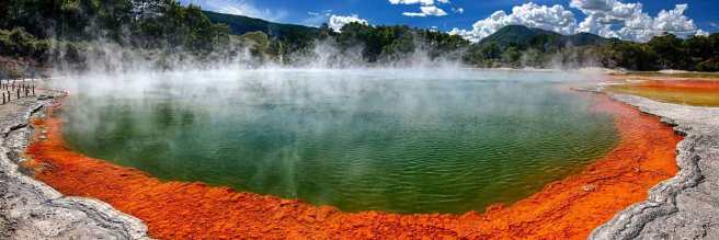
The geothermal wonderlands of Rotorua. Source: Audleytravel

Rotorua is a small city in the central eastern area of the North Island of New Zealand (Aotearoa in the indigenous Māori language).

The name Rotorua comes from the Māori language (‘roto’ meaning lake and rua meaning ‘two’). The full Māori name for the spot is actually Te Rotorua-nui-a-Kahumatamomoe. The early Māori chief and explorer Ihenga named it after his uncle Kahumatamomoe. But given that it was the second major lake found in Aotearoa (after lake Taupo in the centre of the North Island), the name that stuck was Rotorua or ‘Second lake’.

TAM-BLOG-1

Maori culture. Source: TamakiMaoriVillage

Similar to lake Taupo, Rotorua is a caldera resulting from an ancient volcanic eruption (approximately 240,000 years ago). The lake that now fills it is about 22 km (14 mi) in diameter.

p-8709-gns

Lake Rotorua. Source: Teara

The volcano may have disappeared, but the surrounding region is still full of geothermal activity (bubbling mud pools and geysers), providing the region with abundant renewable power and making the city a very popular tourist destination.

FFP_150415-6431-Edit

Tourist playing with mud. Source: Rotoruanz

Before visiting the city, however, travellers should be warned that Rotorua’s other nicknames include “Sulphur City” and “Rotten-rua”, because of the smell that results from the geothermal activity.

And speaking from personal experience, the “rotten eggs” smell is prevalent.

Interesting, but what has this got to do with the science of Parkinson’s disease?

Well, the rotten egg smell is the result of hydrogen sulfide emissions, and recently it has been suggested that this pungent gas may be having positive benefits on people, particularly with regards to Parkinson’s disease.

This idea has been proposed by Dr Yusuf Cakmak at the University of Otago in a recent commentary:

Yusuf

Title: Rotorua, hydrogen sulphide and Parkinson’s disease-A possible beneficial link?
Author: Cakmak Y.
Journal: N Z Med J. 2017 May 12;130(1455):123-125.
PMID: 28494485

In his write up, Dr Cakmak points towards two studies that have been conducted on people from Rotorua. The first focused on examining whether there was any association between asthma and chronic obstructive pulmonary disease and exposure to hydrogen sulfide in Rotorua. By examining air samples and 1,204 participants, the investigators of that study no association (the report of that study is OPEN ACCESS and can be found by clicking here).

The second study is the more interesting of the pair:

roto

Title: Chronic ambient hydrogen sulfide exposure and cognitive function.
Authors: Reed BR, Crane J, Garrett N, Woods DL, Bates MN.
Journal: Neurotoxicol Teratol. 2014 Mar-Apr;42:68-76.
PMID: 24548790                 (This article is OPEN ACCESS if you would like to read it)

In this study, the investigators recruited 1,637 adults (aged 18-65 years) from Rotorua. They conducted neuropsychological tests on the subjects, measuring visual and verbal episodic memory, attention, fine motor skills, psychomotor speed and mood. The average amount of time the participants had lived in the Rotorua region was 18 years (ranging from 3-64 years). The researchers also made measurements of hydrogen sulfide levels at the participants homes and work sites.

While the researchers found no association between hydrogen sulfide exposure and cognitive ability, they did notice something interesting in the measures of fine motor skills: individuals exposed to higher levels of hydrogen sulfide displayed faster motor response times on tasks like finger tapping. Finger tapping speed is an important part of Parkinson’s Motor Rating Scale examination tests.

The investigators behind the study concluded that the levels of hydrogen sulfide in Rotorua do not have any detrimental effect on the individuals living in the area,

Dr Cakmak, however, wondered whether “relatively high, but safe, hydrogen sulfide levels in Rotorua could help protect the degradation of dopaminergic neurons associated with Parkinson’s Disease?” (based on the better performance on the motor response time).

Hang on a second, what exactly is hydrogen sulfide?

Hydrogen sulfide (chemical symbol: H2S) is a colourless gas. Its production often results from the the breaking down of organic material in the absence of oxygen, such as in sewers (this process is called anaerobic digestion. It also occurs in volcanic and geothermal conditions.

Hydrogen_sulfide

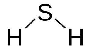
H2S. Source: Wikipedia

About 15 years ago, it was found in various organs in the body and termed a gasotransmitter. A gasotransmitter is a molecule that can be used to transmit chemical signals from one cell to another, which results in certain physiological reactions (oxygen, for example, is a gasotransmitter).

Hydrogen sulfide is now known to be cardioprotective (protection of the heart), and many years of research have demonstrated beneficial aspects of using it in therapy, such as vasodilation and lowering blood pressure, increasing levels of antioxidants, inhibiting inflammation, and activation of anti-apoptotic (anti-cell death) pathways. For a good review of hydrogen sulfide’s cardioprotective properties – click here.
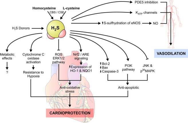
F1.large

Source: Clinsci

The demonstration of the protective properties of hydrogen sulfide in other bodily organs have led neuroscientists to start investigating whether these same benefits could be utilised in treating disorders of the brain.

And the good news is: hydrogen sulfide can have positive benefits in the brain – Click here for a good review of the brain-related research.

Has other research been conducted on hydrogen sulfide regarding Parkinson’s disease?

Yes. And here is where the story starts to get really interesting.

Initially, there were reports that hydrogen sulfide could protect cells grown in culture from exposure to various neurotoxins (Click here and here for examples).

Then hydrogen sulfide was tested in rodent models of Parkinson’s disease:

SH1

Title: Neuroprotective effects of hydrogen sulfide on Parkinson’s disease rat models.
Authors: Hu LF, Lu M, Tiong CX, Dawe GS, Hu G, Bian JS.
Journal: Aging Cell. 2010 Apr;9(2):135-46.
PMID: 20041858           (This article is OPEN ACCESS if you would like to read it)

In this study, the researchers firstly looked at what happens to hydrogen sulfide in the brains of rodent models of Parkinson’s disease. When rats were injected with a neurotoxin (6-OHDA) that kills dopamine neurons, the investigators found a significant drop in the level of hydrogen sulfide in the region where the dopamine cells reside (called the substantia nigra – an area of the brain severely affected in Parkinson’s disease).

Next the researchers gave some rodents the neurotoxin, waited three weeks and then began administering sodium hydrosulfide – which is a hydrogen sulfide donor  – every day for a further 3 weeks. They found that this treatment significantly reduced the dopamine cell loss, motor problems and inflammation in the sodium hydrosulfide treated animals. Interestingly, they saw the same neuroprotective effect when they repeated the study with a different neurotoxin (Rotenone). The investigators concluded that hydrogen sulfide “has potential therapeutic value for treatment of Parkinson’s disease”.

And this first study was followed up one year later by a study investigating inhaled hydrogen sulfide:

SH2
Title: Inhaled hydrogen sulfide prevents neurodegeneration and movement disorder in a mouse model of Parkinson’s disease.
Authors: Kida K, Yamada M, Tokuda K, Marutani E, Kakinohana M, Kaneki M, Ichinose F.
Journal: Antioxid Redox Signal. 2011 Jul 15;15(2):343-52.
PMID: 21050138            (This article is OPEN ACCESS if you would like to read it)

In this study, the investigators gave mice a neurotoxin (MPTP) and then had them breathe air with or without hydrogen sulfide (40 ppm) for 8 hours per day for one week. The mice that inhaled hydrogen sulfide displayed near normal levels of motor behaviour performance and significantly reduced levels of neurodegeneration (dopamine cell loss).

Inhalation of hydrogen sulfide also prevented the MPTP-induced activation of the brain’s helper cells (microglia and astrocytes) and increased levels of detoxification enzymes and antioxidant proteins (including heme oxygenase-1 and glutamate-cysteine ligase). Curiously, hydrogen sulfide inhalation did not significantly affect levels of reduced glutathione (we will come back to this in an upcoming post).

These first two preclinical results have been replicated many times now confirming the initial findings (Click here, here, here and here for examples). The researchers of the second ‘inhalation’ study concluded the study by suggesting that the potential therapeutic effects of hydrogen sulfide inhalation now needed to be examined in more disease relevant models of Parkinson’s disease.

And this is exactly what researchers did next:

HS5

Title: Sulfhydration mediates neuroprotective actions of parkin.
Authors: Vandiver MS, Paul BD, Xu R, Karuppagounder S, Rao F, Snowman AM, Ko HS, Lee YI, Dawson VL, Dawson TM, Sen N, Snyder SH.
Journal: Nat Commun. 2013;4:1626. doi: 10.1038/ncomms2623.
PMID: 23535647          (This article is OPEN ACCESS if you would like to read it)

The researchers conducting this study were interested in the interaction of hydrogen sulfide with the Parkinson’s disease-associated protein Parkin (also known as PARK2). They found that hydrogen sulfide actively modified parkin protein – a process called sulfhydration – and that this enhances the protein’s level of activity.

They also noted that the level of Parkin sulfhydration in the brains of patients with Parkinson’s disease is markedly reduced (a 60% reduction). These finding imply that drugs that increase levels of hydrogen sulfide in the brain may be therapeutic.

Interestingly, cells with genetic mutations in another Parkinson’s disease related gene, DJ-1, also produce less hydrogen sulfide (click here to read more about this).

Has anyone ever looked at hydrogen sulfide and alpha synuclein?

Not that we are aware of.

Alpha synuclein is the Parkinson’s disease associated protein that clusters in the Parkinsonian brain and forms Lewy bodies.

But researchers have looked at hydrogen sulfide and amyloid formation:

HS4
Title: Hydrogen sulfide inhibits amyloid formation
Authors: Rosario-Alomar MF, Quiñones-Ruiz T, Kurouski D, Sereda V, Ferreira EB, Jesús-Kim LD, Hernández-Rivera S, Zagorevski DV, López-Garriga J, Lednev IK.
Journal: J Phys Chem B. 2015 Jan 29;119(4):1265-74.
PMID: 25545790         (This article is OPEN ACCESS if you would like to read it)


Amyloid formations are large clusters of misfolded proteins that are associated with neurodegenerative conditions, like Alzheimer’s disease and Parkinson’s disease. The researchers who conducted this study were interested in the behaviour of these misfolded protein in the presence of hydrogen sulfide. What they found was rather remarkable: the addition of hydrogen sulfide completely inhibited the formation amyloid fibrils (amyloid fibril plaques are found in brains of people with Alzheimer’s disease).

jp-2014-08471v_0008

Source: NCBI

If the addition of hydrogen sulfide can reduce the level of clustered proteins in a model of Alzheimer’s disease, it would be interesting to see what it would do to alpha synuclein.

NOTE: Hydrogen sulfide levels are also reduced in the brains of people with Alzheimer’s disease (click here to read more on this topic)

Has hydrogen sulfide ever been tested in the clinic?

There are currently 17 clinical trials investigating hydrogen sulfide in various conditions (not Parkinson’s disease though).

So where can I get me some of that hydrogen sulfide?

Ok, so here is where we come in with the health warning section.

You see, hydrogen sulfide is a very dangerous gas. It is really not to be played with.

Blakely_June_Hydrogen-Sulfide

Source: Blakely

The gas is both corrosive and flammable. More importantly, at high concentrations, hydrogen sulfide gas can be fatal almost immediately (>1000 parts per milllion – source: OSHA). And the gas only exhibits the “rotten eggs” smell at low concentrations. At higher concentrations it becomes undetectable due to olfactory paralysis (luckily for the folks in Rotorua, the levels of hydrogen sulfide gas there are between 20-25 parts per billion).

Thus, we do not recommend readers to rush out and load up on hydrogen sulfide gas.

There are many foods that contain hydrogen sulfide.

For example, garlic is very rich in hydrogen sulfide. Another rich source is cooked beef, which has about 0.6mg of hydrogen sulfide per pound – cooked lamb has closer to 0.9 milligrams per pound. Heated dairy products, such as skim milk, can have approximately 3 milligrams of hydrogen sulfide per gallon, and cream has slightly more than double that amount.

Any significant change in diet by a person with Parkinson’s disease should firstly be discussed with a trained medical physician as we can not be sure what impact such a change would have on individualised treatment regimes.

What does it all mean?

Summing up: It would be interesting to look at the frequency of Parkinson’s disease in geothermal region of the world (the population of Rotorua is too small for such an analysis – 80,000 people).

Researchers believe that components of the gas emissions from these geothermal areas may be neuroprotective. Of particular interest is the gas hydrogen sulfide. At high levels, it is a very dangerous gas. At lower levels, however, researchers have shown that hydrogen sulfide has many beneficial properties, including in models of neurodegenerative conditions. These findings have led many to propose testing hydrogen sulfide in clinical trials for conditions like Parkinson’s disease.

Dr Cakmak, who we mentioned near the top of this post, goes one step further. He hypothesises that hydrogen sulfide may actually be one of the active components in the neuroprotective affect of both coffee and smoking – and with good reason. It was recently demonstrated that the certain gut bacteria, such as Prevotella, are decreased in people with Parkinson’s disease (see our post on this topic by clicking here). The consumption of coffee has been shown to help improve the Prevotella population in the gut, which may in term increase the levels of Prevotella-derived hydrogen sulfide. Similarly smokers have a decreased risk of developing Parkinson’s disease and hydrogen sulfide is a component of cigarette smoke.

All of these ideas still needs to be further tested, but we are curious to see where this research could lead. An inhaled neuroprotective treatment for Parkinson’s disease may have benefits for other neurodegenerative conditions.

Oh, and if anyone is interested, we are happy to put readers in contact with real estate agents in sunny ‘Rotten-rua’, New Zealand. The locals say that you gradually get used to the smell.


EDITOR’S NOTE: Under absolutely no circumstances should anyone reading this material consider it medical advice. The material provided here is for educational purposes only. Before considering or attempting any change in your treatment regime, PLEASE consult with your doctor or neurologist. While some of the drugs/molecules discussed on this website are clinically available, they may have serious side effects. We therefore urge caution and professional consultation before any attempt to alter a treatment regime. SoPD can not be held responsible for any actions taken based on the information provided here. 


The banner for today’s post was sourced from Trover

New drug approved for ALS

ice-bucket-challenge

The Federal Drug Administration (FDA) in the USA has approved the first drug in 22 years for treating the neurodegenerative condition of Amyotrophic lateral sclerosis (ALS).

The drug is called Edaravone, and it is only the second drug approved for ALS.

In today’s post we’ll discuss what this announcement could mean for Parkinson’s disease.


lou-gehrig

Lou Gehrig. Source: NBC

In 1969, Henry Louis “Lou” Gehrig was voted the greatest first baseman of all time by the Baseball Writers’ Association. He played 17 seasons with the New York Yankees, having signed with his hometown team in 1923.

For 56 years, he held the record for the most consecutive games played (2,130), and he was only prevented from continuing that streak when he voluntarily took himself out of the team lineup on the 2nd May, 1939, after his ability to play became hampered by the disease that now often bears his name. A little more than a month later he retired, and a little less than two years later he passed away.

Amyotrophic lateral sclerosis (or ALS), also known as Lou Gehrig’s disease and motor neuron disease, is a neurodegenerative condition in which the neurons that control voluntary muscle movement die. The condition affects 2 people in every 100,000 each year, and those individuals have an average survival time of two to four years.

als-whats-is-ALS-info

ALS in a nutshell. Source: Walkforals

In addition to Lou Gehrig, you may have heard of ALS via the ‘Ice bucket challenge‘ (see image in the banner of this post). In August 2014, an online video challenge went viral.

By July 2015, the ice bucket campaign had raised an amazing $115 million for the ALS Association.

Another reason you may have heard of ALS is that theoretical physicist, Prof Stephen Hawking also has the condition:

p03dn27d

Source: BBC

He was diagnosed with in a very rare early-onset, slow-progressing form of ALS in 1963 (at age 21) that has gradually left him wheel chair bound.

This is very interesting, but what does it have to do with Parkinson’s disease?

Individuals affected by ALS are generally treated with a drug called Riluzole (brand names Rilutek or Teglutik). Approved in December of 1995 by the FDA, this drug increases survival by approximately two to three months.

Until this last week, Riluzole was the only drug approved for the treatment of ALS.

So what happened this week?

On the 5th May, the FDA announced that they had approved a second drug for the treatment of ALS (Click here for the press release).

It is called Edaravone.

What is Edaravone?

Edaravone is a free radical scavenger – a potent antioxidant – that is marketed as a neurovascular protective agent in Japan by Mitsubishi Tanabe Pharma Corporation.

An antioxidant is simply a molecule that prevents the oxidation of other molecules

Molecules in your body often go through a process called oxidation – losing an electron and becoming unstable. This chemical reaction leads to the production of what we call free radicals, which can then go on to damage cells.

What is a free radical?

A free radical is simply an unstable molecule – unstable because they are missing electrons. They react quickly with other molecules, trying to capture the needed electron to re-gain stability. Free radicals will literally attack the nearest stable molecule, stealing an electron. This leads to the “attacked” molecule becoming a free radical itself, and thus a chain reaction is started. Inside a living cell this can cause terrible damage, ultimately killing the cell.

Antioxidants are thus the good guys in this situation. They are molecules that neutralize free radicals by donating one of their own electrons. The antioxidant don’t become free radicals by donating an electron because by their very nature they are stable with or without that extra electron.

Thus when we say ‘Edaravone is a free radical scavenger’, we mean it’s really good at scavenging all those unstable molecules and stabilising them.

It is an intravenous drug (injected into the body via a vein) and administrated for 14 days followed by 14 days drug holiday.

So, again what has this got to do with Parkinson’s disease?

Well, it is easier to start a clinical trial of a drug if it is already approved for another disease.

And the good news is: Edaravone has been shown to be neuroprotective in several models of Parkinson’s disease.

In this post, we’ll lay out some of the previous research and try to make an argument justifying the clinical testing of Edaravone in Parkinson’s disease

Ok, so what research has been done so far in models of Parkinson’s disease?

The first study to show neuroprotection in a model of Parkinson’s disease was published in 2008:

2008-1

Title: Role of reactive nitrogen and reactive oxygen species against MPTP neurotoxicity in mice.
Authors: Yokoyama H, Takagi S, Watanabe Y, Kato H, Araki T.
Journal: J Neural Transm (Vienna). 2008 Jun;115(6):831-42.
PMID: 18235988

In this first study, the investigators assessed the neuroprotective properties of several drugs in a mouse model of Parkinson’s disease. The drugs included Edaravone (described above), minocycline (antibiotic discussed in a previous post), 7-nitroindazole (neuronal nitric oxide synthase inhibitor), fluvastatin and pitavastatin (both members of the statin drug class).

With regards to Edaravone, the news was not great: the investigators found that Edaravone (up to 30mg/kg) treatment 30 minutes before administering a neurotoxin (MPTP) and then again 90 minutes afterwards had no effect on the survival of the dopamine neurons (compared to a control treatment).

Not a good start for making a case for clinical trials!

This research report, however, was quickly followed by another from an independent group in Japan:

BMC

Title: Neuroprotective effects of edaravone-administration on 6-OHDA-treated dopaminergic neurons.
Authors: Yuan WJ, Yasuhara T, Shingo T, Muraoka K, Agari T, Kameda M, Uozumi T, Tajiri N, Morimoto T, Jing M, Baba T, Wang F, Leung H, Matsui T, Miyoshi Y, Date I.
Journal: BMC Neurosci. 2008 Aug 1;9:75.
PMID: 18671880            (This article is OPEN ACCESS if you would like to read it)

These researchers did find a neuroprotective effect using Edaravone (both in cell culture and in a rodent model of Parkinson’s disease), but they used a much higher dose than the previous study (up to 250 mg/kg in this study). This increase in dose resulted in a graded increase in neuroprotection – interestingly, these researchers also found that 30mg/kg of Edaravone had limited neuroprotective effects, while 250mg/kg exhibited robust dopamine cell survival and rescued the behavioural/motor features of the model even when given 24 hours after the neurotoxin.

The investigators concluded that “Edaravone might be a hopeful therapeutic option for PD, although several critical issues remain to be solved, including high therapeutic dosage of Edaravone for the safe clinical application in the future”

This results was followed by several additional studies investigating edaravone in models of Parkinson’s disease (Click here, here and here to read more on this). Of particular interest in all of those follow up studies was a report in which Edaravone treatment resulted in neuroprotective in genetic model of Parkinson’s disease:

2013-1

Title: Edaravone prevents neurotoxicity of mutant L166P DJ-1 in Parkinson’s disease.
Authors: Li B, Yu D, Xu Z.
Journal: J Mol Neurosci. 2013 Oct;51(2):539-49.
PMID: 23657982

DJ-1 is a gene that has been associated Parkinson’s disease since 2003. The gene is sometimes referred to as PARK7 (there are now more than 20 Parkinson’s associated genomic regions, which each have a number and are referred to as the PARK genes). Genetic mutations in the DJ-1 gene can result in an autosomal recessive (meaning two copies of the mutated gene are required), early-onset form of Parkinson disease. For a very good review of DJ-1 in the context of Parkinson’s disease, please click here.

The exact function of DJ-1 is not well understood, though it does appear to play a role in helping cells deal with ‘oxidative stress’ – the over-production of those free radicals we were talking about above. Now given that edaravone is a potent antioxidant (reversing the effects of oxidative stress), the researchers conducting this study decided to test Edaravone in cells with genetic mutations in the DJ-1 gene.

Their results indicated that Edaravone was able to significantly reduce oxidative stress in the cells and improve the functioning of the mitochondria – the power stations in each cell, where cells derive their energy. Furthermore, Edaravone was found to reduce the amount of cell death in the DJ-1 mutant cells.

More recently, researchers have begun digging deeper into the mechanisms involved in the neuroprotective effects of Edaravone:

2015-1

Title: Edaravone leads to proteome changes indicative of neuronal cell protection in response to oxidative stress.
Authors: Jami MS, Salehi-Najafabadi Z, Ahmadinejad F, Hoedt E, Chaleshtori MH, Ghatrehsamani M, Neubert TA, Larsen JP, Møller SG.
Journal: Neurochem Int. 2015 Nov;90:134-41.
PMID: 26232623             (This article is OPEN ACCESS if you would like to read it)

The investigators who conducted this report began by performing a comparative two-dimensional gel electrophoresis analyses of cells exposed to oxidative stress with and without treatment of Edaravone.

Um, what is “comparative two-dimensional gel electrophoresis analyses”?

Good question.

Two-dimensional gel electrophoresis analyses allows researchers to determine particular proteins within a given solution. Mixtures of proteins are injected into a slab of gel and they are then separated according to two properties (mass and acidity) across two dimensions (left-right side of the gel and top-bottom of the gel).

A two-dimensional gel electrophoresis result may look something like this:

4000716f1

Two-dimensional gel electrophoresis. Source: Nature

As you can see, individual proteins have been pointed out on the image of this slab of gel.

In comparative two-dimensional gel electrophoresis, two samples of solution are analysed by comparing two slabs of gel that have been injected with protein mix solution from two groups of cells treated exactly the same except for one variable. Each solution gets its own slab of gel, and the differences between the gel product will highlight which proteins are present in one condition versus the other (based on the variable being tested).

In this experiment, the variable was Edaravone.

And when the researchers compared the proteins of Edaravone treated cells with those of cells not treated with Edaravone, they found that the neuroprotective effect of Edaravone was being caused by an increase in a protein called Peroxiredoxin-2.

Now this was a really interesting finding.

You see, Peroxiredoxin proteins are a family (there are 6 members) of antioxidant enzymes. And of particular interest with regards to Parkinson’s disease is the close relationship between DJ-1 (the Parkinson’s associated protein discussed above) and peroxiredoxin proteins (Click here, here, here and here to read more about this).

In addition, there are also 169 research reports dealing with the peroxiredoxin proteins and Parkinson’s disease (Click here to see a list of those reports).

So, what do you think about a clinical trial for Edaravone in Parkinson’s disease?

Are you convinced?

Regardless, it an interesting drug huh?

Are there any downsides to the drug?

One slight issue with the drug is that it is injected via a vein. Alternative systems of delivery, however, are being explored.A biotech company in the Netherlands, called Treeway is developing an oral formulation of edaravone (called TW001) and is currently in clinical development.

Edaravone was first approved for clinical use in Japan on May 23, 2001. With almost 17 years of Edaravone clinical use, a few adverse events including acute renal failure have been noted, thus precautions should be taken with individuals who have a history of renal problems. The most common side effects associated with the drug, however, are: fatigue, nausea, and some mild anxiety.

Click here for a good overview of the clinical history of Edaravone.

So what does it all mean?

The announcement from the FDA this week regarding the approval of Edaravone as a new treatment for ALS represents a small victory for the ALS community, but it may also have a significant impact on other neurodegenerative conditions, such as Parkinson’s disease.

Edaravone is a potent antioxidant agent, which has been shown to have neuroprotective effects in various models of Parkinson’s disease and other neurodegenerative conditions. It could be interesting to now test the drug clinically for Parkinson’s disease. Many of the preclinical research reports indicate that the earlier Edaravone treatment starts, the better the outcomes, so any initial clinical trials should focus on recently diagnosed subjects (perhaps even those with DJ-1 mutations).

The take home message of this post is: given that Edaravone has now been approved for clinical use by the FDA, it may be advantageous for the Parkinson’s community to have a good look at whether this drug could be repurposed for Parkinson’s disease.

It’s just a thought.


The banner for today’s post was sourced from Forbes

Old dogs, new tricks – repurposing drugs for Parkinson’s


Drugrepurpose

Exciting news this week from the world of neurodegenerative research. Researchers have identified two clinically available drugs that display neuroprotective properties.

The drugs – Dibenzoylmethane and Trazodone – are currently used to treat cancer and depression, respectively.

In this post, we will review the research and discuss what it could mean for folks with Parkinson’s disease.


Drugs

Old drugs – new tricks? Source: Repurposingdrugs101

As you may have heard from media reports (for examples, click here, here and here), researchers have identified two clinically available drugs that may help in the fight against neurodegenerative conditions, like Parkinson’s disease.

The re-purposing of clinically available drugs is the focus of much attention within the Parkinson’s community as it represents a means of bringing treatments to the clinic faster. The traditional lengthy clinical trial process that is required in the development of new medications means getting a new drug to market for neurodegeneration can take up to 15 years, as the trials run over several years each (and there are three phases to pass through).

ajbtbe-v2-id1047-g001

Shortening the wait. Source: Austinpublishing

In an age of smart phones and instant gratification, who has that kind of patience? ( #Wewontwait ).

Thus, re-purposing of available drugs represents a more rapid means of bringing new treatments/therapies to the Parkinson’s community.

So what is the new research all about?

This is Professor Giovanna Mallucci.

Leicester-2015-05-11-0182_800

Prof Giovanna Mallucci. Source: MRC

She’s awesome.

She led the team from the Medical Research Council’s (MRC) Toxicology Unit (University of Leicester) that conducted the research and she is now based at the University of Cambridge.

Her area of research interest is understanding mechanisms of neurodegeneration, with a particular focus on prion and Alzheimer’s disease.

A few years ago, her group published this report:

Nature

Title: Sustained translational repression by eIF2α-P mediates prion neurodegeneration.
Authors: Moreno JA, Radford H, Peretti D, Steinert JR, Verity N, Martin MG, Halliday M, Morgan J, Dinsdale D, Ortori CA, Barrett DA, Tsaytler P, Bertolotti A, Willis AE, Bushell M, Mallucci GR.
Journal: Nature. 2012 May 6;485(7399):507-11.
PMID: 22622579              (This article is OPEN ACCESS if you would like to read it)

In this study, Prof Mallucci’s group were interested in the biological events that were occurring in the brain following infection of mice with prion disease – another neurodegenerative condition. They found that a sudden loss of protein associated with the connections between neurons (those connections being called synapses) occurred at 9 weeks post infection. This led them to investigate the production of protein and they found that an increase in the levels of phosphorylation of a protein called eIF2alpha was associated with the reduction in protein synthesis occurring at 9 weeks post infection.

What is Phosphorylation?

Phosphorylation of a protein is basically the process of turning it on or off – making it active or inactive – for a particular function.

U2CP5-4_Phosphorylation_revised

Phosphorylation of a kinase protein. Source: Nature

And what is eIF2alpha?

Eukaryotic Translation Initiation Factor 2 Alpha is (as the label on the can suggests) a translation initiation factor. This means that this particular protein functions in the early steps of the production of protein. That is to say, eIF2alpha has important roles in the first steps – the initiation – of making other proteins.

Roles of eIF2 kinases in the pathogenesis of Alzheimer's disease

eIF2alpha’s role in neurodegeneration. Source: Frontiers

The increased phosphorylation of eIF2alpha results in the inactivation of eIF2alpha and therefore the transient shutdown of protein production.

This shutdown in protein production can serve as an important ‘checkpoint’ when a cell is stressed. By blocking general protein production, a damaged or stressed cell can have the opportunity to either recuperate or be eliminated (if the damage is beyond repair).

The shutdown can also be caused by the unfolded protein response (or UPR). The unfolded protein response is a protective mechanism triggered by rising levels of misfolded proteins.

What are misfolded proteins?

When proteins are being produced, they need to be folded into the correct shape to do their job. Things can turn ugly very quickly for a cell if protein are being misfolded or only partially folded.

prions

Two proteins. Guess which is the misfolded protein. Source: Biogeekery

In fact, misfolded proteins are suspected of being responsible for many of the neurodegenerative conditions we know of (including Parkinson’s, Alzheimer’s, etc). Thus the unfolded protein response gives a cell time to stop protein production, degrade & dispose of any misfolded proteins, and then re-activate proteins involved with increasing the production again.

And Prof Mallucci’s group found an increase in the phosphorylation of eIF2alpha?

Yes. At 9 weeks post infection with prions, there is a decrease in the proteins required for maintaining the connections between neurons and an increase in the phosphorylation of eIF2alpha.

The interesting thing is that the researchers found that levels of phosphorylated eIF2alpha increased throughout the course of study.

So, the researchers asked themselves if promoting a recovery in protein production in the cells in neuroprotective. To test this they used a protein called GADD34, which is a specific eIF2alpha phosphatase (a phosphatase is a protein that dephosphates a protein). By introducing a lot of GADD34 in the cells, the researchers were able to re-activate eIF2alpha, rescue the connectivity between neurons and protect the cells from dying.

A cool trick, huh?

This report established the importance of eIF2alpha in the early stages of neurodegeneration, and Prof Mallucci and her group next decided to conduct a massive screening study of currently available medications to see which could be used to target eIF2alpha levels.

And that research gave rise to the report that caused so much excitement this week. This report here:

Brain
Title: Repurposed drugs targeting eIF2α-P-mediated translational repression prevent neurodegeneration in mice
Authors: Halliday M, Radford H, Zents KAM, Molloy C, Moreno JA, Verity NC, Smith E, Ortori CA, Barrett DA, Bushell M, Mallucci GR.
Journal: Brain, 2017 Epub early online publication
PMID: N/A         (This article is OPEN ACCESS if you would like to read it)

The investigators began by testing 1,040 compounds (that represent many of the clinically available drugs we have) on tiny microscopic worms (called C.elegans). These worms represent a useful experimental model for screening drugs as many aspects of biology can be examined. These worms were exposed to both a chemical (called tunicamycin, which induces the unfolded protein response we were talking about above) and one of the 1040 compounds.

Of the 1040 compounds tested, the investigators selected the 20 that provided the best protection to the worms. They next analysed those top 20 compounds for their ability to reduce levels of phosphorylated eIF2alpha in cells. Cells were engineered to produce a bioluminescent signal when eIF2alpha was phosphorylated. The researchers used a potent blocker of the unfolded protein response (called GSK2606414) and a drug called ISRIB (which is an experimental drug which reverses the effects of eIF2alpha phosphorylation) as controls for the experiment.

Their results were interesting:

Figure1

The results of the top 20 drugs screened. Source: Brain

As you can see from the graph above, there were five compounds (highlighted with ***) that provided a similar level of reduction as the ISRIB (control) drug:

  • Azadirachtin – which is the active ingredient in many pesticides.
  • Dibenzoylmethane – a cancer treatment.
  • Proguanil – a medication used to treat and prevent malaria.
  • Trazodone – an antidepressant used to treat depression and anxiety disorders.
  • Trifluoperazine – an antipsychotic of the phenothiazine chemical class.

The investigators decided not to further investigate Azadirachtin as it is a pesticide and displays a poor ability to penetrant the blood-brain-barrier – the protective layer surrounding the brain. They also rejected Proguanil because while it is safe to use in humans, it is toxic in mice. This detail limited the amount of preclinical testing for neurodegeneration that the researchers could do. And finally Trifluoperazine was eliminated as it should not be used in the elderly populations (apparently it ‘increases the risk of death’!), which obviously limited it’s further utility given that age is a major determinant of neurodegeneration.

This selection process left the researchers with Dibenzoylmethane and Trazodone.

The researchers found that both of these drugs can cross the blood-brain-barrier and were able to prevent neurodegeneration and rescue behavioural deficits in prion-infected mice. And they observed no toxic effects of these treatments in other organs (such as the pancreas). The drugs restore correct protein production and increased the survival of the prion-infected mice.

Taking the study one step further, Prof Mallucci and her group asked if the drugs could be effective in a model of another neurodegenerative condition, such as Alzheimer’s disease. To investigated this, they treated rTg4510 mice with both of the drugs. rTg4510 mice produce a lot of a human protein (called tau) that has a particular mutation (known as P301L), which results in the onset of Alzheimer’s like pathology at an early age. The rTg4510 mice received either trazodone or Dibenzoylmethane on a daily basis from 4 months of age and were examined at 8 months of age. The researchers found significantly less cell loss and shrinkage in the brains of the mice treated with one of the two drugs when compared to rTg4510 mice that received no treatment.

The researchers concluded that “these compounds therefore represent potential new disease-modifying treatments for dementia. Trazodone in particular, a licensed drug, should now be tested in clinical trials in patients”.

As Professor Mallucci suggested to the press: “We know that trazodone is safe to use in humans, so a clinical trial is now possible to test whether the protective effects of the drug we see on brain cells in mice with neurodegeneration also applies to people in the early stages of Alzheimer’s disease and other dementias. We could know in 2-3 years whether this approach can slow down disease progression, which would be a very exciting first step in treating these disorders. Interestingly, trazodone has been used to treat the symptoms of patients in later stages of dementia, so we know it is safe for this group.  We now need to find out whether giving the drug to patients at an early stage could help arrest or slow down the disease through its effects on this pathway.”

This is great for Alzheimer’s disease, but what about Parkinson’s?

Well, the researchers did not test the drugs in models of Parkinson’s disease. But we can assume that several research groups are going to be testing this drug in the near future… if they aren’t already!

But have increased levels of eIF2alpha been seen in Parkinson’s disease?

Great question. And the answer is: Yes.

ParkUPS

Title: Activation of the unfolded protein response in Parkinson’s disease.
Authors: Hoozemans JJ, van Haastert ES, Eikelenboom P, de Vos RA, Rozemuller JM, Scheper W.
Journal: Biochem Biophys Res Commun. 2007 Mar 16;354(3):707-11.
PMID: 17254549

In this study the investigators analysed the levels of Unfolded Protein Response activation in the postmortem brains of people who passed away with or without Parkinson’s disease. Specifically, they focused their analysis on the substantia nigra (the region where the dopamine neurons reside and which is most severely affected in Parkinson’s).

The researchers found that both eIF2alpha and a protein called PERK (also known as protein kinase-like ER kinase – which phosphalates eIF2alpha) are present in the dopamine neurons in the substantia nigra of brains from people with Parkinson’s disease, but not in healthy control brains. And as the graph below shows, the investigators noted that there was a trend towards the levels of these proteins peaking within the first five years after diagnosis.

graph

eIF2alpha & PERK levels in the brain. Source: ScienceDirect

Similar postmortem analysis studies have also highlighted the increased levels of Unfolded Protein Response activation in the Parkinsonian brain (Click here to read more on this).

The increase in Unfolded Protein Response activation could be a common feature across different neurodegenerative conditions, suggesting that trazodone and dibenzoylmethane could be used widely to slow the progress of various conditions.

Another connection to Parkinson’s disease is the finding that high levels of the Parkinson’s associated protein alpha synuclein can cause the Unfolded Protein Response:

Activation
Title: Induction of the unfolded protein response by α-synuclein in experimental models of Parkinson’s disease.
Authors: Bellucci A, Navarria L, Zaltieri M, Falarti E, Bodei S, Sigala S, Battistin L, Spillantini M, Missale C, Spano P.
Journal: J Neurochem. 2011 Feb;116(4):588-605.
PMID: 21166675       (This article is OPEN ACCESS if you would like to read it)

The researchers in this study found that introducing large amounts of alpha synuclein into cell cultures results in the initiation of the unfolded protein response. They also observed this phenomenon in genetically engineered mice that produce large amounts of alpha synuclein.

Thus, there is some evidence for eIF2alpha and unfolded protein response-related activities in Parkinson’s disease

So is there are evidence that Dibenzoylmethane might be neuroprotective for Parkinson’s disease?

Yes there is (sort of):

Basic RGB

Title: A dibenzoylmethane derivative protects dopaminergic neurons against both oxidative stress and endoplasmic reticulum stress.
Authors: Takano K, Kitao Y, Tabata Y, Miura H, Sato K, Takuma K, Yamada K, Hibino S, Choshi T, Iinuma M, Suzuki H, Murakami R, Yamada M, Ogawa S, Hori O.
Journal: Am J Physiol Cell Physiol. 2007 Dec;293(6):C1884-94. Epub 2007 Oct 3.
PMID: 17913843             (This article is OPEN ACCESS if you would like to read it)

The investigators of this study found a derivative of dibenzoylmethane which they called 14-26 (chemical name 2,2′-dimethoxydibenzoylmethane) displayed neuroprotective functions both in cell culture and animal models of Parkinson’s disease. The researchers did not look at the unfolded protein response or eIF2alpha and PERK levels, nor did they determine if dibenzoylmethane itself exhibits neuroprotective properties.

This may now need to be re-addressed.

And is there any evidence trazodone having neuroprotective effects in other neurodegenerative conditions?

Yes.

For a review of the neuroprotective effects of trazodone (and other anti-psychotic/anti-depressant drugs) in Huntington’s Disease – Click here.

This sounds very positive for Parkinson’s disease then, no?

Weeeeeell, there is a word of caution to be thrown in here:

There have been reports in the past of trazodone causing motor-related issues in the elderly. Such as this one:

Trazodone

Title: Can trazodone induce parkinsonism?
Authors: Albanese A, Rossi P, Altavista MC.
Journal: Clin Neuropharmacol. 1988 Apr;11(2):180-2.
PMID: 3378227

This report was a single case study of a 74 year old lady who developed depression after losing her sister with whom she lived. She was prescribed trazodone, which was effective in improving her mood. Just several months later, however, she began presenting Parkinsonian symptoms.

Firstly the onset of a resting tremor in the left arm, then a slowing of movement and a masking of the face. The attending physician withdrew the trazodone treatment and within two months the symptoms began to disappear, with no symptoms apparent 12 months later.

And unfortunately this is not an isolated case – other periodic reports of trazodone-induced motor issues have been reported (Click here and here for examples). And this is really strange as Trazodone apparently has no dopaminergic activity that we are aware of. It is a serotonin antagonist and reuptake inhibitor (SARI); it should not affect the re-uptake of norepinephrine or dopamine within the brain.

Thus, we may need to proceed with caution with the use of Trazodone for Parkinson’s disease.

So what does it all mean?

The repurposing of old drugs to treat alternative conditions is a very good idea. It means that we can test treatments that we usually know a great deal about (with regards to human usage) on diseases that they were not initially designed for, in a rapid manner.

Recently, scientists have identified two clinically available drugs that have displayed neuroprotection in two different models of neurodegeneration. Without doubt there will now be follow up investigations, before rapid efforts are made to set up clinical trials to test the efficacy of these drugs in humans suffering from dementia.

Whether these two treatments are useful for Parkinson’s disease still needs to be determined. There is evidence supporting the idea that they may well be, but caution should always be taken in how we proceed. This does not mean that other clinically available drugs can not be tested for Parkinson’s disease, however, and there are numerous clinical trials currently underway testing several of them (Click here to read more on this).

We’ll let you know when we hear anything about these efforts.


EDITOR’S NOTE: Under absolutely no circumstances should anyone reading this material consider it medical advice. The material provided here is for educational purposes only. Before considering or attempting any change in your treatment regime, PLEASE consult with your doctor or neurologist. While some of the drugs discussed on this website are clinically available, they may have serious side effects. We urge caution and professional consultation before altering any treatment regime. SoPD can not be held responsible for any actions taken based on the information provided here. 


The banner for today’s post was sourced from Linkedin

The biology of immortality and Parkinson’s disease


live-forever

A research paper was published in the prestigious journal Cell this week that has some people excitedly suggesting that we are one step closer to living longer.

Age is the no. 1 correlate with neurodegenerative conditions like Parkinson’s disease. A better understanding of the ageing process would greatly aid us in understanding these conditions.

In today’s post we will review the research paper in question and discuss what it will mean for Parkinson’s disease.


o-hela-cells-facebook

Henrietta Lacks with her husband David. Source: HuffingtonPost

The lady in this photo is basically immortal.

Henrietta Lacks was an African American woman who died in October, 1951, but (it could be argued) lives in almost every research institute in the world. Henrietta died with cervical cancer, and during her treatment, she had a (unethical) biopsy conducted on her tumor which give rise to the first human immortalised cell line that is named after her: Hela cells (Henrietta Lacks).

And I kid you not when I say that there are samples of Hela cells in a freezer in almost every research institute in the world. Since the 1950s, scientists have grown 20 tons (and counting) of her cells (Source). And some of our greatest advances have been made with Hela cells, for example in 1954, Jonas Salk used HeLa cells in his research to develop the polio vaccine.

Unwittingly, Henrietta achieved immortality.

tumblr_lyubehxk1h1qfcyz0o1_1280

Hercules fighting off death. Source: ProactionaryTranshumanist

We humans have a strange fascination with immortality. It could be argued that it underlies our religions, and also drives us to achieve great things during our live times.

During the last three decades, science has begun probing the biology of ageing with the goal of helping us to understand how and why our bodies degrade over time in the hope that it will lead to better treatments for disease that predominantly affect the elderly.

Last week research groups from Spain and the Salk institute in California published the results of a study that may be considered the first tentative steps towards actually extending life and perhaps reversing the ageing process.

This is the article:

cell-title-5

Title: In Vivo Amelioration of Age-Associated Hallmarks by Partial Reprogramming.
Authors: Ocampo A, Reddy P, Martinez-Redondo P, Platero-Luengo A, Hatanaka F, Hishida T, Li M, Lam D, Kurita M, Beyret E, Araoka T, Vazquez-Ferrer E, Donoso D, Roman JL, Xu J, Rodriguez Esteban C, Nuñez G, Nuñez Delicado E, Campistol JM, Guillen I, Guillen P, Izpisua Belmonte JC.
Journal: Cell. 2016 Dec 15;167(7):1719-1733.
PMID: 27984723

In their study, the researchers used something called the ‘Yamanaka factors’ to try and reverse the ageing process in mice.

What are the Yamanaka factors?

This is Prof Shinya Yamanaka:

yamanaka-s

Source: Glastone Institute

He’s a dude.

In 2012 he and Prof John Gurdon (University of Cambridge) were awarded the Nobel prize for Physiology and Medicine for the discovery that mature cells can be converted back to stem cells. Prof Gurdon achieved this feat by transplanting a young nucleus into an old cell, while Prof Yamanaka did the same thing with the ‘Yamanaka factors’.

The Yamanaka factors are a set of 4 genes (named Myc, Oct3/4, Sox2 and Klf4) that when turned on in a mature cells (like a skin cell) can force the cell to ‘de-differentiate’ back into an immature cell that is capable of becoming any kind of cell. This induced un-programmed state means that the once adult cell has changed into something similar to an embryonic stem cell.

This new cell is called an induced pluripotent stem (IPS) cell – ‘pluripotent’ meaning capable of any fate.

So what did the researchers in the Cell paper do with the Yamanaka factors?

150206-mouseresearch-stock

A lab mouse. Source: USNews

They genetically engineered mice that produced the Yamanaka factors in every cell in the body. They could turn on the Yamanaka factors by adding a special chemical to the drinking water of the mice for 2 out of every 7 days.

By turning on the 4 genes in this manner, the researchers found that they could extend the life span of the mice by 30%. In human terms, this would increase the average age of death from the current 80 years to 105 years!

Not only did they not the increase in the life span of the mice, but the researchers also found that the reprogramming of cells improved the regenerative ability of the cells in the aged mice. They even demonstrated this in human cells carrying the Yamanaka factors.

The study is very artificial – the mice and the human cell lines were engineered to produce the Yamanaka factors, which does not happen in normal nature – and we won’t be seeing any clinical trial for this kind of approach anytime soon. But the results will have big implications for the field of neurodegeneration.

Ok, but what has all this got to do with Parkinson’s disease?

Remember that the no. 1 correlate (association) with Parkinson’s disease is age. That is to say, your risk of developing Parkinson’s disease increases with age.

So this begs the question: if we had a better understanding of the ageing process (or even just a concept of what ageing is), could we beat off Parkinson’s disease?

Until the research paper reviewed above was published last week, this was all just silly hypothetical stuff. Now that we can extend the lives of mice by 30%, however, do we need to start actually considering the hypothetical as possible?

Science has brought us along way and provided us with many wonderful things (I would be lost without my Apple ipod). But we are now interesting a strange new world where science is going beyond normal basic biology and this may allow us to reverse what was previously un-negotiable facts. We can argue till the cows come home over the ethics of letting people live longer, but there is tremendous potential to use this technology to deal with the neurodegenerative conditions currently affecting us.

This is all very speculative, but it will be interesting to see where this leads. Stay tuned.


The banner for today’s post was sourced from TechieKids

PAMs for Parkinson’s?

clinicaltrials

In today’s post we are going to review the results of a phase 1 trial for new kind of drug being oriented at Parkinson’s disease. The results were announced in late September, and they indicate that the drug was well tolerated by subjects taking part in the study.


4912prexton

Source: Prexton

Here at the Science of Parkinson’s disease we are always on the look out for novel drug therapies. Many of the treatments currently being tested in the clinic are simply different versions of L-dopa or a dopamine agonist.

So when Prexton Therapeutics recently  announced the results of their phase 1 clinical trial for their lead drug, PXT002331, we sat up and took notes. PXT002331 (formerly called DT1687) is the first drug of its kind to be tested in Parkinson’s disease.

It is a mGluR4 positive allosteric modulator.

What on earth is mGluR4 positive allosteric modulator?
The metabotropic glutamate receptors (mGluR) are an abundant family of receptors in the brain. Proteins bind to these receptors and activate (or block) an associated function. There are many different types of these receptors and mGluR4 is simply a small subset. The mGluR4s, however, are present in the areas affected by Parkinson’s disease, and this is why this particular family of receptors has been the focus of much research attention.

But what about the positive allosteric modulator part of ‘mGluR4 positive allosteric modulator’

Yes, good question.

This is the key part of this new approach. Allosteric modulators are a new class of orally available small molecule therapeutic agents. Traditionally, most marketed drugs bind directly to the same part of receptors that the body’s own natural occurring proteins attach to. This means that those drugs are competing with those endogenous proteins, thus limiting the potential effect of the drug.

Allosteric modulators get around this problem by binding different parts of the receptor. And instead of simply turning on or off the receptor, allosteric modulators can either turn up the volume of the signal being sent by the receptor or decrease the signals. This means that when the body’s naturally occurring protein binds in the receptor, allosteric modulators can either amplify the effect or reduce it depending on which type of allosteric modulators is being administered.

allosteric_modulation_mechanism

How Allosteric modulators work. Source: Addrex Thereapeutics

There are two different types of allosteric modulators: positive and negative. And as the label suggests, positive allosteric modulators (or PAMs) increase the signal from the receptor while negative allosteric modulators (or NAMs) reduce the signal. Thus, mGluR4 PAMS are amplifying the signal of the mGluR4 receptors.

Why do we want an amplification of a particular signal?

That is a hard question to answer.

Here’s the short explanation:

When you are planning to make a movement with your body, the process of actually initiating that movement begins in the cortex, specifically the primary motor cortex:

decisionmaking

A cross section of the human brain illustrating the primary motor cortex. Source: Droso4schools

The primary motor cortex receives information from other regions of the brain (such as the prefrontal cortex where you make a lot of your decisions), and it will then send a signal down into the brain and down the spinal cord telling the limbs to move. On the way down through the brain, the signal will pass through a series of check points that will filter the signal and determine the final strength of it.

basal-ganglia-dbs-figure-1

A schematic of the feedback loop of check points. Source: Parkinson’s Biology

EDITOR’S NOTE: We have borrowed this image from the Parkinson’s biology blog, which we are huge fans of. We highly recommend people visit that site as well as our lovely site. They also provide easy to understand explanations of the biology of Parkinson’s disease.

These checkpoints represent a large feedback loop. The critical step in this process is the processing being conducted in the basal ganglia, which can be broken down into different subregions:

basal-ganglia-dbs-figure-5

A schematic of the components of the basal ganglia. Source: Parkinson’s Biology

The globus pallidus (GPi) is the last area of the basal ganglia that the signal will pass through on it’s way to the thalamus (the ultimate decider of whether you will move or not), so if there is anything going wrong between these two structures the initiation of movement will be disrupted.

In a normal brain, the chemical dopamine is being produced in an area called the substantia nigra pars compacta (say that three times really fast). That dopamine is released in the striatum and other areas of the basal ganglia, and it has a mediating effect on the signal passing through these structures.

basal-ganglia-dbs-figure-3

A schematic of the source of dopamine. Source: Parkinson’s Biology

In Parkinson’s disease, however, the dopamine producing cells of the pars compacta are loss – 60% by the time a person starts to have the clinical motor features appearing. The loss of this dopamine leaves the whole system ‘unmediated’. The feedback loop becomes extremely inhibited, resulting in problems initiating movement.

Deep brain stimulation can un-inhibit the globus pallidus, by mediating the signal passing through that structure. But this requires surgery and the implanting of probes deep inside the brain.

basal-ganglia-dbs-figure-7

A schematic of deep brain stimulation of the globus pallidus. Source: Parkinson’s Biology (great website!)

A better way of reducing the inhibition in this feedback loop is the replacement of dopamine (which we do via the taking of treatments like L-dopa). This has been the standard approach for more than 50 years.

A new method of reducing the inhibition in the feedback loop would be to chemically targeting the globus pallidus, and this is what scientists are trying to do with the mGluR4 PAMS. By amplifying the signal of mGluR4s in the globus pallidus, the scientists believe that they can reduce the level of inhibition in the feedback loop.

The hope is that this approach is a less Parkinson’s disease-affected treatment. That is to say, the globus pallidus is structurally less affected by Parkinson’s disease than the substantia nigra pars compacta, and thus any treatment of the globus pallidus should be more stable over time (as the disease progresses).

That said, it is acknowledged that mGluR4 PAMS are NOT a potential cure for Parkinson’s disease, but rather a better way of treating the condition.

What research has been done on mGluR4 PAMS and Parkinson’s disease?

In August of 2003, some researchers at the pharmaceutical company Merck published a study which indicated that activation of mGluR4 could decrease the excessive levels of inhibition in the  globus pallidus.

jns

Title: Group III metabotropic glutamate receptor-mediated modulation of the striatopallidal synapse.
Authors: Valenti O, Marino MJ, Wittmann M, Lis E, DiLella AG, Kinney GG, Conn PJ.
Journal: Journal of Neuroscience. 2003 Aug 6;23(18):7218-26.
PMID: 12904482      (This article is OPEN ACCESS if you would like to read it)

The researchers found that an mGluR4 agonist (a protein that binds to the receptor directly, encouraging the associated action) reduced inhibitory signal being produced in the globus pallidus (through a presynaptic mechanism of action). They next demonstrated that the effect did not happen in mice which do not have mGluR4s, illustrating the specificity of the effect. They finished the study by injecting the mGluR4 agonist into a rodent model of Parkinson’s disease and found beneficial effects – that were equivalent to L-dopa.

Based on this research, the scientists at Merck next turned their attention to modulating the mGluR4s in the globus pallidus using allosteric modulators:

pnastitle

Title: Allosteric modulation of group III metabotropic glutamate receptor 4: a potential approach to Parkinson’s disease treatment.
Authors: Marino MJ, Williams DL Jr, O’Brien JA, Valenti O, McDonald TP, Clements MK, Wang R, DiLella AG, Hess JF, Kinney GG, Conn PJ.
Journal: Proc Natl Acad Sci U S A. 2003 Nov 11;100(23):13668-73.
PMID: 14593202           (This article is OPEN ACCESS if you would like to read it)

In this article, the same researchers introduce a positive allosteric modulator called ‘PHCCC’ which has a preference for binding to mGluR4. They found that when they put PHCCC – in combination with the mGluR4 agonist used in the previous study – onto cells in petri dishes, they got an amplification of the reduction in inhibition in the cells. Administered alone, PHCCC also produced a marked reversal of the motor deficit observed in a rodent model of Parkinson’s disease.

With these results, the scientists could begin building the justification for taking mGluR4 PAMs to the clinic. They were interested, however, in what impact mGluR4 PAMs could have on the involuntary motor problems associated with long-term L-dopa use, called dyskinesias (we have previously written about these – click here to read that post). So they decided to investigate whether mGluR4 PAMs may have an impact on dyskinesias:

dyskotitle

Title: Pharmacological stimulation of metabotropic glutamate receptor type 4 in a rat model of Parkinson’s disease and L-DOPA-induced dyskinesia: Comparison between a positive allosteric modulator and an orthosteric agonist.
Authors: Iderberg H, Maslava N, Thompson AD, Bubser M, Niswender CM, Hopkins CR, Lindsley CW, Conn PJ, Jones CK, Cenci MA.
Journal: Neuropharmacology. 2015 Aug;95:121-9.
PMID: 25749357          (This article is OPEN ACCESS if you would like to read it)

In this study, the investigators compared a mGluR4 PAM with a mGluR4 agonist (similar to that used in the previous studies) in rodent models of L-dopa induced dyskinesias. They found that the neither of the two drugs modified the development of dyskinetic behaviours, nor could they modify the behaviours when given together with L-dopa. In fact, when a low dose of L-dopa was given to the animals (resulting in only mild dyskinesias), the researchers found that by adding mGluR4 PAM the dyskinetic behaviours became more exaggerated. The investigators concluded that stimulation of mGluR4 does not have anti-dyskinetic activity. This is an important characteristic to determine before taking a drug to the clinic for Parkinson’s disease.

So what were the results of the phase 1 clinical trial?

In July of 2012, Merck spun off the research into a new company called Prexton Therapeutics. The company almost immediately started setting up a phase 1 safety clinical trial for its lead compound, the mGluR4 PAM: PXT002331. A total of 64 healthy volunteers were enrolled to evaluate the safety and tolerability of several different doses of orally taken PXT002331. The study was completed on time and demonstrated that PXT002331 is safe and well tolerated (at doses well above those that produce robust effects in Parkinson’s disease animal models).

Very positive news.

The planning of a phase 2 clinical trial in people with Parkinson’s disease is now underway. It will take place in the first half of 2017, and this study will provide the first indications as to whether this new treatment approach will be effective in human at treating the features of Parkinson’s disease. We will keep you posted on the success of that study when the results become available.

Are other biotech companies using this approach?

Yes, PAM-based therapies for Parkinson’s disease are very much in vogue at the moment.

Just this month, the biotech company Asceneuron received a grant from The Michael J. Fox Foundation for Parkinson’s Research for the development of positive allosteric modulators of the M1 muscarinic acetylcholine receptor (M1 PAMs). So we can hopefully expect more from this approach to therapies.

Interesting times. And hopefully positive results to come.

EDITOR’S NOTE: It is important to remember that any clinical trial research discussed on this blog is of an educational nature. Nothing written here can or should be mistaken as medical advice. All of these drugs are still experimental and require extensive testing before being offered to the general population. Please speak with a certified clinician before attempting any change to your current medical treatment regime.


The image used in the banner of today’s post was sourced from MedTechBoston

The curious case of Bulgarian Gypsies and the incidence of Parkinson’s disease

balkan-music-night-background-images-i

In 2000, a research paper investigating the incidence of Parkinson’s disease in Bulgaria was published in the journal Neuroepidemiology.

The results were rather startling.

In their study, the researchers included a subpopulation of over 6,000 gypsies. In a population of that size they had expected to find 10-30 cases of Parkinson’s disease (based on the incidence in other populations of people).

What they actually found didn’t make any sense.

In this post we will look at the incidence of Parkinson’s disease around the world and why the Bulgarian gypsies are unique in the data.


maxresdefault

Bulgarian gypsies. Source: Youtube

Trying to determine how frequently a particular phenomenon occurs within a given population sounds like a pretty straightforward task, right?

In practise, however, it proves to be very difficult. In some cases, almost impossible. In the western/developed world – where the medical records databases exist – the task of determining certain medical characteristics within a population of interest is slightly easier, but most experts will agree that most measures of incidence still include a pinch of error and a smidgen of guesstimating.

Beyond the developed world, determining incidence in a population is a ‘door-knocking’ job. Researchers literally have to go from house to house and asking for a survey to be filled in, or conduct doorstep evaluations of the inhabitants. A much harder task and cultural characteristics begin to play a role in the outcomes (such as lower incidence of a particular disease in communities that don’t  like to ‘lose face’).

Additional problems with measuring incidence 

Other problems with measuring incidence within a population include:

  • Unimpeded access to the population (eg. some people live in isolated locations/communities)
  • Accurate measures/criteria of the disease (eg. remember we don’t have an accurate diagnostic test for Parkinson’s disease)
  • No response bias (posted surveys receive a limited response, and many affected individuals within a community will live with a condition without alerting their doctor)
  • The size of the effect (if only one or two people are affected by a characteristic, the task of determining incidence becomes much harder – consider the very low incidence of juvenile onset Parkinson’s disease – Click here for more on this)

With all of that said, many efforts have been made in trying to determine the incidence of Parkinson’s disease. Some consensus has become apparent, but there are some interesting differences.

The incidence of Parkinson’s disease

The incidence of Parkinson’s disease varies around the world and there are some interesting differences.

Most studies agree, however, that the incidence of Parkinson’s disease is approximately 0.3% of the general population in industrialized countries. That is, 1 person in every 2-300. As we are all aware, Parkinson’s disease is more common in the elderly, and as such the incidence rises to about 1% (or 1 in 100) in those over 60 years of age. The incidence rate continues to rise with age to 4% of the population over 80 years of age (almost 1 in every 20 people over 80 year of age).

In 2009, Parkinson’s UK published their report on the incidence of Parkinson’s disease within the UK and their numbers are very similar to those summarised above (Click here for a PDF file of that report).

Disease burden – another way of measuring a disease

Many epidemiologists (the people who measure all of this incidence stuff) now incorporate a different kind of population-disease measurement into their analysis: ‘Disease burden’.

Below is a map of ‘hotspot’ countries (in red) around the world that have the disease burden due to Parkinson’s disease according to the World Health Organisation (WHO) (click here for their raw data – Microsoft Excel file).

2000px-parkinson_disease_world_map_-_daly_-_who2004-svg

A world map of Parkinson’s disease burden (red = high incidence). Source: Wikipedia

 The map illustrates the disability-adjusted life year (DALY) rates from Parkinson disease by country (per 100,000 inhabitants).

Yeah I know. It sound complicated, but it isn’t really.

The DALY is simply a measure of the overall disease burden that a population experiences, and it is expressed as the number of years lost due to ill-health, disability or early death. Put another way, the DALY for any given country is calculated by taking the total number of the years of life lost due to dying early and adding it to the number of years lost due to disability. So for the map above, the Maldives (dark red dot in the Indian Ocean) exhibits the highest burden with the country loses 557 years per 100,000 inhabitants.

And importantly these measures are ‘age adjusted’, so that countries with a higher proportion of elderly people (such as Japan) do not appear to have a higher burden due to Parkinson’s disease than a country with a younger population. The WHO numbers are provided by the government health services in each country.

The highest incidence of Parkinson’s disease

Ok, so if we leave the global/macro world of Parkinson’s disease incidence and focus on particular nations/communities of people, what does the research literature tell us about the incidence of Parkinson’s disease?

Well, one of the highest incidence occurs in the Amish community of the US midwest.

disc_parkinsons_01

The Amish communities of the American midwest. Source: DartMed

The Amish community started in Switzerland in the 17th century. In the 18th and 19th centuries, many adherents
immigrated to the USA in an attempt to flee religious persecution. They now live in communities rather culturally isolated from society – maintaining a traditional way of life, ignoring the modern conveniences, and
marrying strictly within their religion (maintaining strict endogamy). They are not completely isolated, however, as they are work/conduct business with mainstream society. From a scientific standpoint, the Amish are a wonderful cases study. They have diligently kept meticulous family records dating far back in history. In addition, they forbid consumption of alcohol or use of tobacco.

Many years ago, researchers began to notice a high incidence of Parkinson’s features within the community. Several population studies have been conducted on the Amish, including this one:

amishtitle

Title: A population-based study of parkinsonism in an Amish community.
Authors: Racette BA, Good LM, Kissel AM, Criswell SR, Perlmutter JS.
Journal: Neuroepidemiology. 2009;33(3):225-30.
PMID: 19641327       (This article is OPEN ACCESS if you would like to read it)

The researcher in this study tried to recruit all of the individuals over the age of 60 (total 262 people) in an Old-Order Amish community of 4,369 people. Of the 213 subjects who agreed to participate, 15 had Parkinson’s disease while a further 73 individuals had a UPDRS (Unified Parkinson’s Disease Rating Scale) motor score of >9 (indicating early stages of Parkinson’s). The researchers calculated the prevalence of Parkinson’s disease in this population of people at 5,703/100,000 or 5% of the population over 60 years of age. This was far higher than the 1% of the 60+ years population in the rest of the world.

There are over 200,000 Amish in North America, and they have played a prominent historical role in Parkinson’s disease research – the first Parkinson’s-related genetic mutations were identified in genetically isolated Amish populations (Click here for more on this). The genetics of Parkinson’s disease in the Amish is not clear, however, as a recent large population analysis demonstrated:

amish2

Title: Parkinson disease loci in the mid-western Amish.
Authors: Davis MF, Cummings AC, D’Aoust LN, Jiang L, Velez Edwards DR, Laux R, Reinhart-Mercer L, Fuzzell D, Scott WK, Pericak-Vance MA, Lee SL, Haines JL.
Journal: Hum Genet. 2013 Nov;132(11):1213-21.
PMID: 23793441     (This article is OPEN ACCESS if you would like to read it)

The scientists behind this study collected DNA samples from 798 individuals (31 with diagnosed Parkinson’s disease) who are part of a 4,998 individuals living in the Amish communities of Indiana and Ohio. Although there were a couple of areas of DNA that may confer susceptibility towards Parkinson’s disease, the researchers did not find any major/significant regions (or loci) suggesting that even within the Amish the genetics of Parkinson’s disease may be more extensive than previously appreciated.

Is there a gender bias in the incidence of Parkinson’s disease?

Yes there is.

On average women have a later onset of Parkinson’s disease than men. In addition, around the world, men are more likely to be affected by Parkinson’s disease than women by a ratio of approximately 2:1.

Curiously, there is one country that bucks this trend: Japan

c2e9ac90-75a3-459d-987d-dfa037582d6d

Source: Emaze

There are now several studies that find the incidence of Parkinson’s disease in Japan is higher in females than males (Click here for more on this), and we have previously looked at this curious difference in a previous post (Click here to read that post)

Is there any evidence that the incidence of Parkinson’s disease is increasing?

Interesting question, and yes there is:

jama1

Title: Time Trends in the Incidence of Parkinson Disease
Authors: Savica R, Grossardt BR, Bower JH, Ahlskog JE, Rocca WA.
Journal: JAMA Neurol. 2016 Aug 1;73(8):981-9.
PMID: 27323276

This very recent study analysed the incidence of Parkinson’s disease by using medical records from the Rochester Epidemiology Project to identify incidence cases of Parkinson’s disease and other types of parkinsonism in Olmsted County (Minnesota) between 1976 to 2005. And the researchers made an interesting discovery: between 1976 and 2005, the incidence of Parkinson’s disease has increased, particularly in men 70 years and older. The researchers speculate as to whether this increase is associated with a dramatic decrease in the rates of smoking or other environmental/life styles changes.

We should add that there is some research that refutes this finding and we are waiting to see what follow up analysis shows us – we will report that when it is available.

So what about the Bulgarian gypsies?

Oh yeah, almost forgot.

gyspy

Title: Prevalence of Parkinson’s disease in Bulgarian Gypsies.
Authors: Milanov I, Kmetski TS, Lyons KE, Koller WC.
Journal: Neuroepidemiology. 2000 Jul-Aug;19(4):206-9.
PMID: 10859500

So between January and November of 1997, the Bulgarian scientists sent out their questionnaire, and they conducted door-to-door visits, eventually collecting a pool of over 6,000 people of gypsy descent. They were trying to determine the incidence of Parkinson’s disease within this community, but what they discovered was not what they expected:

Just one case of Parkinson’s disease.

A 61 year old man.

Given the incidence in most other communities, in a population of 6,000 people one might expect to see maybe 20 cases. Not just one!

The researchers concluded that the prevalence of Parkinson’s disease in the Gypsies was found to be 16/100,000 (based on that 1 case out of 6163 people), compared to 137/100,000 for Caucasians (based on 119 cases from 87,025 people). This means that Bulgarian gypsies have the lowest incidence of Parkinson’s disease in the world.

What? How?

Our answer: ????

We really do not know. No one does.

The authors of the research paper suggest that gypsies are believed to originate from North India, and given that the inhabitants of Asia have a lower rate of Parkinson’s disease than their western counterparts, this may partly explain the low frequency in the Bulgarian gypsies. This is only applicable, however, if similar low rates of Parkinson’s disease are found in other gypsy populations. To our knowledge, these studies have not been done (please feel free to correct us on this matter).


The banner for today’s post was sourced from BalkanMusicNight

Cannabis and Parkinson’s disease

medimarijuana

This is the kind of post that can really get someone in quite a bit of trouble.

Both the legal kind of trouble and the social media type of trouble.

Given the online excitement surrounding a particular video that appeared on the internet last week, however, we thought that it would be useful to have a look at the research that has been done on the medicinal use of Cannabis and Parkinson’s disease.

In addition, we will assess the legal status regarding the medicinal use of Cannabis (in the UK at least).


medicinal

Cannabis being grown for medicinal use. Source: BusinessWire

This week a video appeared online that caused a bit of interest (and hopefully not too many arrests) in the Parkinson’s community.

Here is the video in question:

The video was posted by Ian Frizell, a 55 year old man with early onset Parkinson’s disease. He has recently had deep brain stimulation (DBS) surgery to help control his tremors and he has also posted a video regarding that DBS surgery which people might find useful (Click here to see this).

In the video, Ian turns off his DBS stimulator and his tremors quickly become apparent. He then ‘self medicates’ with cannabis off camera and begins filming again some 20-30 minutes later to show the difference. The change with regards to his tremor are very clear and quite striking.

Here at the SoPD, we find the video very interesting, but we have two immediate questions:

  1. How is this reduction in tremors working?
  2. Would everyone experience the same effect?

We have previously seen many miraculous treatments online (such as coloured glasses controlling dyskinesias video from a few years ago) which have failed when tested under controlled conditions (the coloured glasses did not elicit any effect in the clinical setting – click here to read more). Some of these amazing results can simply be put down to the notorious placebo effect (we have previously discussed this in relation to Parkinson’s disease – click here to read the post), while others may vary on a person to person basis.

Thus, while we applaud Mr Frizell for sharing his finding with the Parkinson’s community, we are weary that the effect may not be applicable to everyone. For this reason, we have made a review of the scientific literature surrounding Cannabis and Parkinson’s disease.

But first:

What exactly is Cannabis?

cannabis

Drawings of the Hemp plant, from  Franz Eugen Köhler’s ‘Medizinal-Pflantzen’. Source: Wikipedia

Cannabis (also known as marijuana) is a family of flowering plants that can be found in three types: sativa, indica, and ruderalis. Cannabis is widely used as a recreational drug, behind only alcohol, caffeine and tobacco in its usage. It typically consumed as dried flower buds (marijuana), as a resin (hashish), or as various extracts which are collectively known as hashish oil.

While the three varieties of cannabis (sativa, indica, and ruderalis) may look very similar, pharmacologically they have very different properties. Cannabis sativa is often reported to cause a “spacey” or heady feeling, while Cannabis indica causes more of a “body high”.  Cannabis ruderalis, by contrast, is less well used due to its low Tetrahydrocannabinol levels.

What is Tetrahydrocannabinol?

thc-structure

Tetrahydrocannabinol (or THC) is one of the principle psychoactive components in Cannabis. It a chemical that is believed to be a plant defensive mechanism against herbivores. THC is a cannabinoid, a type of chemical that attaches to the cannabinoid receptors in the body, and it is this pathway that many scientists are exploring for future neuroprotective therapies for Parkinson’s disease (For a good review on the potential cannabinoid-based therapies for Parkinson’s disease, click here).

A second type of cannabinoid is Cannabidiol (or CBD). CBD is considered to have a wider scope for potential medical applications. This is largely due to clinical reports suggesting reduced side effects compared to THC, in particular a lack of psychoactivity.

So what research has been done regarding Cannabis and Parkinson’s disease?

In 2004, a group of scientists in Prague (Czech Republic) were curious to determine cannabis use in people with Parkinson’s disease, so they conducted a study and published their results:

survey

Title: Survey on cannabis use in Parkinson’s disease: subjective improvement of motor symptoms.
Authors: Venderová K, Růzicka E, Vorísek V, Visnovský P.
Journal: Mov Disord. 2004 Sep;19(9):1102-6.
PMID: 15372606

The researchers posted out 630 questionnaires to people with Parkinson’s disease in Prague.  In total, 339 (53.8%) completed questionnaires were returned to them. Of these, 85 people reported Cannabis use (25.1% of returned questionnaires). They usually consumed it with meals (43.5%), and most of them were taking it once a day (52.9%).

After consuming cannabis, 39 responders (45.9%) described mild or substantial alleviation of their Parkinson’s symptoms in general, 26 (30.6%) improvement of rest tremor, 38 (44.7%) alleviation of rigidity (bradykinesia), 32 (37.7%) alleviation of muscle rigidity, and 12 (14.1%) improvement of L-dopa-induced dyskinesias.

Importantly, half of the people who consumed cannabis experience no effect on their Parkinson’s disease features, and four responders (4.7%) reported that cannabis actually worsened their symptoms. So while this survey suggested some positive effects of cannabis in the treatment of Parkinson’s disease, it is apparent that the effect is different between people.

Additional surveys have been conducted around the world, with similar results (Click here to read more on this).

Have there been any clinical trials?

Yes, there have.

In the 1990s, there was a very small clinical study of cannabis use as a treatment option for Parkinson’s disease, and this study failed to demonstrate any positive outcome. In the study, none of the 5 people with Parkinson’s disease experienced any effect on their Parkinson’s motor features after a week of smoking cannabis (click here for more on this).

This study was followed up by a larger study:

cannabistitle

Title: Cannabis for dyskinesia in Parkinson disease: a randomized double-blind crossover study.
Authors: Carroll CB, Bain PG, Teare L, Liu X, Joint C, Wroath C, Parkin SG, Fox P, Wright D, Hobart J, Zajicek JP.
Journal: Neurology. 2004 Oct 12;63(7):1245-50.
PMID: 15477546

In this randomized, double-blind, placebo-controlled study, 19 people with Parkinson’s disease randomly received either oral cannabis extract or a placebo (twice daily) for 4 weeks. They then took no treatment for an intervening 2-week ‘washout’ period, before they were given the opposite treatment for 4 weeks (so if they received the cannabis extract during the first 4 weeks, they would be given the placebo during the second 4 weeks). In all cases, the participants and the researchers were ‘blind’ to (unaware of) which treatment was being given.

The results indicated that cannabis was well tolerated by all of the participants in the study, but that it had no pro- or anti-Parkinsonian actions. The researchers found no evidence for a treatment effect on levodopa-induced dyskinesia.

In addition to this study, there has been a recent double-blind clinical study of cannabidiol (CBD, mentioned above) in the treatment of Parkinson’s disease:

cbd

Title: Effects of cannabidiol in the treatment of patients with Parkinson’s disease: an exploratory double-blind trial.
Authors: Chagas MH, Zuardi AW, Tumas V, Pena-Pereira MA, Sobreira ET, Bergamaschi MM, dos Santos AC, Teixeira AL, Hallak JE, Crippa JA.
Journal: J Psychopharmacol. 2014 Nov;28(11):1088-98.
PMID: 25237116

The Brazilian researchers who conducted the study took 21 people with Parkinson’s disease and assigned them to one of three groups which were treated with placebo, small dose of CBD (75 mg/day) or high dose of CBD (300 mg/day). They found that there was no positive effects by administering CBD to people with Parkinson’s disease, except in their self-reported measures on ‘quality of life’.

So what does all of this mean?

Firstly, let us be clear that we are not trying to discredit Mr Frizell or suggest that what he is experiencing is not a real effect. The video he has uploaded suggests that he is experiencing very positive benefits by consuming cannabis to help treat his tremors.

Having said that, based on the studies we have reviewed above we (here at the SoPD) have to conclude that the clinical evidence supporting the idea of cannabis as a treatment for Parkinson’s disease is inconclusive. There does appear to be some individuals (like Mr Frizell) who may experience some positive outcomes by consuming the drug, but there are also individuals for whom cannabis has no effect.

One of the reasons that cannabis may not be having an effect on everyone with Parkinson’s disease is that many people with Parkinson’s disease actually have a reduction in the cannabis receptors in the brain (click here for more on this). This reduction is believed to be due to the course of the disease. If there are less receptors for cannabis to bind to, there will be less effect of the drug.

Ok, but how might cannabis be having a positive effect on the guy in the video?

Cannabis is known to cause the release of dopamine in the brain – the chemical classically associated with Parkinson’s disease (Click here and here for more on this). Thus the positive effects that Mr Frizell is experiencing may simply be the result of more dopamine in his brain, similar to taking an L-dopa tablet. Whether enough dopamine is being released to explain the full effect is questionable, but this is still one possible explanation.

There could be questions regarding the long term benefits of Mr Frizell’s cannabis use, as long term users of cannabis generally have reduced levels of dopamine being released in the brain (Click here for more on this). Although the drug initially causes higher levels of dopamine to be released, over time (with long term use) the levels of dopamine in the brain gradually reduce.

I live in the UK. Is it legal for me to try using Cannabis for my Parkinson’s disease?

legality_of_cannabis_for_medical_purposes_new

National status on Cannabis possession for medical purposes. Source: Wikipedia

The map above is incorrect, with regards to the UK at least (and may be incorrect for other regions as well).

According to the Home Office, it is illegal for UK residents to possess cannabis in any form (including medicinal).

Cannabis is illegal to possess, grow, distribute or sell in the UK without the appropriate licences. It is a Class B drug, which carries penalties for unlicensed dealing, unlicensed production and unlicensed trafficking of up to 14 years in prison (Source: Wikipedia; and if you don’t trust Wikipedia, here is the official UK Government website).

In 1999, a major House of Lords inquiry made the recommendation that cannabis should be made available with a doctor’s prescription. The government of the U.K., however, has not accepted the recommendations. Cannabis is not recognised as having any therapeutic value under the law in England and Wales.

Having said all of this, there has recently been an all-party group calls for the legalisation regarding cannabis for medicinal uses to be changed (click here for more on this). Whether this will happen is yet to be seen.

So the answer is “No, you are not allowed to use cannabis to treat your Parkinson’s disease”.

Except…

(And here is where things get a really grey)

There is a cannabis-based product – Sativex – which can be legally prescribed and supplied under special circumstances. Sativex is a mouth spray developed and manufactured by GW Pharmaceuticals in the UK. It is derived from two strains of cannabis leaf and flower, cultivated for their controlled proportions of the active compounds
THC and CBD.

In 2006, the Home Office licensed Sativex so that:

  • Doctors could privately prescribe it (at their own risk)
  • Pharmacists could possess and dispense it
  • Named patients with a prescription could possess

In June 2010 the Medicines Healthcare Regulatory products Agency (MHRA) authorised Sativex as an extra treatment for patients with spasticity due to Multiple Sclerosis (MS). Importantly, doctors can also prescribe it for other things outside of the authorisation, but (again) this is at their own risk.


EDITORIAL NOTE: Given that possessing cannabis is illegal and that more research into the medicinal benefits of cannabis for Parkinson’s disease is required, we here are the SoPD can not endorse the use of cannabis for treating Parkinson’s disease. 

While we are deeply sympathetic to the needs of many individuals within the Parkinson’s community and agree with a reconsideration of the laws surrounding the medicinal use of cannabis, we are also aware of the negative consequences of cannabis use (which can differ from person to person).

If a person with Parkinson’s disease is considering a change in their treatment regime for any reason, we must insist that they first discuss the matter with their trained medical physician before undertaking any changes.

The information provided here is strictly for educational purposes only.


The banner for today’s post was sourced from the IBTimes.

The mystery deepens – Melanoma and Parkinson’s disease

o-melanoma-facebook

Interesting new data published today regarding the curious connection between Parkinson’s disease and melanoma.

It was interesting because the data suggests that there is no genetic connection. No obvious connection that is.

In this post we will review the study and discuss what it all means.


Melanoma

Melanoma. Source: Wikipedia

Question 1.: why are people with Parkinson’s disease are 2-8 times more likely to develop melanoma – the skin cancer – than people without Parkinson’s?

Question 2.: why are people with melanoma almost 3 times more likely to develop Parkinson’s disease than someone without melanoma?

This topic is an old favourite here at the SoPD, and we have discussed it several times in previous posts (Click here and here to read those posts). It is a really good mystery. A lot of people have looked at this issue and the connection between the two conditions has not been immediately forthcoming.

When the genetics mutations of both conditions were previously analysed, it was apparent that none of the known Parkinson’s mutations make someone more susceptible to melanoma, and likewise none of the melanoma-associated genetic mutations make a person vulnerable to Parkinson’s disease (Meng et al 2012;Dong et al 2014; Elincx-Benizri et al 2014).

So what was published today?

New genetic data! Rather than simplifying things, however, this new data has simply made the mystery….well, more of a mystery. The publication in question is:

skin

Title: Rare variants analysis of cutaneous malignant melanoma genes in Parkinson’s disease.
Authors: Lubbe SJ, Escott-Price V, Brice A, Gasser T, Pittman AM, Bras J, Hardy J, Heutink P, Wood NM, Singleton AB, Grosset DG, Carroll CB, Law MH, Demenais F, Iles MM; Melanoma Meta-Analysis Consortium, Bishop DT, Newton-Bishop J, Williams NM, Morris HR; International Parkinson’s Disease Genomics Consortium.
Journal: Neurobiol Aging. 2016 Jul 28.
PMID: 27640074             (This article is OPEN ACCESS if you would like to read it)

Given that previous studies have suggested that there are no obvious genetic mutations connecting Parkinson’s disease with melanoma, the researchers in this study looked for very rare genetic variations in 29 melanoma-associated genes. They did this analysis on a very large pool of genetic data, from 6875 people with Parkinson’s disease and 6065 normal healthy control subjects.

What the researchers found was only very weak connections between two conditions, based on only a few of these genetic mutations (none of which were statistically significant, which means that they could be due to chance).

One very rare genetic mutation in a gene called TYR p.V275F is very interesting. It was found to be more common in people with Parkinson’s disease than controls in 3 independent groups of data. The gene TYR produces a protein called Tyrosinase, which an important ‘rate-limiting enzyme’ in biological production in both neuromelanin and dopamine (the chemical critically associated with Parkinson’s disease).

So what does this mean?

This data suggests that the connection between Parkinson’s disease and melanoma is not due to a known shared genetic mutation. This conclusion, however, leaves open many alternative possibilities. One such possibility involves the vast pieces of human DNA that are described as ‘non-coding‘. These are sections of DNA that will produce a piece of RNA, but that RNA will not be used to produce a protein (as is normal in biology 101). That non-coding RNA will, however, have functions in regulating the activity on sections of DNA or other RNAs (yeah, I know. It’s complicated. Even for me!). Importantly, these non-coding RNA can play a role in diseases. For example, it was discovered a few years ago that a non-coding RNA called BACE1-AS is produced in very large amounts in patients with Alzheimer’s disease (Click here for more on this). We are simply speculating here though.

The scientists who published the research today suggest that they will further investigate and better characterise the interesting link between the gene TYR and Parkinson’s disease, and they will now broaden their analysis of genetic regions that could be influencing the curious connection between Parkinson’s disease and melanoma. Rather than simply focusing on known genetic mutations (common or rare), they will start to dig deeper into our DNA to see what else may underlie the connection between these two conditions.

Watch this space.


The banner for today’s post was sourced from the Huffington Post

First film footage of Parkinson’s disease

f4-large

Something different today.

I have recently been made aware of the work of Arthur Van Gehuchten (1861–1914), a Belgian anatomist who provided the world with some of the earliest films of Parkinson’s disease. The collection of footage is very precious.

In this post we share a couple of the films, which should be of interest to scientists, clinicians, historian and layman alike.


dopamine2016_webbanner_v6

Dopamine 2016 conference in Vienna. Source: Dopamine 2016

Two weeks ago, I attended a 4 days conference in Vienna (Austria) where everyone in the world of Dopamine (the chemical in the brain that plays such a critical role in Parkinson’s disease) collected and compared notes. It was a very interesting meeting (set in a spectacular city), with lots of fascinating new discoveries.

During the third afternoon, I was particularly intrigued by something in the introduction of a presentation. The speaker (Dr Eugene Mosharov from Columbia University) displayed some historical film footage and spoke briefly of the contribution of Arthur Van Gehuchten. I was mesmerised and I asked Dr Mosharov after the talk about the source of his film. He kindly shared the information.

The footage is part of an article published in the journal ‘Lancet’ which reviewed the work of Van Gehuchten, but also provides a nice bit of historical background which gives the films some context:

art1

Title: Moving pictures of Parkinson’s disease.
Authors: Jeanjean A, Aubert G.
Journal: Lancet. 2011 Nov 19;378(9805):1773-4.
PMID: 22106466     (This article is OPEN ACCESS if you would like to read it)

The films can be downloaded from the Lancet website, or they can be found on Youtube. Here is the first film:

And the second film:

And the third film:

Six film clips are presented with the article, but Arthur Van Gehuchten actually filmed and collected over 3 hours of short sequences, which are now housed at the Cinematek (Royal Belgian Film Archive) in Brussels.

It is remarkable to consider that there were very few treatment options available when these films were made – L-dopa was still 50 years away! While some of the content is difficult to watch considering this fact, I thought it would be of interest to acknowledge Gehuchten’s contribution and to share the films here today.


The banner of today’s post was sourced from the journal ‘Neurology

Update: Stem cells trial for Parkinson’s disease

Deep-Brain-Stimulation-60pghfsukanm4j4bljb8mbq9hyafm3pj0e6t4iuyndm

Last night surgeons at the Royal Melbourne Hospital, conducted an 8 hour surgery during which stem cells were injected into 14 sites in the brain of a 64 year old person with Parkinson’s disease. This was the first of 12 surgeries being conducted in a phase 1 clinical trial that will assess the safety of this particular type of stem cell in human.

6380900-3x2-940x627

Surgeons at work. Source: Reuters

Some media outlets have reported the surgery as taking us ‘one step closer to a cure for Parkinson’s disease’ (Click here, here, and here to see their reports). We here at the SoP.com are less excited by this new development, having previously expressed serious concerns about this trial (Click here for that post). We believe that the preclinical data presented thus far does not support going forward to the clinic prematurely with this particular type of stem cell.

Our primary concerns arise from some of the most recent preclinical work published by the company – International Stem Cell Corporation (ISCO) – behind the trial, particularly their preclinical non-human primate work:

stem

Title: Neural Stem Cells Derived from Human Parthenogenetic Stem Cells Engraft and Promote Recovery in a Nonhuman Primate Model of Parkinson’s Disease.
Authors: Gonzalez R, Garitaonandia I, Poustovoitov M, Abramihina T, McEntire C, Culp B, Attwood J, Noskov A, Christiansen-Weber T, Khater M, Mora-Castilla S, To C, Crain A, Sherman G, Semechkin A, Laurent LC, Elsworth JD, Sladek J, Snyder EY, Jr DE, Kern RA.
Journal: Cell Transplant. 2016, 25 (11), 1945-1966.
PMID: 27213850     (This article is OPEN ACCESS if you would like to read it)

In this study, 12 African Green monkeys with induced Parkinson’s disease (caused by the neurotoxin MPTP) were injected in the brain with the ISCO’s stem cells (called hpNSCs). The cells are injected into two areas of the brain: the midbrain (where the dopamine cell that are lost in Parkinson’s disease normal reside) and the striatum (where the dopamine cells release their dopamine). Six additional monkeys with induced Parkinson’s disease received saline as a control condition. Behavioural testing was conducted and the brains were inspected at 6 and 12 months.

When the brains were analysed at 12 months post surgery, the researchers found that less than 2% of the transplanted cells actually developed into dopamine neurons. While this is a very low number of dopamine neurons, of greater concern is that we don’t know what became of the remaining transplanted cells.

More disturbing, however, is that the authors noted extensive migration of the cells into other areas of the brain. They have also reported this phenomena in their previous study involving mice. This is represents a major concern regarding the move to the clinic. The goal of the surgery is to inject the cells into a specific region of the brain for a specific reason  – localised production of dopamine. The surgeons want the cells to stay where they are placed and for them to produce dopamine in that location. If cells are migrating away from that location and the dopamine is being produced in different areas of the brain, the therapeutic effect of the cell transplantation procedure may be reduced and there could also be unexpected side-effects (for example, dopamine being produced in the wrong areas of the brain – areas where dopamine should not be produced). Based on these findings, we still believe that proceeding to the clinic with these particular types of stem cells is premature and unwise.

ISCO is yet to make a press release about this overnight surgery (we can hopefully expect it later today given US time zones). The surgeons who conducted the surgery, however, have reiterated that this study is just a phase 1 trial to determine the safety of these cells in human. The transplanted subjects will be monitored for 12 months.

We will follow the proceedings here at the Science of Parkinson’s and keep you updated.


FULL DISCLOSURE – The author of this blog is associated with research groups conducting the current Transeuro transplantation trials and the proposed G-Force embryonic stem cell trials planned for 2018. He shares the concerns of the Parkinson’s scientific community that the research supporting the current Australian trial does not support the trial moving into the clinic. 

EDITORIAL NOTE – It is important for all readers of this post to appreciate that cell transplantation for Parkinson’s disease is still experimental. Anyone declaring otherwise (or selling a procedure based on this approach) should not be trusted. While we appreciate the desperate desire of the Parkinson’s community to treat the disease ‘by any means possible’, bad or poor outcomes at the clinical trial stage for this technology could have serious consequences for the individuals receiving the procedure and negative ramifications for all future research in the stem cell transplantation area. 


The banner for today’s post is of a scan of a brain after surgery. Source: Bionews-tx