The Antibiotic and Parkinson’s: Oppsy, they got doxy!

maxresdefault

The general population are wrong to look up to scientists as the holders of the keys to some kind of secret knowledge that allows them to render magic on a semi-irregular basis.

All too often, the great discoveries are made by accident.

A while back, some researchers from Germany and Brazil made an interesting discovery that could have important implications for Parkinson’s disease. But they only made this discovery because their mice were feed the wrong food.

Today we’ll review their research and discuss what it could mean for Parkinson’s disease.


image-title1

Sir Alexander Fleming. Source: Biography

Sir Alexander Fleming is credited with discovering the antibiotic properties of penicillin.

But, as it is often pointed out, that the discovery was a purely chance event – an accident, if you like.

After returning from a two week holiday, Sir Fleming noticed that many of his culture dishes were contaminated with fungus, because he had not stored them properly before leaving. One mould in particular caught his attention, however, as it was growing on a culture plate with the bacteria staphylococcus. Upon closer examination, Fleming noticed that the contaminating fungus prevented the growth of staphylococci.

In an article that Fleming subsequently published in the British Journal of Experimental Pathology in 1929, he wrote, “The staphylococcus colonies became transparent and were obviously undergoing lysis … the broth in which the mould had been grown at room temperature for one to two weeks had acquired marked inhibitory, bactericidal and bacteriolytic properties to many of the more common pathogenic bacteria.”

photograph_from_1929_paper_by_fleming

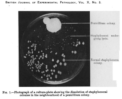
Penicillin in a culture dish of staphylococci. Source: NCBI

Fleming isolated the organism responsible for prohibiting the growth of the staphylococcus, and identified it as being from the penicillium genus.

He named it penicillin and the rest is history.

Fleming himself appreciated the serendipity of the finding:

“When I woke up just after dawn on Sept. 28, 1928, I certainly didn’t plan to revolutionise all medicine by discovering the world’s first antibiotic, or bacteria killer. But I guess that was exactly what I did.” (Source)

And this gave rise to his famous quote:

“One sometimes finds what one is not looking for” (Source)

While Fleming’s discovery of the antibiotic properties of penicillin was made as he was working on a completely different research problem, the important thing to note is that the discovery was made because the evidence came to prepared mind.

Louis-Pasteur-Quotes-1

Pasteur knew the importance of a prepared mind. Source: Thequotes

And this is the purpose of all the training in scientific research – not acquiring ‘the keys to some secret knowledge’, but preparing the investigator to notice the curious deviation.

That’s all really interesting. But what does any of this have to do with Parkinson’s disease?

Three things:

  1. Serendipity
  2. Prepared minds
  3. Antibiotics.

Huh?

Five years ago, a group of Brazilian and German Parkinson’s disease researchers made a serendipitous discovery:

While modelling Parkinson’s disease in some mice, they noticed that only two of the 40 mice that were given a neurotoxic chemical (6-OHDA) developed the motor features of Parkinson’s disease, while the rest remained healthy. This result left them scratching their heads and trying to determine what had gone wrong.

Then it clicked:

“A lab technician realised the mice had mistakenly been fed chow containing doxycycline, so we decided to investigate the hypothesis that it might have protected the neurons.” (from the press release).

The researchers had noted the ‘curious deviation’ and decided to investigate it further.

They repeated the experiment, but this time they added another group of animals which were given doxycycline in low doses (via injection) and fed on normal food (not containing the doxycycline).

And guess what: both group demonstrated neuroprotection!

Hang on a second. Two questions: 1. What exactly is 6-OHDA?
6-hydroxydopamine (or 6-OHDA) is one of several chemicals that researchers use to cause dopamine cells to die in an effort to model the cell death seen in Parkinson’s disease. It shares many structural similarities with the chemical dopamine (which is so severely affected in the Parkinson’s disease brain), and as such it is readily absorbed by dopamine cells who unwittingly assume that they are re-absorbing excess dopamine.

Once inside the cell, 6-OHDA rapidly transforms (via oxidisation) into hydrogen peroxide (H2O2 – the stuff folk bleach their hair with) and para-quinone (AKA 1,4-Benzoquinone). Neither of which the dopamine neurons like very much. Hydrogen peroxide in particular quickly causes massive levels of ‘oxidative stress’, resulting in the cell dying.
6OHDA

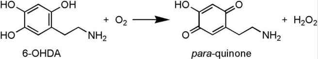
Transformation of the neurotoxin 6-OHDA. Source: NCBI

Think of 6-OHDA as a trojan horse, being absorbed by the cell because it looks like dopamine, only for the cell to work out (too late) that it’s not.

Ok, and question 2. What is doxycycline?

Doxycycline is an antibiotic that is used in the treatment of a number of types of infections caused by bacteria.

doxycycline100-1-1k_1

Remind me again, what is an antibiotic?

Antibiotics are a class of drugs that either kill or inhibit the growth of bacteria. They function in one of several ways, either blocking the production of bacterial proteins, inhibiting the replication of bacterial DNA (nuclei acid in the image below), or by rupturing/inhibiting the repair of the bacteria’s outer membrane/wall.

2018_1

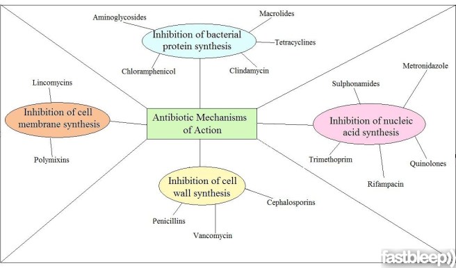
The ways antibiotics function. Source: FastBleep

So the researchers accidentally discovered that the a bacteria-killing drug called doxycycline prevented a trojan horse called 6-OHDA from killing dopamine cells?

Basically, yeah.

And then these prepared minds followed up this serendipitous discovery with a series of experiments to investigate the phenomenon further, and they published the results recently in the journal ‘Glial’:

Glial

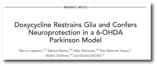
Title: Doxycycline restrains glia and confers neuroprotection in a 6-OHDA Parkinson model.
Authors: Lazzarini M, Martin S, Mitkovski M, Vozari RR, Stühmer W, Bel ED.
Journal: Glia. 2013 Jul;61(7):1084-100. doi: 10.1002/glia.22496. Epub 2013 Apr 17.
PMID: 23595698

In the report of their research, the investigators noted that doxycycline significantly protected the dopamine neurons and their nerve branches (called axons) in the striatum – an area of the brain where dopamine is released – when 6-OHDA was given to mice. Both oral administration and peripheral injections of doxycycline were able to have this effect.

They also reported that doxycycline inhibited the activation of astrocytes and microglial cells in the brains of the 6-OHDA treated mice. Astrocytes and microglial cells are usually the helper cells in the brain, but in the context of disease or injury these cells can quickly take on the role of judge and executioner – no longer supporting the neurons, but encouraging them to die. The researchers found that doxycycline reduced the activity of the astrocytes and microglial cells in this alternative role, allowing the dopamine cells to recuperate and survive.

The researchers concluded that the “neuroprotective effect of doxycycline may be useful in preventing or slowing the progression of Parkinson’s disease”.

Wow, was this the first time this neuroprotective effect of doxycycline has been observed?

Curiously, No.

We have known of doxycycline’s neuroprotective effects in different models of brain injury since the 1990s (Click here, here and here for more on this). In fact, in their research report, the German and Brazilian researchers kindly presented a table of all the previous neuroprotective research involving doxycycline:

table1

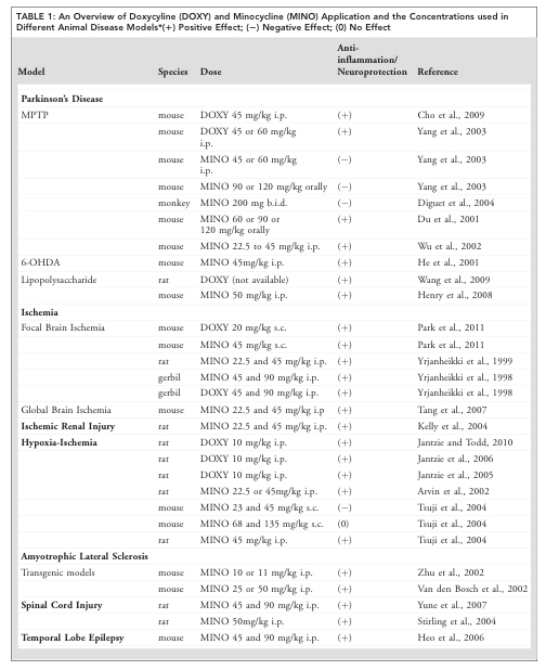
And there was so much of it that the table carried on to a second page:
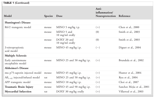
Table2

Source: Glia

And as you can see from the table, the majority of these reports found that doxycycline treatment had positive neuroprotective effects.

Is doxycycline the only antibiotic that exhibits neuroprotective properties?

No.

Doxycycline belongs to a family of antibiotics called ‘tetracyclines‘ (named for their four (“tetra-“) hydrocarbon rings (“-cycl-“) derivation (“-ine”)), and other members of this family have also been shown to display neuroprotection in models of Parkinson’s disease:

MPTP

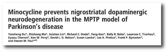
Title: Minocycline prevents nigrostriatal dopaminergic neurodegeneration in the MPTP model ofParkinson’s disease.
Authors: Du Y, Ma Z, Lin S, Dodel RC, Gao F, Bales KR, Triarhou LC, Chernet E, Perry KW, Nelson DL, Luecke S, Phebus LA, Bymaster FP, Paul SM.
Journal: Proc Natl Acad Sci U S A. 2001 Dec 4;98(25):14669-74.
PMID: 11724929                    (This article is OPEN ACCESS if you would like to read it)

In this study, the researchers treated mice with an antibiotic called minocycline and it protected dopamine cells from the damaging effects of a toxic chemical called MPTP (or 1-methyl-4-phenyl-1,2,3,6-tetrahydropyridine). MPTP is also used in models of Parkinson’s disease, as it specifically affects the dopamine cells, while leaving other cells unaffected.

The researchers found that the neuroprotective effect of minocycline is associated a reduction in the activity of proteins that initiate cell death (for example, Caspace 1). This left the investigators concluding that ‘tetracyclines may be effective in preventing or slowing the progression of Parkinson’s disease’.

Importantly, this result was quickly followed by two other research papers with very similar results (Click here and here to read more about this). Thus, it would appear that some members of the tetracycline class of antibiotics share some neuroprotective properties.

So what did the Brazilian and German researchers do next with doxycycline?

They continued to investigate the neuroprotective effect of doxycycline in different models of Parkinson’s disease. They also got some Argentinians and Frenchies involved in the studies. And these lines of research led to their recent research report in the journal Scientific Reports:

Doxy1
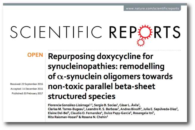
Title: Repurposing doxycycline for synucleinopathies: remodelling of α-synuclein oligomers towards non-toxic parallel beta-sheet structured species.
Authors: González-Lizárraga F, Socías SB, Ávila CL, Torres-Bugeau CM, Barbosa LR, Binolfi A, Sepúlveda-Díaz JE, Del-Bel E, Fernandez CO, Papy-Garcia D, Itri R, Raisman-Vozari R, Chehín RN.
Journal: Sci Rep. 2017 Feb 3;7:41755.
PMID: 28155912                (This article is OPEN ACCESS if you would like to read it)

In this study, the researchers wanted to test doxycycline in a more disease-relevant model of Parkinson’s disease. 6-OHDA is great for screening and testing neuroprotective drugs. But given that 6-OHDA is not involved with the underlying pathology of Parkinson’s disease, it does not provide a great measure of how well a drug will do against the disease itself. So, the researchers turned their attention to our old friend, alpha synuclein – the protein which forms the clusters of protein (called Lewy bodies) in the Parkinsonian brain.

What the researchers found was fascinating: Doxycycline was able to inhibit the disease related clustering of alpha synuclein. In fact, by reshaping alpha synuclein into a less toxic version of the protein, doxycycline was able to enhance cell survival. The investigators also conducted a ‘dosing’ experiment to determine the most effect dose and they found that taking doxycycline in sub-antibiotic doses (20–40 mg/day) would be enough to exert neuroprotection. They concluded their study by suggesting that these novel effects of doxycycline could be exploited in Parkinson’s disease by “repurposing an old safe drug”.

Wow, has doxycycline ever been used in clinical trials for brain-related conditions before?

Yes.

From 2005-12,there was a clinical study to determine the safety and efficacy of doxycycline (in combination with Interferon-B-1a) in treating Multiple Sclerosis (Click here for more on this trial). The results of that study were positive and can be found here.

More importantly, the other antibiotic to demonstrate neuroprotection in models of Parkinson’s disease, minocycline (which we mentioned above), has been clinically tested in Parkinson’s disease:

title1

Title: A pilot clinical trial of creatine and minocycline in early Parkinson disease: 18-month results.
Authors: NINDS NET-PD Investigators..
Journal: Clin Neuropharmacol. 2008 May-Jun;31(3):141-50.
PMID: 18520981                (This article is OPEN ACCESS if you would like to read it)

This research report was the follow up of a 12 month clinical study that can be found by clicking here. The researchers had taken two hundred subjects with Parkinson’s disease and randomly sorted them into the three groups: creatine (an over-the-counter nutritional supplement), minocycline, and placebo (control). All of the participants were diagnosed less than 5 years before the start of the study. At 12 months, both creatine and minocycline were noted as not interfering with the beneficial effects of symptomatic therapy (such as L-dopa), but a worrying trend began with subjects dropping out of the minocycline arm of the study.

At the 18 month time point, approximately 61% creatine-treated subjects had begun to take additional treatments (such as L-dopa) for their symptoms, compared with 62% of the minocycline-treated subjects and 60% placebo-treated subjects. This result suggested that there was no beneficial effect from using either creatine or minocycline in the treatment of Parkinson’s disease, as neither exhibited any greater effect than the placebo. In addition, the investigators suggested that the decreased tolerability of minocycline was a concern.

Ok, so where do I sign up for the next doxy clinical trial?

Well, the researchers behind the Scientific reports research (discussed above) are hoping to begin planning clinical trials soon.

But theoretically speaking, there shouldn’t be a trial.

Huh?!?

There’s a good reason why not.

In fact, if you look at the comments section under the research article, a cautionary message has been left by Prof Paul M. Tulkens of the Louvain Drug Research Institute in Belgium. He points out that:

“…using antibiotics at sub-therapeutic doses is the best way to trigger the emergence of resistance (supported by many in vitro and in vivo studies). Using an antibiotic for other indications than an infection caused by a susceptible bacteria is something that should be discouraged”

And he is correct.

We recklessly over use antibiotics all over the world at the moment and they are one of the few lines of defence that we have against the bacterial world. Long term use (which Parkinson’s disease would probably require) of an antibiotic at sub-therapeutic levels will only encourage the rise of antibiotic resistant bacteria (possibly within individuals).

The resistance of bacteria to antibiotics can occur spontaneously via several means (for example, through random genetic mutations during cell division). With the right mutation (inferring antibiotic resistance), an individual bacteria would then have a natural advantage over their friends and it would survive our attempts to kill it with antibiotics. Being resistant to antibiotic would leave that bacteria to wreak havoc upon us.

Its the purest form of natural selection.

natural-selection_140211

How bacteria become resistant to antibiotics. Source: Reactgroup

And antibiotic resistant bacteria are fast becoming a major health issue for us, with the number of species of bacteria developing resistance increasing every year (Click here for a good review on factors contributing to the emergence of resistance, and click here for a review of the antibiotic resistant bacteria ‘crisis’).

But don’t be upset on the Parkinson’s disease side of things. Prof Tulken adds that:

“If doxycycline really acts as the authors propose, the molecular targets are probably very different from those causing antibacterial activity. it should therefore be possible to dissociate these effect from the antibacterial effects and to get active compounds devoid of antibacterial activity This is where research must go to rather than in trying to use doxycycline itself.”

And he is correct again.

Rather than tempting disaster, we need to take the more prudent approach.

Independent researchers must now attempt to replicate the neuroprotective results in carefully controlled conditions. At the same time, chemists should conduct an analysis of the structure of doxycycline to determine which parts of it are having this neuroprotective effect.

Doxycycline_structure.svg

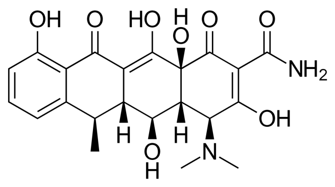
The structure of doxycycline. Source: Wikipedia

If researchers can isolate those neuroprotective elements and those same parts are separate from the antibiotic properties, then we may well have another experimental drug for treating Parkinson’s disease.

And the good news is that researchers are already reasonably sure that the mechanisms of the neuroprotective effect of doxycycline are distinct from its antimicrobial action.

So what does it all mean?

Researchers have once again identified an old drug that can perform a new trick.

The bacteria killing antibiotic, doxycycline, has a long history of providing neuroprotection in models of brain disease, but recently researchers have demonstrated that doxycycline may have beneficial effects on particular aspects of Parkinson’s disease.

Given that doxycycline is an antibiotic, we must be cautious in our use of it. It will be interesting to determine which components of doxycycline are neuroprotective, and whether other antibiotics share these components. Given the number of researchers now working in this area, it should not take too long.

We’ll let you know when we hear something.


EDITOR’S NOTE: Under absolutely no circumstances should anyone reading this material consider it medical advice. The material provided here is for educational purposes only. Before considering or attempting any change in your treatment regime, PLEASE consult with your doctor or neurologist. While some of the drugs discussed on this website are clinically available, they may have serious side effects. We therefore urge caution and professional consultation before any attempt to alter a treatment regime. SoPD can not be held responsible for any actions taken based on the information provided here. 


The banner for today’s post was sourced from Youtube

Trying to digest gut research

2015-02-16-BrainvsStomachImage.png

Our first ever posting here on the SoPD dealt with the curious relationship between the gut and Parkinson’s disease (Click here to see that post). Since then, there have been a string of interesting research reports adding to the idea that the gastrointestinal system may be somehow influencing the course of Parkinson’s disease.

In today’s post we will review the most recent helpings and discuss how they affect our understanding of Parkinson’s disease.


Qz

Source: Qz

Interesting fact: The human digestive system is about 26 feet long – approximately 8 meters – from mouth to anus.

Recent research indicates that our brains are heavily influenced by the activities of this food consuming tract. Not just the nutrients that it takes in, but also by the bugs that live within those 26 feet.

Another interesting fact: The human gut hosts tens of trillions of microorganisms, including at least 1000 species of bacteria (which is a guess-timate as we are not really sure how many species there are). They make up as much as 2 kg of your total weight.

And those bacteria have influence!

In December of last year, we reviewed a study in which the researchers demonstrated that mice genetically engineered to display features of Parkinson’s disease performed as well as normal mice if they were raised with reduced levels of bacteria in their gut (either in a germ-free environment or using antibiotics). That study also showed that transplanting bacteria from the gut of people with Parkinson’s disease into mice raised in a germ-free environment resulted in those mice performing worse on the behavioural tasks than mice injected with gut samples from healthy human subjects (Click here to read that post).

Wow, so what new gut research has been reported?

A little bit of history first:

Two years ago, some Danish researchers published this research report:

Gut3

Title: Vagotomy and Subsequent Risk of Parkinson’s Disease.
Authors: Svensson E, Horváth-Puhó E, Thomsen RW, Djurhuus JC, Pedersen L, Borghammer P, Sørensen HT.
Journal: Annals of Neurology, 2015, May 29. doi: 10.1002/ana.24448.
PMID: 26031848

In their report, the researchers highlighted the reduced risk of Parkinson’s disease following a truncal vagotomy.

So what’s a truncal vagotomy?

A vagotomy is a surgical procedure in which the vagus nerve is cut. It is typically due to help treat stomach ulcers.

The vagus nerve runs from the lining of the stomach to the brain stem, near the base of the brain.

gut_aid_in_PD
A diagram illustrating the vagal nerve connection with the enteric nervous system which lines the stomach. Source: NCBI

A vagotomy comes in two forms: it can be ‘truncal‘ (in which the main nerve is cut) or ‘superselective’ (in which specific branches of the nerve are cut, which the main nerve is left in tact).

Vagotomy

A schematic demonstrating the vagal nerve surrounding the stomach. Image A. indicates a ‘truncal’ vagotomy, where the main vagus nerves are cut above the stomach; while image B. illustrates the ‘superselective’ vagotomy, cutting specific branches of the vagus nerve connecting with the stomach. Source: Score

And what did the Danish scientists find?

Exploring the public health records, the Danish researcher found that between 1975 and 1995, 5339 individuals had a truncal vagotomy and 5870 had superselective vagotomy. Using the Danish National registry (which which stores all of Denmark’s medical information), they then looked for how many of these individuals went on to be diagnosed with Parkinson’s disease. They compared these vagotomy subjects with more than 60,000 randomly-selected, age-matched controls.

They found that subjects who had a superselective vagotomy had the same chance of developing Parkinson’s disease as anyone else in the general public (a hazard ratio (or HR) of 1 or very close to 1).

But when they looked at the number of people in the truncal vagotomy group who were later diagnosed with Parkinson’s disease, the risk had dropped by 35%. Furthermore, when they followed up the truncal group 20 years later, checking to see who had been diagnosed with Parkinson’s in 2012, they found that their rate was half that of both the superselective group and the control group (see table below; HR=0.53). The researchers concluded that a truncal vagotomy reduces the risk of developing Parkinson’s disease.

Svensson_Table2

Source: Svensson et al (2015) Annals of Neurology – Table 2.

Then last year, at the meeting in Berlin, data was presented that failed to replicate the findings in a separate group of people (Sweds).

Vagotomy

Title: Vagotomy and Parkinson’s disease risk: A Swedish register-based matched cohort study
Authors: B. Liu, F. Fang, N.L. Pedersen, A. Tillander, J.F. Ludvigsson, A. Ekbom, P. Svenningsson, H. Chen, K. Wirdefeldt
Abstract Number: 476 (click here to see the original abstract – OPEN ACCESS)

The Swedish researchers collected information regarding 8,279 individuals born in Sweden between 1880 and 1970 who underwent vagotomy between 1964 and 2010 (3,245 truncal and 5,029 selective). For each vagotomized individual, they  collected medical information for 40 control subjects matched for sex and year of birth (at the date of surgery). They found that vagotomy was not associated with Parkinson’s disease risk.

Truncal vagotomy was associated with a lower risk more than five years after the surgery, but that result was not statistically significant. The researcher suggested that the findings needs to be verified in larger samples.

The results of that study have now been published (this week):

Swedish
Title: Vagotomy and Parkinson disease: A Swedish register-based matched-cohort study
Authors: Liu B, Fang F, Pedersen NL, Tillander A, Ludvigsson JF, Ekbom A, Svenningsson P, Chen H, Wirdefeldt K.
Journal: Neurology. 2017 Apr 26. pii: 10.1212/WNL.0000000000003961.
PMID: 28446653             (This article is OPEN ACCESS if you would like to read it)

In this report, the researchers suggest that “there was a suggestion of lower risk among patients with truncal vagotomy” and they note that the hazard ratio (or HR) is 0.78 for this group (ranging between 0.55-1.09), compared to the HR of 0.96 (ranging between 0.78-1.17) for all of the vagotomy group combined. And they not that this trend is further apparent when the truncal vagotomy was conducted at least 5 years before Parkinson’s disease diagnosis (HR = 0.59, ranging between 0.37-0.93). These numbers are not statistically significant, so the investigators could only suggest that there was a trend towards truncal vagotomy lowering the risk of Parkinson’s disease.

What are the differences between the studies?

The Danish researcher analysed medical records between 1975 and 1995 from 5339 individuals had a truncal vagotomy and 5870 had superselective vagotomy. The Sweds on the other hand, looked over a longer period (1964 – 2010) but at a smaller sample size for the truncal group (3,245 truncal and 5,029 selective). Perhaps if the truncal group in the Swedish study was higher, the trend may have become significant.

So should we all rush out and ask our doctors for a vagotomy?

No.

That would not be advised (though I’d love to be a fly on the wall for that conversation!).

It is important to understand that a vagotomy can have very negative side-effects, such as vomiting and diarrhoea (Click here to read more on this).

Plus, while the results are interesting, we really need a much larger study for definitive conclusions to be made. You see, in the Danish study (the first report above) the number of people that received a truncal vagotomy (total = 5339) who then went on develop Parkinson’s disease 20 years later was just 10 (compared with 29 in the superselective group). And while that may seem like a big difference between those two numbers, the numbers are still too low to be truly conclusive. We really need the numbers to be in the hundreds.

Plus, it is important to determine whether this result can be replicated in other countries. Or is it simply a Scandinavian trend?

Mmm, interesting. So what does it all mean?

No, stop. We’re not summing up yet. This is one of those ‘but wait there’s more!’ moments.

It has been a very busy week for Parkinson’s gut research.

A German research group published a report about their analysis of the microbes in the gut and how they differ in Parkinson’s disease (when compared to normal healthy controls).

maxresdefault

Microbes. Source: Youtube

Regular readers of this blog will realise that we have discussed this kind of study before in a previous post (Click here for that post).

This type of study – analysing the bacteria of the gut – has now been done not just once:

biota-title

Title: Gut microbiota are related to Parkinson’s disease and clinical phenotype.
Authors: Scheperjans F, Aho V, Pereira PA, Koskinen K, Paulin L, Pekkonen E, Haapaniemi E, Kaakkola S, Eerola-Rautio J, Pohja M, Kinnunen E, Murros K, Auvinen P.
Journal: Mov Disord. 2015 Mar;30(3):350-8.
PMID: 25476529

Nor twice:

Fecal3

Title: Short chain fatty acids and gut microbiota differ between patients with Parkinson’s disease andage-matched controls.
Authors: Unger MM, Spiegel J, Dillmann KU, Grundmann D, Philippeit H, Bürmann J, Faßbender K, Schwiertz A, Schäfer KH.
Journal: Parkinsonism Relat Disord. 2016 Nov;32:66-72.
PMID: 27591074

Not three times:

Kesh1

Title: Colonic bacterial composition in Parkinson’s disease
Authors: Keshavarzian A, Green SJ, Engen PA, Voigt RM, Naqib A, Forsyth CB, Mutlu E, Shannon KM.
Journal: Mov Disord (2015) 30, 1351-1360.
PMID: 26179554

Not even four times:

Plos1

Title: Intestinal Dysbiosis and Lowered Serum Lipopolysaccharide-Binding Protein in Parkinson’s Disease.
Authors: Hasegawa S, Goto S, Tsuji H, Okuno T, Asahara T, Nomoto K, Shibata A, Fujisawa Y, Minato T, Okamoto A, Ohno K, Hirayama M.
Journal: PLoS One. 2015 Nov 5;10(11):e0142164.
PMID: 26539989                    (This article is OPEN ACCESS if you would like to read it)

But FIVE times now (all the results published in the 2 years):

gut-title

Title: Parkinson’s disease and Parkinson’s disease medications have distinct signatures of the gut microbiome.
Authors: Hill-Burns EM, Debelius JW, Morton JT, Wissemann WT, Lewis MR, Wallen ZD, Peddada SD, Factor SA, Molho E, Zabetian CP, Knight R, Payami H.
Journal: Mov Disord. 2017 Feb 14. [Epub ahead of print]
PMID: 28195358

(And we apologies to any researchers not mentioned here – these are simply the studies we are aware of).

The researchers in the study published this week, however, did something different to these previous studies:

Gut1
Title: Functional implications of microbial and viral gut metagenome changes in early stage L-DOPA-naïve Parkinson’s disease patients
Authors: Bedarf JR, Hildebrand F, Coelho LP, Sunagawa S, Bahram M, Goeser F, Bork P, Wüllner U.
Journal: Genome Med. 2017 Apr 28;9(1):39.
PMID: 28449715            (This article is OPEN ACCESS if you would like to read it)

The researchers in this study focused their analysis on 31 people with early stage Parkinson’s disease. In addition, all of those subjects were not taking any L-DOPA. The fecal samples collected from these subjects was compared with samples from 28 age-matched controls.

And what did they find?

In the early-stage, L-dopa-naïve Parkinson’s disease fecal samples, the researchers found increased levels of two families of microbes (Verrucomicrobiaceae and unclassified Firmicutes) and lower levels of two other familes (Prevotellaceae and Erysipelotrichaceae). And these differences could be used to reliably differentiate between the two groups (PD and control) to an accuracy of 84%.

In addition, the investigators found that the total virus abundance was decreased in the Parkinsonian participants. The researchers concluded that their study provides evidence of differences in the microbiome of the gut in Parkinson’s disease at a very early stage in the course of the condition, and that exploration of the Parkinson’s viral populations “is a promising avenue to follow up with more specific research” (we here at SoPD are particularly intrigued with this statement!).

So is there a a lot of consensus between the studies? Any new biomarkers?

(Big sigh) Yes….. and no on the consensus question.

The good news is that all of the studies agree that there is a difference between the abundance of different groups of bacteria in the Parkinsonian gut.

BUT only three of the six studies studies demonstrate any agreement as to which groups of bacteria. And those three studies could only agree on one family of bacteria. The recent study (Bedarf et al) agreed with the Scheperjans et al and Unger et al studies in that they all observed found reduced levels of Prevotellaceae bacteria in the gut of people with Parkinson’s disease.

216-5198-1-pb

The Prevotellaceae family of bacteria. Source: MindsofMalady

Unfortunately, the reduction in abundance of this particular bacteria does not appear to be specific to Parkinson’s disease, as similar reduced levels have been observed in Japanese multiple sclerosis patients and in autistic children (Click here and here to read more about those studies).

This lack of agreement between the studies with regards to the difference in the abundance of the families of bacteria may reflect the complexity of the gut microbiome. Alternatively, it could also reflect regional differences (the Keshavarzian et al. study was conducted in Chicago, the Bedarf et al and Unger et al studies were in Germany, Scheperjans et al was in Finland, Hill-Burn et al in Alabama, and the Hasegawa et al study was in conducted in Japan).

Either way, it leaves the field lacking agreement as to which families of bacteria should be followed up in future research.


So what does it all mean?

Right, so summing up, researchers are trying to determine what role the gut may play the course of Parkinson’s disease. There is evidence that the nerves connecting the digestive organ to the brain may act as some kind of gate way for an unknown agent or simply a provocative element in the condition. Severing those nerves to the gut appears to reduce the risk of developing Parkinson’s disease.

And the bacteria populating the gut appears to be different in people with Parkinson’s disease, but there does not seem to be consistency between studies, leaving the search for biomarkers in this organ sadly lacking. Maybe it reflects regional differences, perhaps it reflects the complexity of Parkinson’s disease. Hopefully as follow up research into this particular field continues, a consensus will begin to appear. Admittedly, most of these studies are based on single fecal samples collected from individuals at just one time point. A better experimental design would be to collect multiple samples over time, allowing for variability within and between individuals to be ironed out.

Despite all of these cautionary comments, there does appear to be some smoke here. And we will be watching the gut with great interest as more research comes forward.


The banner for today’s post was sourced from the HuffingtonPost

Improving Patient Education – Introducing Eirwen Malin

1352_Patient_Education_1140x400

Today’s post is something a bit different from our usual fodder.

Here at SoPD HQ, we like to throw our support behind worthy projects. And we were recently contacted by Eirwen Malin regarding an idea that we were genuinely passionate about: Improving patient education

Eirwen is being supported by the Winston Churchill Memorial Trust and in the second half of the year she will travel to the USA and then later to Argentina to find new ideas for Patient Education.

In today’s post, we are handing over the keys to the car to Eirwen and we will let her explain the project that she is about to undertake. The goal of this post is to get feedback, ideas and thoughts about the plans for her project. We also encourage all our readers to follow Eirwen (contact details at the bottom of the post) as she undertakes this exciting endeavour.


UntitledHello, this is me – Eirwen Malin. I’m not prepared to own up to quite how many years I worked in the Third sector in Wales but take it from me (and my photo) quite a lot. I worked mostly trying to influence policy and practice, advocating on behalf of a range of different groups and issues, researching, running demonstration projects, that type of thing. Trying to get the issues heard above the clamour and competing for funding which would hopefully make a difference.

In 2014 I was diagnosed with Parkinson’s and life changed a lot.

It took me a while to realise it but apart from threatening my physical voice, (it’s suggested that 75-90% of people with Parkinson’s have some sort of voice, speech or communication difficulties, see here for more information) having Parkinson’s gave me a new and more powerful voice. I could now speak with the authority of “lived experience”, which might help me make a difference for me and my fellow Parkies.

My own experience of diagnosis was not good. I’d been referred to a neurologist to “put my mind at rest”, so it was completely unexpected. I was not diagnosed by a PD specialist and had to be referred on to a clinic, I was told what I needed was information and then sent off to wait for an appointment with no phone number, website address, fact sheet, nothing!

While I waited, for nearly 6 months, I found masses of information, some of it well expressed and clear, some incomprehensible, some coming from authoritative sources, some from people who were living with the condition, some contradictory, some pseudo-scientific, some completely off the wall yet plausible, in short a potential minefield! Now I am a reasonably competent person, who quite enjoys and is able to read and make sense of research papers, understand the statistics, weigh up the arguments and generally make sense of what’s available. However lots of people with Parkinson’s will not be like me.

So, now I thought maybe I could use my new voice to shout out for the need for Patient Education.

I signed up as a volunteer facilitator for Parkinson’s UK’s Self-Management Programme.

parkinsonsuk_logolong_4cp

It’s an excellent course, based on work long championed by Dr Kate Lorig at Stanford:

static1.squarespace

I have seen it help people to gain confidence and regain a measure of control over their lives. I would thoroughly recommend it, however, 6 half day sessions can’t provide the on-going information about medical, social and lifestyle adjustments that are required to live as well as possible with Parkinson’s. Sometimes one needs almost daily updates to manage the complex and peskily changing symptoms. After all the patient is the only one who is there 24/7/365! They must know where to find information and importantly what questions to ask. I meet far too many people desperately seeking guidance.

The opportunity arose to apply for a Winston Churchill Travel Fellowship, as the strapline says the idea is “Travel to learn – return to inspire”.

Logo + image resized_11

Established in 1965, following the death of Sir Winston Churchill, the Winston Churchill Memorial Trust hands out 150 fellowships each year providing a unique opportunity for UK citizens to travel overseas with the goal of bringing back fresh ideas and new solutions to today’s issues, for the benefit of others in the UK.

At the end of a quite lengthy application process which has whittled over 1000 application down to about 150 Fellowships I feel very honoured, “Yippee”, to have been successful in the Medical Practice and Education category alongside a CEO of an NHS Trust, Doctors, Nurses, Researchers etc, Yikes!

My quest is to find new ideas for Patient Education. I am focussing particularly in the field of degenerative neurological conditions. This area is particularly tricky because I think it’s reasonable to say that even the experts still have much to learn and the multi-faceted nature of the conditions and their symptoms result in the need for a team of medical practitioners to support the patient making it even more difficult to provide consistent information. The patient has to know whats going on.

For my fellowship, I will be travelling to USA and Argentina.

map-canada-usa-argentina

Yippee, exciting trips, but once again Yikes, the journeys are long, I’ll get even stiffer, and I don’t do well in crowds or queues. It’s a good job that my partner can come and help with the stressful bits.

I’ve tried to cover as many perspectives as I can think of but I’d welcome ideas from readers. I’ll try to fit them in.

A bit more detail on the US trip July-August 2017.

New York

A meeting with the Michael J Fox Foundation. I found lots of information on their website, it seems like a good place to start.

MJFFLogo

I’m hoping to visit New York University’s Electronic Media Patient Education Initiative but still waiting for confirmation

I’m excited to be going to visit Dance for PD in Brooklyn. They do some fantastic work getting people moving and I’m sure there is much to learn.

DFPDlogo400x129

I’ll be talking with the charismatic David Leventhal and other staff to ask how they see their educational role. Most important I want to get feedback from their participants. I have developed a one-woman storytelling performance Sorting the Sock Drawer which I will use to stimulate discussion.

San Francisco area

I’m going to see Dr Kate Lorig (mentioned above) and hoping to talk to some people who will be at Stanford on a one-week course.

University of California San Francisco Parkinson’s Disease Clinic and Research Centre. Really clearly written information on their website that I wish I had found earlier.

UCSF_sublogo_ParkinsonsDiseaseClinic_navy_RGB

By the way I’ll be packing my cheesecloth tunic and flares – it’s the 50th anniversary of the “Summer of Love” with apologies to younger readers who don’t remember!

Denver

I will meet Professor Cynthia McRae, a behavioural psychologist who focusses on the impact of non-medical symptoms such as quality of life, depression, loneliness, and other psychological factors that are often associated with Parkinson’s disease, but are not always included within medical research.

cMcRae_000

Professor Cynthia McRae

She said “If there is such a thing as a good place to have Parkinson’s then Denver is it!” and introduced me to the Parkinson’s Association of the Rockies. They have so much going on I shall spend some time with them. Once again I’ll be using my performance to stimulate a discussion.

Dallas

A flying visit to spend a day with the Parkinson’s Voice Project, their mantra about people with Parkinson’s living “with intent” really speaks to me.

logoxx

Pittsburgh and around

University of Pittsburgh Institute for Neurodegenerative Diseases to be confirmed

I will visit the Wheeling Hospital Parkinson’s Education Centre. Also at Wheeling I will be filling a gap in the schedule to speak with general medical practitioners to ask about issues for them in helping patients with neurodegenerative conditions

Finally Health Plan, in St Clairesville Ohio. Health Plan is a not-for profit health insurance provider that calls itself a health maintenance organisation. It will be interesting to get a business perspective on Patient Education.

url

Alongside the formal meetings I hope to just talk to people I meet, tell them what I am doing and get their views. In the end that might be as important as the planned programme.

Planning for Argentina in Oct-Nov still to be finalised.

So once again Yippee it’s going to be fascinating and exciting but Yikes I really do hope I can come home with the goods and help influence the provision of a better system of educating patients than I encountered. I feel the sense of responsibility to the Winston Churchill Memorial Trust who have invested faith and funds in my idea and my ability to deliver and even more so to my fellow Parkies diagnosed or not for whom I’d like to make a difference.

I’ll be posting activities, photos, videos of the formal meetings and the more touristy parts of the trips on facebook https://www.facebook.com/EirwenWCTF you can follow my activities, send me messages there os send messages via e-mail eirwenwctf@gmail.com

Wearable Tech 4 Parkinson’s

wt-cover-crop

We live in an increasingly interconnected technological world.

One can chose to embrace it or ignore it, but I don’t think anyone can do anything to stop it – the masses seem to desire it.

The benefits of all this technology are many, however, for people with Parkinson’s disease. In today’s post we will look at some of the ways wearable technology can be used to improve the lives of people with Parkinson’s disease.


wpid-photo-jan-1-2014-817-pm2

Does anyone still talk to each other? Source: Teachingwithipad

The great Albert Einstein once said that he feared “the day that technology will surpass our human interaction. The world will have a generation of idiots”.

While there are certainly many examples of this situation playing out in our modern society today, the quote misses the mark with regards to the application and benefits of such technology.

For example, people with Parkinson’s disease can now communicate with people in the Parkinson’s community (like ourselves) from anywhere the world. They can reach out and share not only their experiences, but also what treatments and remedies have worked for them.

And then there are the other less obvious applications of an interconnected world:

selfcare-infographic-english

A schematic illustrating the limited monitoring of Parkinson’s. Source: Riggare

On her fantastic blog, engineer and ‘proud mother’ Sara Riggare posted the image above to illustrate the ridiculous current situation regarding the monitoring of Parkinson’s disease. In 2014, she spent 8,765 hours in self care, applying her own knowledge and experience to managing her Parkinson’s disease (8,765 being the number of hours in a year) and had just 1 hour with her physician.

The schematic perfectly illustrates perfectly how little monitoring people with Parkinson’s receive in the standard healthcare system.

People like Sara, however, are taking matters into their own hands. She has become an enthusiastic proponent of ‘self tracking’:

Self tracking represents a fantastic opportunity not only for people with Parkinson’s disease to track their progress, but also for researchers to build up large databases of information relating to the disease from which new theories/hypotheses/treatment approaches could be generated.

And this is possible on a global scale, only because we are a generation of idiots living in a fully interconnected world.

So what opportunities exist for me to self track?

2015-04-21-1429651896-9470404-awatch

Apple Watch. Source: Huffington Post

Recently the technology company Apple announced that it is working on new devices to help track Parkinson’s disease (Click here and here for more on this). The company already offers ResearchKit – a platform available on their iphone.

Apple, however, is actually coming to this party rather late. The Michael J Fox foundation and computer giant Intel formed a partnership back in 2014 to look at wearable technology (Click here to read more about this).

And there are already dozens of applications that can be used on smart phones and tablets. We have previously discussed one such app from a company called uMotif – Click here to read that post.

umotif

umotif. Source: ParkinsonsMovement

In addition, there are other smart phone apps available that readers could try (such as MyTherapyApp) and you can even support new applications as they are being developed (such as Progress Recorder).


What if I don’t have time for entering all the details on the smart phone app?

Not a problem.

Why not just wear a recording sensor? The same way you may wear a piece of jewellery. Simple, easy approach and you can just forget that it is even there.

Would you like an interesting example?

This is Utkarsh Tandon.

photo

He’s a 17 years old student at Cupertino High School. He is also the Founder and CEO of OneRing, an intelligent tool for monitoring Parkinson’s

Yes, you read that correctly – he is just 17 years old. Smart kid, we’ll be watching him.

Why is this technology important?

Until recently out understanding of Parkinson’s has relied entirely on what occurs in the lab and clinic based settings. Now information is being collected 24 hours a day. From sleep quality apps to measuring tremor, all of this technology has several very positive features from the view point of research scientists:

  1. Objective monitoring – rather than subjective measures (eg. clinician’s opinion or subject survey) definitive, replicatable data can be generated.
  2. Continuous monitoring – rather than brief periods of monitoring in an artificial research clinic environment, data can be collected in real world settings on a continuous basis
  3. Data accessibility – rather than pencil and paper collection of results, data can be collected electronically and converted to different formats.
  4. Participant engagement – this included benefits such as getting the community involved with the research, getting feedback about the technology throughout the study, and being able to provide subjects with performance reports on a regular basis.

Is wearable tech only for measuring Parkinson’s disease?

No.

Recently it has also started to aid people with the condition. The best example of this is the story that has most recently captured the attention of the Parkinson’s community here in the UK:

Emma Lawton was diagnosed with Parkinson’s disease at just 29 years of age. Working with Haiyan Zhang (Director of Innovation at Microsoft Research) and colleagues, a bracelet was created that counteracted the tremor in Emma’s wrist.

It’s a good story.

Other tech is helping to make life easier for people with Parkinson’s disease – just have a look at what LiftWare is doing.

liftware560

The Liftware stablising spoon. Source: The Verge

In a clinical study, the Liftware spoons reduced shaking of the spoon bowl by an average of 76 per cent (Click here to read more about this).

meet-anupam-pathak-lift-labs-founder

Anupam Pathak – founder of LiftWare. Source: ET

Liftware has now been aquired by Verily Life Sciences – an independent subsidiary of Alphabet Inc (formerly of Google).

So what does it all mean?

The point of this post was to make readers aware of some of the technological resources that are available to them in this modern age. Using these tools, we can quickly collect a vast amount of information regarding all aspects of life for people with Parkinson’s disease. And it also offers folks the opportunity to get involved with research indirectly (if they have a fear of university hospitals!).

There is also another element to all of this recording of information about Parkinson’s disease that is not immediately apparent: we are potentially (and hopefully) the last generations of human being that will be affected by Parkinson’s disease. If current research efforts allow us to block or dramatically slow the condition in the near future, there may not be a disease for our descendants to worry about. While this is a very worthy goal, there is also a responsibility on the current generation to record, document and learn as much as we can about the condition so that those future generations will have information at hand regarding a forgotten medical condition.

Some folks are already doing this in their own creative ways. For example, we recommend all readers subscribe to PD365 –  a fantastic project in which David Sangster and Emma Lawton (her of the bracelet described above) will be making one short video each day about life with Parkinson’s disease. Raising awareness about the condition and providing intimate insight into basic daily life with PD.

Here is Emma’s first video:

And here is David’s first video:

And this idea is really important.

Consider the great fire of London in 1666. It is estimated that the fire destroyed the homes of 70,000 of the City’s 80,000 inhabitants (Source: Wikipedia), and yet our best sources of information regarding the events of that catastrophe are limited to just a few books like the diary of Samuel Pepys.

This may seem like a silly example, but the premise stands. Given all of the technology we have available today, it would be a great failure for our generation not to be able to provide a thorough source of information regarding this disease.

That said, have a think about getting involved.


The banner for today’s post was sourced from Raconteur

Stress and Parkinson’s disease

stress_general_shutterstock-ollyy_0

Stress.

We all suffer it. Whether it is work related, relationship related, or simply self-induced, we humans foolishly put a great deal of pressure on our bodies.

Many pieces of research suggest that this pressure takes a toll on our health, which could lead to long-term conditions like Parkinson’s disease.

Recently some Korean researchers have identified a stress-related hormone that could have beneficial effects for Parkinson’s disease.

In today’s post, we will review their recently published research and look at what it means for people with Parkinson’s disease.


timthumb

Source: Islamicity

Shortly before leaving the role of President of the United States of America, ex-President Barrack Obama was asked about the stress that comes with the job, and his answer was interesting. He suggested that it is important to take a ‘long view’ of events and not to get bogged down by the weight of everything going on around you:

Despite these sage words, it is difficult not to notice the impact that his previous job has had on the man:

mfxiqpy

What a stress can do to a person. Source: Reddit

Stress seems to be a major part of modern life for many people – some people even indicate that they need it and that they thrive on it. But this pressure that we put on our bodies tends to have a damaging effect on our general health. And there is evidence that that stress may even lead to long term consequences such as cancer and neurodegenerative conditions such as Parkinson’s disease.

Causality, however, is very difficult to determine in science.

The best we can do is suggest that a particular variable (such as stress) may increase one’s risk of developing a particular condition (such as Parkinson’s disease).

So what do we know about stress and Parkinson’s disease?

This is Professor Bas Bloem.

0903tzbasbloem

Prof Bloem – no stress here. Source: NRC

He’s awesome.

Professor Bloem is a consultant neurologist at the Department of Neurology, Radboud University Nijmegen Medical Centre (the Netherlands). He is also one of the researchers behind ParkinsonNet – an innovative healthcare concept that now consists of 64 professional networks for people with Parkinson’s disease covering all of the Netherlands.

In 2010, his research group noticed something interesting:

Bloem

Title: Artistic occupations are associated with a reduced risk of Parkinson’s disease.
Authors: Haaxma CA, Borm GF, van der Linden D, Kappelle AC, Bloem BR.
Journal: J Neurol. 2015 Sep;262(9):2171-6.
PMID: 26138540               (This article is OPEN ACCESS if you would like to read it)

In their study, Prof Bloem and his colleagues conducted a case–controlled analysis of 750 men with Parkinson’s disease (onset ≥40 years) and 1300 healthy men, which involved the participants completing a questionnaire about their occupational history. As expected (based on previous reports), they found that farming was associated with an increased risk of developing Parkinson’s disease (click here for more on this).

Interestingly, artistic occupations late in life were associated with a reduced risk of subsequent Parkinson’s disease. Another interesting observation from the study was that no initial occupation (early in life) predicted Parkinson’s disease, which the researchers proposed indicated that the premotor phase of the disease starts later in life.

One interpretation of this finding is that creative people are less likely to develop Parkinson’s disease. An alternative theory, however, may be that artist jobs are associated with a less stressful, more relaxed lifestyle.

Could it be that the lower levels of stress associated with artistic occupations may be having an impact on the risk of developing Parkinson’s disease?

This idea is not as crazy as it sounds.

Consider different kinds of stress. Research suggests that people who undergo tremendous emotional stress have a higher risk of developing Parkinson’s disease. For example, there is the case of ex-prisoners of war:

Prisoner

Title:Neurological disease in ex-Far-East prisoners of war
Authors: Gibberd FB, Simmonds JP.
Journal: Lancet. 1980 Jul 19;2(8186):135-7.
PMID: 6105303

At the end of World war II, a neurological unit was set up at Queen Mary’s Hospital (Roehampton) to treat Ex-Far East prisoners of war. 4684 individuals were referred to the unit, of these 679 were found to have neurological disease (most of these – 593 cases – were loss of sight and peripheral nerve damage).

In follow up work in the 1970s, however, it was found that many of these individuals had gone on to develop other neurological conditions (dementia, multiple sclerosis, etc). Of interest to us, though was the finding that across the entire group of ex-prisoners investigated (4684 individuals), Parkinson’s disease was apparent in 24 of them – this is a frequency 5x that of the general population!

Even in animal models of Parkinson’s disease, emotional stress seems to exaccerbate the neurodegeneration that is being modelled:

Canada
Title: Stress accelerates neural degeneration and exaggerates motor symptoms in a rat model ofParkinson’s disease.
Authors: Smith LK, Jadavji NM, Colwell KL, Katrina Perehudoff S, Metz GA.
Journal: Eur J Neurosci. 2008 Apr;27(8):2133-46.
PMID: 18412632                  (This article is OPEN ACCESS if you would like to read it)

The investigators in this study demonstrated that chronic stress exaggerates the motor/behavioural deficits in a rat model of Parkinson’s disease. In addition, the stress resulted in a greater loss of dopamine neurons in the brains of these rats.

For an interesting review of the effect of stress in Parkinson’s disease – Click here.

Interesting. So what did the Korean researchers – you mentioned above – find this week?

Something interesting.

This is Dr Yoon-Il Lee.

Lee


Source: Dgist

He’s a dude.

He is a senior research scientists at the Daegu Gyeongbuk Institute of Science and Technology (DGIST) in Daegu Metropolitan City, South Korea.

Recently, his group has collaborated with Professor Yunjong Lee’s research team published this research report:

Lee

Title: Hydrocortisone-induced parkin prevents dopaminergic cell death via CREB pathway inParkinson’s disease model
Authors: Ham S, Lee YI, Jo M, Kim H, Kang H, Jo A, Lee GH, Mo YJ, Park SC, Lee YS, Shin JH, Lee Y.
Journal: Sci Rep. 2017 Apr 3;7(1):525. doi: 10.1038/s41598-017-00614-w.
PMID: 28366931         (This article is OPEN ACCESS if you would like to read it)

Dr Lee and his colleagues began this study with cells were engineered to produce a bioluminescent signal when a gene called Parkin was activated. Parkin is a Parkinson’s associated gene as genetic mutations in this gene can result in carriers developing a juvenile-onset/early-onset form of Parkinson’s disease.

The researchers then conducted an enormous screening experiment to find agents that turn on the Parkin gene. They applied a library of 1172 FDA-approved drugs (from Selleck Chemicals) to these cells – one drug per cell culture – and looked at which cell cultures began to produce a bioluminescent signal. They found 5 drugs that not only made the cells bioluminescent, but also resulted in Parkin protein being produced at levels 2-3 times higher than normal. Those drugs were:

  • Deferasirox – an iron chelator (interesting considering our previous post)
  • Vorinostat – a cancer drug (for treating lymphoma)
  • Metformin – a diabetes medication
  • Clindamycin – an antibiotic
  • Hydrocortisone

Hydrocortisone produced the highest levels of Parkin (Interestingly, hydrocortisone also did not increase the activity of PERK, an indicator of endoplasmic reticulum stress, while the other drugs did).

What is Hydrocortisone?

Hydrocortisone is the name for the hormone ‘cortisol’ when supplied as a medication.

Ok, so what is cortisol?

Cortisol is a glucocorticoid (a type of hormone) produced from cholesterol by enzymes in the cortex of the adrenal gland, which sits on top of the kidneys. It is produced in response to stress (physical or emotional)

CDR739009

The location of the adrenal glands. Source: Cancer

Cortisol helps us to deal with physical or emotional stress by reducing the activity of certain bodily functions – such as the immune system – so that the body can focus all of it’s energies toward dealing with the stress at hand.

Now generally, the functions of cortisol are supposed to be short-lived – long enough for the body to deal with the offending stressor and then levels go back to normal. But the normal levels of cortisol also fluctuate across the span of the day, with levels peaking around 8-9am:


open-uri20120801-11642-h5dnby

A graph of cortisol levels over the day. Source: HealthTap


Ok, so what did the Korean researchers do next?

Dr Lee and his colleagues gave the hydrocortisone drug to cell cultures which they then stressed (causing cell death). Hydrocortisone protected the cells from dying, and (importantly) it achieved this feat in a manner that was dependent on parkin activation. In cells that do not naturally have parkin, hydrocortisone was found to have no effect on cell survival.

Next the researchers treated mice with hydrocortisone before they then modelled Parkinson’s disease using the neurotoxin 6-OHDA. Hydrocortisone treatment resulted in approximate a two-fold increase in levels of parkin within particular areas of the brain. Without hydrocortisone treatment, the mice suffered the loss of approximately 45% of their dopamine neurons. Mice pre-treated with hydrocortisone, however, demonstrated enhanced dopamine neuron survival.

The researchers concluded that a sufficient physiological supply of hydrocortisone was required for protection of the brain, and that hydrocortisone treatment could be further tested as a means of maintaining high levels of parkin in the brain.

So what do we know about cortisol in Parkinson’s disease?

So this is where the story gets interesting;

Dobbs

Title: Cortisol is higher in parkinsonism and associated with gait deficit.
Authors: Charlett A, Dobbs RJ, Purkiss AG, Wright DJ, Peterson DW, Weller C, Dobbs SM.
Journal: Acta Neurol Scand. 1998 Feb;97(2):77-85.
PMID: 9517856

The researchers who conducted this study were interested in the role of cortisol in Parkinson’s disease. They measured cortisol levels in the blood of 96 subjects with Parkinson’s disease and 170 control subjects.  They found that cortisol levels were 20% higher in the subjects with Parkinson’s disease, and that MAO-B inhibitor treatment for Parkinson’s (Selegiline) reduced cortisol levels.

And MAO-B inhibitors are not the only Parkinson’s medication associated with reduced levels of cortisol:

Muller

Title: Acute levodopa administration reduces cortisol release in patients with Parkinson’s disease.
Authors: Müller T, Welnic J, Muhlack S.
Journal: J Neural Transm (Vienna). 2007 Mar;114(3):347-50.
PMID: 16932991

In this study the researchers found that cortisol levels started to decrease significantly just 30 minutes after L-dopa was taken.

Whether this lowering of cortisol levels may have any kind of detrimental effect on Parkinson’s disease is yet to be determined and required further investigation.

Is hydrocortisone or cortisol used in the clinic?

Yes it is.

Hydrocortisone is used to treat rheumatism, skin diseases, and allergies.

white-tablet-pills-on-reflective-surface

Hydrocortisone tablets. Source: Wisegeeks

Thus, there is the potential for another example of drug repurposing here. But the drug is not without side effects, which include:

  • Sleep problems (insomnia)
  • Mood changes
  • Acne, dry skin, thinning skin, bruising or discoloration;
  • Slow wound healing
  • Increased sweating
  • Headache, dizziness, spinning sensation;
  • nausea, stomach pain

For the full list of potential side effects – click here.

So what does it all mean?

Researchers in Korea have recently found that hydrocortisone (cortisol) can increase levels of Parkinson’s associated protein Parkin in cells, which in turn has a positive, neuroprotective effect on models of Parkinson’s disease.

We will now wait to see if the results can be independently replicated before attempting to take this drug to clinical trials for Parkinson’s disease. Any replication of the study should involve a range of treatment regimes so that we can determine if delayed administration can also be beneficial (this would involve delaying hydrocortisone treatment until after the neurotoxin has been given). Those studies could also look at the inflammatory effect in the brains as hydrocortisone has previously been demonstrated to have anti-inflammatory effects.

Interesting times. Stay tuned.


EDITOR’S NOTE: Under absolutely no circumstances should anyone reading this material consider it medical advice. The material provided here is for educational purposes only. Before considering or attempting any change in your treatment regime, PLEASE consult with your doctor or neurologist. While some of the drugs discussed on this website are clinically available, they may have serious side effects. We urge caution and professional consultation before altering any treatment regime. SoPD can not be held responsible for any actions taken based on the information provided here. 


The banner for today’s post was sourced from ZetaYarwood

Old dogs, new tricks – repurposing drugs for Parkinson’s


Drugrepurpose

Exciting news this week from the world of neurodegenerative research. Researchers have identified two clinically available drugs that display neuroprotective properties.

The drugs – Dibenzoylmethane and Trazodone – are currently used to treat cancer and depression, respectively.

In this post, we will review the research and discuss what it could mean for folks with Parkinson’s disease.


Drugs

Old drugs – new tricks? Source: Repurposingdrugs101

As you may have heard from media reports (for examples, click here, here and here), researchers have identified two clinically available drugs that may help in the fight against neurodegenerative conditions, like Parkinson’s disease.

The re-purposing of clinically available drugs is the focus of much attention within the Parkinson’s community as it represents a means of bringing treatments to the clinic faster. The traditional lengthy clinical trial process that is required in the development of new medications means getting a new drug to market for neurodegeneration can take up to 15 years, as the trials run over several years each (and there are three phases to pass through).

ajbtbe-v2-id1047-g001

Shortening the wait. Source: Austinpublishing

In an age of smart phones and instant gratification, who has that kind of patience? ( #Wewontwait ).

Thus, re-purposing of available drugs represents a more rapid means of bringing new treatments/therapies to the Parkinson’s community.

So what is the new research all about?

This is Professor Giovanna Mallucci.

Leicester-2015-05-11-0182_800

Prof Giovanna Mallucci. Source: MRC

She’s awesome.

She led the team from the Medical Research Council’s (MRC) Toxicology Unit (University of Leicester) that conducted the research and she is now based at the University of Cambridge.

Her area of research interest is understanding mechanisms of neurodegeneration, with a particular focus on prion and Alzheimer’s disease.

A few years ago, her group published this report:

Nature

Title: Sustained translational repression by eIF2α-P mediates prion neurodegeneration.
Authors: Moreno JA, Radford H, Peretti D, Steinert JR, Verity N, Martin MG, Halliday M, Morgan J, Dinsdale D, Ortori CA, Barrett DA, Tsaytler P, Bertolotti A, Willis AE, Bushell M, Mallucci GR.
Journal: Nature. 2012 May 6;485(7399):507-11.
PMID: 22622579              (This article is OPEN ACCESS if you would like to read it)

In this study, Prof Mallucci’s group were interested in the biological events that were occurring in the brain following infection of mice with prion disease – another neurodegenerative condition. They found that a sudden loss of protein associated with the connections between neurons (those connections being called synapses) occurred at 9 weeks post infection. This led them to investigate the production of protein and they found that an increase in the levels of phosphorylation of a protein called eIF2alpha was associated with the reduction in protein synthesis occurring at 9 weeks post infection.

What is Phosphorylation?

Phosphorylation of a protein is basically the process of turning it on or off – making it active or inactive – for a particular function.

U2CP5-4_Phosphorylation_revised

Phosphorylation of a kinase protein. Source: Nature

And what is eIF2alpha?

Eukaryotic Translation Initiation Factor 2 Alpha is (as the label on the can suggests) a translation initiation factor. This means that this particular protein functions in the early steps of the production of protein. That is to say, eIF2alpha has important roles in the first steps – the initiation – of making other proteins.

Roles of eIF2 kinases in the pathogenesis of Alzheimer's disease

eIF2alpha’s role in neurodegeneration. Source: Frontiers

The increased phosphorylation of eIF2alpha results in the inactivation of eIF2alpha and therefore the transient shutdown of protein production.

This shutdown in protein production can serve as an important ‘checkpoint’ when a cell is stressed. By blocking general protein production, a damaged or stressed cell can have the opportunity to either recuperate or be eliminated (if the damage is beyond repair).

The shutdown can also be caused by the unfolded protein response (or UPR). The unfolded protein response is a protective mechanism triggered by rising levels of misfolded proteins.

What are misfolded proteins?

When proteins are being produced, they need to be folded into the correct shape to do their job. Things can turn ugly very quickly for a cell if protein are being misfolded or only partially folded.

prions

Two proteins. Guess which is the misfolded protein. Source: Biogeekery

In fact, misfolded proteins are suspected of being responsible for many of the neurodegenerative conditions we know of (including Parkinson’s, Alzheimer’s, etc). Thus the unfolded protein response gives a cell time to stop protein production, degrade & dispose of any misfolded proteins, and then re-activate proteins involved with increasing the production again.

And Prof Mallucci’s group found an increase in the phosphorylation of eIF2alpha?

Yes. At 9 weeks post infection with prions, there is a decrease in the proteins required for maintaining the connections between neurons and an increase in the phosphorylation of eIF2alpha.

The interesting thing is that the researchers found that levels of phosphorylated eIF2alpha increased throughout the course of study.

So, the researchers asked themselves if promoting a recovery in protein production in the cells in neuroprotective. To test this they used a protein called GADD34, which is a specific eIF2alpha phosphatase (a phosphatase is a protein that dephosphates a protein). By introducing a lot of GADD34 in the cells, the researchers were able to re-activate eIF2alpha, rescue the connectivity between neurons and protect the cells from dying.

A cool trick, huh?

This report established the importance of eIF2alpha in the early stages of neurodegeneration, and Prof Mallucci and her group next decided to conduct a massive screening study of currently available medications to see which could be used to target eIF2alpha levels.

And that research gave rise to the report that caused so much excitement this week. This report here:

Brain
Title: Repurposed drugs targeting eIF2α-P-mediated translational repression prevent neurodegeneration in mice
Authors: Halliday M, Radford H, Zents KAM, Molloy C, Moreno JA, Verity NC, Smith E, Ortori CA, Barrett DA, Bushell M, Mallucci GR.
Journal: Brain, 2017 Epub early online publication
PMID: N/A         (This article is OPEN ACCESS if you would like to read it)

The investigators began by testing 1,040 compounds (that represent many of the clinically available drugs we have) on tiny microscopic worms (called C.elegans). These worms represent a useful experimental model for screening drugs as many aspects of biology can be examined. These worms were exposed to both a chemical (called tunicamycin, which induces the unfolded protein response we were talking about above) and one of the 1040 compounds.

Of the 1040 compounds tested, the investigators selected the 20 that provided the best protection to the worms. They next analysed those top 20 compounds for their ability to reduce levels of phosphorylated eIF2alpha in cells. Cells were engineered to produce a bioluminescent signal when eIF2alpha was phosphorylated. The researchers used a potent blocker of the unfolded protein response (called GSK2606414) and a drug called ISRIB (which is an experimental drug which reverses the effects of eIF2alpha phosphorylation) as controls for the experiment.

Their results were interesting:

Figure1

The results of the top 20 drugs screened. Source: Brain

As you can see from the graph above, there were five compounds (highlighted with ***) that provided a similar level of reduction as the ISRIB (control) drug:

  • Azadirachtin – which is the active ingredient in many pesticides.
  • Dibenzoylmethane – a cancer treatment.
  • Proguanil – a medication used to treat and prevent malaria.
  • Trazodone – an antidepressant used to treat depression and anxiety disorders.
  • Trifluoperazine – an antipsychotic of the phenothiazine chemical class.

The investigators decided not to further investigate Azadirachtin as it is a pesticide and displays a poor ability to penetrant the blood-brain-barrier – the protective layer surrounding the brain. They also rejected Proguanil because while it is safe to use in humans, it is toxic in mice. This detail limited the amount of preclinical testing for neurodegeneration that the researchers could do. And finally Trifluoperazine was eliminated as it should not be used in the elderly populations (apparently it ‘increases the risk of death’!), which obviously limited it’s further utility given that age is a major determinant of neurodegeneration.

This selection process left the researchers with Dibenzoylmethane and Trazodone.

The researchers found that both of these drugs can cross the blood-brain-barrier and were able to prevent neurodegeneration and rescue behavioural deficits in prion-infected mice. And they observed no toxic effects of these treatments in other organs (such as the pancreas). The drugs restore correct protein production and increased the survival of the prion-infected mice.

Taking the study one step further, Prof Mallucci and her group asked if the drugs could be effective in a model of another neurodegenerative condition, such as Alzheimer’s disease. To investigated this, they treated rTg4510 mice with both of the drugs. rTg4510 mice produce a lot of a human protein (called tau) that has a particular mutation (known as P301L), which results in the onset of Alzheimer’s like pathology at an early age. The rTg4510 mice received either trazodone or Dibenzoylmethane on a daily basis from 4 months of age and were examined at 8 months of age. The researchers found significantly less cell loss and shrinkage in the brains of the mice treated with one of the two drugs when compared to rTg4510 mice that received no treatment.

The researchers concluded that “these compounds therefore represent potential new disease-modifying treatments for dementia. Trazodone in particular, a licensed drug, should now be tested in clinical trials in patients”.

As Professor Mallucci suggested to the press: “We know that trazodone is safe to use in humans, so a clinical trial is now possible to test whether the protective effects of the drug we see on brain cells in mice with neurodegeneration also applies to people in the early stages of Alzheimer’s disease and other dementias. We could know in 2-3 years whether this approach can slow down disease progression, which would be a very exciting first step in treating these disorders. Interestingly, trazodone has been used to treat the symptoms of patients in later stages of dementia, so we know it is safe for this group.  We now need to find out whether giving the drug to patients at an early stage could help arrest or slow down the disease through its effects on this pathway.”

This is great for Alzheimer’s disease, but what about Parkinson’s?

Well, the researchers did not test the drugs in models of Parkinson’s disease. But we can assume that several research groups are going to be testing this drug in the near future… if they aren’t already!

But have increased levels of eIF2alpha been seen in Parkinson’s disease?

Great question. And the answer is: Yes.

ParkUPS

Title: Activation of the unfolded protein response in Parkinson’s disease.
Authors: Hoozemans JJ, van Haastert ES, Eikelenboom P, de Vos RA, Rozemuller JM, Scheper W.
Journal: Biochem Biophys Res Commun. 2007 Mar 16;354(3):707-11.
PMID: 17254549

In this study the investigators analysed the levels of Unfolded Protein Response activation in the postmortem brains of people who passed away with or without Parkinson’s disease. Specifically, they focused their analysis on the substantia nigra (the region where the dopamine neurons reside and which is most severely affected in Parkinson’s).

The researchers found that both eIF2alpha and a protein called PERK (also known as protein kinase-like ER kinase – which phosphalates eIF2alpha) are present in the dopamine neurons in the substantia nigra of brains from people with Parkinson’s disease, but not in healthy control brains. And as the graph below shows, the investigators noted that there was a trend towards the levels of these proteins peaking within the first five years after diagnosis.

graph

eIF2alpha & PERK levels in the brain. Source: ScienceDirect

Similar postmortem analysis studies have also highlighted the increased levels of Unfolded Protein Response activation in the Parkinsonian brain (Click here to read more on this).

The increase in Unfolded Protein Response activation could be a common feature across different neurodegenerative conditions, suggesting that trazodone and dibenzoylmethane could be used widely to slow the progress of various conditions.

Another connection to Parkinson’s disease is the finding that high levels of the Parkinson’s associated protein alpha synuclein can cause the Unfolded Protein Response:

Activation
Title: Induction of the unfolded protein response by α-synuclein in experimental models of Parkinson’s disease.
Authors: Bellucci A, Navarria L, Zaltieri M, Falarti E, Bodei S, Sigala S, Battistin L, Spillantini M, Missale C, Spano P.
Journal: J Neurochem. 2011 Feb;116(4):588-605.
PMID: 21166675       (This article is OPEN ACCESS if you would like to read it)

The researchers in this study found that introducing large amounts of alpha synuclein into cell cultures results in the initiation of the unfolded protein response. They also observed this phenomenon in genetically engineered mice that produce large amounts of alpha synuclein.

Thus, there is some evidence for eIF2alpha and unfolded protein response-related activities in Parkinson’s disease

So is there are evidence that Dibenzoylmethane might be neuroprotective for Parkinson’s disease?

Yes there is (sort of):

Basic RGB

Title: A dibenzoylmethane derivative protects dopaminergic neurons against both oxidative stress and endoplasmic reticulum stress.
Authors: Takano K, Kitao Y, Tabata Y, Miura H, Sato K, Takuma K, Yamada K, Hibino S, Choshi T, Iinuma M, Suzuki H, Murakami R, Yamada M, Ogawa S, Hori O.
Journal: Am J Physiol Cell Physiol. 2007 Dec;293(6):C1884-94. Epub 2007 Oct 3.
PMID: 17913843             (This article is OPEN ACCESS if you would like to read it)

The investigators of this study found a derivative of dibenzoylmethane which they called 14-26 (chemical name 2,2′-dimethoxydibenzoylmethane) displayed neuroprotective functions both in cell culture and animal models of Parkinson’s disease. The researchers did not look at the unfolded protein response or eIF2alpha and PERK levels, nor did they determine if dibenzoylmethane itself exhibits neuroprotective properties.

This may now need to be re-addressed.

And is there any evidence trazodone having neuroprotective effects in other neurodegenerative conditions?

Yes.

For a review of the neuroprotective effects of trazodone (and other anti-psychotic/anti-depressant drugs) in Huntington’s Disease – Click here.

This sounds very positive for Parkinson’s disease then, no?

Weeeeeell, there is a word of caution to be thrown in here:

There have been reports in the past of trazodone causing motor-related issues in the elderly. Such as this one:

Trazodone

Title: Can trazodone induce parkinsonism?
Authors: Albanese A, Rossi P, Altavista MC.
Journal: Clin Neuropharmacol. 1988 Apr;11(2):180-2.
PMID: 3378227

This report was a single case study of a 74 year old lady who developed depression after losing her sister with whom she lived. She was prescribed trazodone, which was effective in improving her mood. Just several months later, however, she began presenting Parkinsonian symptoms.

Firstly the onset of a resting tremor in the left arm, then a slowing of movement and a masking of the face. The attending physician withdrew the trazodone treatment and within two months the symptoms began to disappear, with no symptoms apparent 12 months later.

And unfortunately this is not an isolated case – other periodic reports of trazodone-induced motor issues have been reported (Click here and here for examples). And this is really strange as Trazodone apparently has no dopaminergic activity that we are aware of. It is a serotonin antagonist and reuptake inhibitor (SARI); it should not affect the re-uptake of norepinephrine or dopamine within the brain.

Thus, we may need to proceed with caution with the use of Trazodone for Parkinson’s disease.

So what does it all mean?

The repurposing of old drugs to treat alternative conditions is a very good idea. It means that we can test treatments that we usually know a great deal about (with regards to human usage) on diseases that they were not initially designed for, in a rapid manner.

Recently, scientists have identified two clinically available drugs that have displayed neuroprotection in two different models of neurodegeneration. Without doubt there will now be follow up investigations, before rapid efforts are made to set up clinical trials to test the efficacy of these drugs in humans suffering from dementia.

Whether these two treatments are useful for Parkinson’s disease still needs to be determined. There is evidence supporting the idea that they may well be, but caution should always be taken in how we proceed. This does not mean that other clinically available drugs can not be tested for Parkinson’s disease, however, and there are numerous clinical trials currently underway testing several of them (Click here to read more on this).

We’ll let you know when we hear anything about these efforts.


EDITOR’S NOTE: Under absolutely no circumstances should anyone reading this material consider it medical advice. The material provided here is for educational purposes only. Before considering or attempting any change in your treatment regime, PLEASE consult with your doctor or neurologist. While some of the drugs discussed on this website are clinically available, they may have serious side effects. We urge caution and professional consultation before altering any treatment regime. SoPD can not be held responsible for any actions taken based on the information provided here. 


The banner for today’s post was sourced from Linkedin

Iron, life force, and Parkinson’s disease

pranaLogo

‘Prana’ is a Hindu Sanskrit word meaning “life force”.

An Australian biotech company has chosen this word for their name.

Recently Prana Biotechnology Ltd announced some exciting results from their Parkinson’s disease research programme.

In today’s post we will look at what the company is doing, the science underlying the business plan, and review the results they have so far.


adpd2017

Source: ADPD2017

At the end of March, over 3000 researchers in the field of neurodegeneration gathered in the Austrian capital of Vienna for the 13th International Conference on Alzheimer’s and Parkinson’s Diseases and Related Neurological Disorders (also known as ADPD2017).

crop

The Vienna city hall. Source: EUtourists

A lot of interesting new research in the field of Parkinson’s disease was presented at the conference (we will look at some other presentation in future posts), but one was of particular interest to us here at SoPD HQ.

The poster entitled: Abstract: 104 – PBT434 prevents neuronal loss, motor function and cognitive impairment in preclinical models of movement disorders by modulation of intracellular iron’, was presented by Associate Professor David Finkelstein, of the Florey Institute of Neuroscience and Mental Health (Melbourne, Australia).

Unfortunately the ADPD2017 conference’s scientific programme search engine does not allow for individual abstracts to be linked to on the web so if you would like to read the abstract, you will need to click here for the search engine page and search for ‘PBT434’ or ‘Finkelstein’ in the appropriate boxes.

Prof Finkelstein was presenting preclinical research that had been conducted by an Australian biotech company called Prana Biotechnology Ltd.

promo1

Source: Prana Biotechnology Ltd

What does the company do?

Prana Biotechnology Ltd has a large portfolio of over 1000 small chemical agents that they have termed ‘MPACs’ (or Metal Protein Attenuating Compounds). These compounds are designed to interrupt the interactions between particular metals and target proteins in the brain. The goal of this interruption is to prevent deterioration of brain cells in neurodegenerative conditions.

For Parkinson’s disease, the company is proposing a particular iron chelator they have called PBT434.

What is an iron chelator?

Iron chelator therapy involves the removal of excess iron from the body with special drugs. Chelate is from the Greek word ‘chela’ meaning “claw”.

chelationtherapy_edited-01

Chelator therapy. Source: Stanford

Iron overload in the body is a common medical problem, sometimes arising from disorders of increased iron absorption such as hereditary haemochromatosis. Iron chelator therapy represents one method of reducing the levels of iron in the body.

But why is iron overload a problem?

iron

Iron. Source: GlobalSpec

Good question. It involves the basic properties of iron.

Iron is a chemical element (symbol Fe). It has the atomic number 26 and by mass it is the most common element on Earth (it makes up much of Earth’s outer and inner core). It is absolutely essential for cellular life on this planet as it is involved with the interactions between proteins and enzymes, critical in the transport of oxygen, and required for the regulation of cell growth and differentiation.

So why then – as Rosalind asked in Shakespeare’s As You Like It – “can one desire too much of a good thing?”

Well, if you think back to high school chemistry class you may recall that there are these things called electrons. And if you have a really good memory, you will recall that the chemical hydrogen has one electron, while iron has 26 (hence the atomic number 26).

atoms

The electrons of iron and hydrogen. Source: Hypertonicblog

Iron has a really interesting property: it has the ability to either donate or take electrons. And this ability to mediate electron transfer is one of the reasons why iron is so important in the body.

Iron’s ability to donate and accept electrons means that when there is a lot of iron present it can inadvertently cause the production of free radicals. We have previously discussed free radicals (Click here for that post), but basically a free radical is an unstable molecule – unstable because they are missing electrons.

imgres

How free radicals and antioxidants work. Source: h2miraclewater

In an unstable format, free radicals bounce all over the place, reacting quickly with other molecules, trying to capture the much needed electron to re-gain stability. Free radicals will literally attack the nearest stable molecule, to steal an electron. This leads to the “attacked” molecule becoming a free radical itself, and thus a chain reaction is started. Inside a living cell this can cause terrible damage, ultimately killing the cell.

Antioxidants can help try and restore the balance, but in the case of iron overload iron doctors will prescribe chelator treatment to deal with the situation more efficiently. By soaking up excess iron, we can limit the amount of damage caused by the surplus of iron.

So what research has been done regarding iron content and the Parkinsonian brain?

Actually, quite a lot.

In 1968, Dr Kenneth Earle used an X-ray based technique to examine the amount of iron in the substantia nigra of people with Parkinson’s disease (Source). The substantial nigra is one of the regions in the brain most badly damaged by the condition – it is where most of the brain’s dopamine neurones resided.

d1ea3d21c36935b85043b3b53f2edb1f87ab7fa6

The dark pigmented dopamine neurons in the substantia nigra are reduced in the Parkinson’s disease brain (right). Source:Memorangapp

Earle examined 11 samples and compared them to unknown number of control samples and his results were a little startling:

The concentration of iron in Parkinsonian samples was two times higher than that of the control samples.

Since that first study, approximately 30 investigations have been made into levels of iron in the Parkinsonian brain. Eleven of those studies have replicated the Earle study by looking at postmortem tissue. They have used different techniques and the results have varied somewhat:

  • Sofic et al. (1988)                             1.8x increase in iron levels
  • Dexter et al. (1989)                         1.3x increase in iron levels
  • Uitti et al. (1989)                              1.1x increase in iron levels
  • Riederer et al 1989                         1.3x increase in iron levels
  • Griffiths and Crossman (1993)     2.0x increase in iron levels
  • Mann et al. (1994)                           1.6x increase in iron levels
  • Loeffler et al. (1995)                       0.9   (lower)
  • Galazka-Friedman et al., 1996     1.0   (no difference)
  • Wypijewska et al. (2010)               1.0   (no difference)
  • Visanji et al, 2013                            1.7x increase in iron levels

Overall, however, there does appear to be a trend in the direction of higher levels of iron in the Parkinsonian brains. A recent meta-analysis of all this data confirmed this assessment as well as noting an increase in the caudate putamen (the region of the brain where the dopamine neuron branches release their dopamine – Click here for that study).

Brain imaging of iron (using transcranial sonography and magnetic resonance imaging (MRI)) has also demonstrated a strong correlation between iron levels in the substantia nigra region and Parkinson’s disease severity/duration (Click here and here to read more on this).

Thus, there appears to be an increase of iron in the regions most affected by Parkinson’s disease and this finding has lead researchers to ask whether reducing this increase in iron may help in the treatment of Parkinson’s disease.

How could iron overload be bad in Parkinson’s disease?

Well in addition to causing the production of free radicals, there are many possible ways in which iron accumulation could be aggravating cell loss in Parkinson’s disease.

983245.fig.001

Possible causes and consequences of iron overload in Parkinson’s disease. Source: Hindawi

High levels of iron can cause the oxidation of dopamine, which results in the production of hydrogen peroxide (H2O– a reactive oxygen species – the stuff that is used to bleach hair and is also used as a propellant in rocketry!). This reaction can cause further oxidative stress that can then lead to a range of consequences including protein misfolding, lipid peroxidation (which can cause the accumulation of the Parkinson’s associated protein alpha synuclein), mitochondrial dysfunction, and activation of immune cells in the brain.

And this is just a taster of the consequences.

For further reading on this topic we recommend two very good reviews – click here and here.

Ok, so iron overload is bad, but what was the research presented in Austria?

The abstract:

Title: PBT434 prevents neuronal loss, motor function and cognitive impairment in preclinical models of movement disorders by modulation of intracellular iron
Authors: D. Finkelstein, P. Adlard, E. Gautier, J. Parsons, P. Huggins, K. Barnham, R. Cherny
Location: C01.a Posters – Theme C – Alpha-Synucleinopathies

The researchers at Prana Biotechnology Ltd assessed the potential of one of their candidate drugs, PBT434, in both cell culture and animal models of Parkinson’s disease. The PBT434 drug was selected for further investigation based on its performance in cell culture assays designed to test the inhibition of oxidative stress and iron-mediated aggregation of Parkinson’s associated proteins like alpha synuclein.

PBT434 significantly reduced the accumulation of alpha synuclein and markers of oxidative stress, and prevented neuronal loss.

The investigators also demonstrated that orally administered PBT434 readily crossed the blood brain barrier and entered the brain. In addition the drug was well-tolerated in the experimental animals and improved motor function in toxin-induced (MPTP and 6-hydroxydopamine) and transgenic mouse models of Parkinson’s disease (alpha synuclein -A53T and tau – rTg4510).

These results are in agreement with previous studies that have looked at iron chelator therapy in models of Parkinson’s disease (Click here, here and here for some examples)

Interestingly, PBT434 also demonstrated neuroprotective properties in animal models of multiple systems atrophy (or MSA). Suggesting that perhaps iron chelation could be a broad neuroprotective approach.

The researchers concluded that this preclinical data demonstrates the efficacy of PBT434 as a clinical candidate for Parkinson’s disease. PBT434 shows a strong toxicology profile and favourable therapeutic activity.  Prana is preparing its pre-clinical development package for PBT434 to initiate human clinical trials.

Does Prana have any other drugs in clinical trials?

Yes, they do.

pipeline-assets07-1024x571

Source: Prana

Prana Biotechnology has another product called PBT2.

The company currently has two clinical trial programs for PBT2 focused on two other neurodegenerative diseases: Alzheimer’s disease and Huntington’s disease.

The Alzheimer’s study was called the IMAGINE Trial, but (there is always a ‘but’) recently PBT2 failed to meet its primary endpoint (significantly reducing levels of beta-amyloid  – the perceived bad guy in Alzheimer’s disease) in a phase III trial of mild Alzheimer’s disease. PBT2 was, however, shown to be safe and very well tolerated over the 52 week trial, with no difference in the occurrence of adverse events between the placebo and treated groups.

In addition, there was less atrophy (shrinkage) in the brains of those patients treated with PBT2 when compared to control brains, 2.6% and 4.0%, respectively (based on brain imaging).  The company is tracking measures of brain volume and cognition in a 12 month extension study. It could be interesting to continue that follow up long term to evaluate the consequences of long term use of this drug on Alzheimer’s disease – even if the effect is minimal, any drug that can slow the disease down is useful and could be used in conjunction with other neuroprotective medications.

For Huntington’s disease, the company is also using the PBT2 drug and this study has had a bit more success. The study, called Reach2HD, was a six month phase II clinical trial in 109 patients with early to mid-stage Huntington’s disease, across 20 sites in the US and Australia. The company was aiming to assess the safety profile of this drug in this particular condition, as well as determining the motor and behavioural benefits.

In the ReachHD study, PBT2 showed signs of improving some aspects of cognitive function in the study, which potentially represents a major event for a disease for which there is very little in the way of medical treatments.

For a full description of the PBT2 trials, see this wikipedia page on the topic.

Is Prana the only research group working on iron chelators technology for Parkinson’s disease?

No.

There is a large EU-based consortium called FAIR PARK II, which is running a five year trial (2015 – 2020) of the iron chelator deferiprone (also known as Ferriprox). The study is a multi-centre, placebo-controlled, randomised clinical trial involving 338 people with recently diagnosed Parkinson’s disease.

LOGO_FAIR_PARK_TIME1

The population will be divided into two group (169 subjects each). They will then be assigned either deferiprone (15 mg/kg twice a day) or a placebo. Each subject will be given 9-months of treatment followed by a 1-month post-treatment monitoring period, in order to assess the disease-modifying effect of deferiprone (versus placebo).

Product-14303066240

Deferiprone. Source: SGPharma

As far as we are aware, this FAIR PARK II clinical trial is still recruiting participants – please click here to read more about this – thus it will most likely be some time before we hear the results of this study.

Are there natural sources of chelators?

Yes there are. In fact, many natural antioxidants exert some chelating activities.

Prominent among the natural sources of chelators: Green tea has components of plant extracts, such as epigallocatechin gallate (EGCG – which we have previously discussed in regards to Parkinson’s disease, click here to read that post) which possess structures which infer metal chelating properties.

As we have said before people, drink more green tea!

cup and teapot of linden tea and flowers isolated on white

Anyone fancy a cuppa? Source: Expertrain

So what does it all mean?

Summing up: We do not know what causes Parkinson’s disease. Most of our experimental treatments are focused on the biological events that occur in the brain around and after the time of diagnosis. These include an apparent accumulation of iron in affected brain regions.

Research groups are currently experimenting with drugs that reduce the levels of iron in the brain as a potential treatment for Parkinson’s disease. Preclinical data certainly look positive. We will now have to wait and see if those results translate into the human.

Previous clinical trials of metal chelators in neurodegeneration have had mixed success in demonstrating positive benefits. It may well be, however, that this treatment approach should be used in conjunction with other neuroprotective approaches – as a supplement. It will be interesting to see how Prana Biotechnology’s drug PBT434 fares in human clinical trials for Parkinson’s disease.

Stay tuned for more on this.


UPDATE – 3rd May 2017

Today the results of a double-blind, phase II clinical trial of iron chelator deferiprone in Parkinson’s disease were published. The results of the study indicate a mildly positive effect (though not statistically significant) after 6 months of daily treatment.

Iron1
Title: Brain iron chelation by deferiprone in a phase 2 randomised double-blinded placebo controlled clinical trial in Parkinson’s disease
Authors: Martin-Bastida A, Ward RJ, Newbould R, Piccini P, Sharp D, Kabba C, Patel MC, Spino M, Connelly J, Tricta F, Crichton RR & Dexter DT
Journal: Scientific Reports (2017), 7, 1398.
PMID: 28469157        (This article is OPEN ACCESS if you would like to read it)

In this Phase 2 randomised, double-blinded, placebo controlled clinical trial, the researchers recruited 22 people with early stage Parkinson’s disease (disease duration of less than 5 years; 12 males and 10 females; aged 50–75 years). They were randomly assigned to either a placebo group (8 participants), or one of two deferiprone treated groups: 20mg/kg per day (7 participants) or 30mg/kg per day (7 participants). The treatment was two daily oral doses (taken morning and evening), and administered for 6 months with neurological examinations, brain imaging and blood sample collections being conducted at 0, 3 and 6 months.

Deferiprone therapy was well tolerated and brain imaging indicated clearance of iron from various parts of the brain in the treatment group compared to the placebo group. Interestingly, the 30mg/kg deferiprone treated group demonstrated a trend for improvement in motor-UPDRS scores and quality of life (although this was not statistically significance). The researchers concluded that “more extensive clinical trials into the potential benefits of iron chelation in PD”.

Given the size of the groups (7 people) and the length of the treatment period (only 6 months) in this study it is not really a surprise that the researchers did not see a major effect. That said, it is very intriguing that they did see a trend towards motor score benefits in the  30mg/kg deferiprone group – remembering that this is a double blind study (so even the investigators were blind as to which group the subjects were in).

We will now wait to see what the FAIR PARK II clinical trial finds.


UPDATE: 28th June 2017

Today, the research that Prana biotechnology Ltd was presenting in Vienna earlier this year was published:

Prana

Title: The novel compound PBT434 prevents iron mediated neurodegeneration and alpha-synuclein toxicity in multiple models of Parkinson’s disease.
Authors: Finkelstein DI, Billings JL, Adlard PA, Ayton S, Sedjahtera A, Masters CL, Wilkins S, Shackleford DM, Charman SA, Bal W, Zawisza IA, Kurowska E, Gundlach AL, Ma S, Bush AI, Hare DJ, Doble PA, Crawford S, Gautier EC, Parsons J, Huggins P, Barnham KJ, Cherny RA.
Journal: Acta Neuropathol Commun. 2017 Jun 28;5(1):53.
PMID: 28659169             (This article is OPEN ACCESS if you would like to read it)

The results suggest that PBT434 is far less potent than deferiprone or deferoxamine at lowering cellular iron levels, but this weakness is compensated by the reduced levels of alpha synuclein accumulation in models of Parkinson’s disease. PBT434 certainly appears to be neuroprotective demonstrating improvements in motor function, neuropathology and biochemical markers of disease state in three different animal models of Parkinson’s disease.

The researchers provide little information as to when the company will be exploring clinical trials for this drug, but in the press release associated with the publication, Dr David Stamler (Prana’s Chief Medical Officer and Senior Vice President, Clinical Development) was quoted saying that they “are eager to begin clinical testing of PBT434”. We’ll keep an eye to the ground for any further news.


FULL DISCLOSURE: Prana Biotechnology Ltd is an Australasian biotechnology company that is publicly listed on the ASX. The information presented here is for educational purposes. Under no circumstances should investment decisions be made based on the information provided here. The SoPD website has no financial or beneficial connection to either company. We have not been approached/contacted by the company to produce this post, nor have we alerted them to its production. We are simply presenting this information here as we thought the science of what the company is doing might be of interest to other readers. 

In addition, under absolutely no circumstances should anyone reading this material consider it medical advice. The material provided here is for educational purposes only. Before considering or attempting any change in your treatment regime, PLEASE consult with your doctor or neurologist. Metal chelators are clinically available medications, but it is not without side effects (for more on this, see this website). We urge caution and professional consultation before altering a treatment regime. SoPD can not be held responsible for any actions taken based on the information provided here. 


The banner for today’s post was sourced from Prana

On astrocytes and neurons – reprogramming for Parkinson’s

NG2+-flare

Last week scientists in Sweden published research demonstrating a method by which the supportive cells of the brain (called astrocytes) can be re-programmed into dopamine neurons… in the brain of a live animal!

It was a really impressive trick and it could have major implications for Parkinson’s disease.

In today’s post is a long read, but in it we will review the research leading up to the study, explain the science behind the impressive feat, and discuss where things go from here.


human-body-cells-25962548

Different types of cells in the body. Source: Dreamstime

In your body at this present moment in time, there is approximately 40 trillion cells (Source).

The vast majority of those cells have developed into mature types of cell and they are undertaking very specific functions. Muscle cells, heart cells, brain cells – all working together in order to keep you vertical and ticking.

Now, once upon a time we believed that the maturation (or the more technical term: differentiation) of a cell was a one-way street. That is to say, once a cell became what it was destined to become, there was no going back. This was biological dogma.

Then a guy in Japan did something rather amazing.

Who is he and what did he do?

This is Prof Shinya Yamanaka:

yamanaka-s

Prof Shinya Yamanaka. Source: Glastone Institute

He’s a rockstar in the scientific research community.

Prof Yamanaka is the director of Center for induced Pluripotent Stem Cell Research and Application (CiRA); and a professor at the Institute for Frontier Medical Sciences at Kyoto University.

But more importantly, in 2006 he published a research report demonstrating how someone could take a skin cell and re-program it so that was now a stem cell – capable of becoming any kind of cell in the body.

Here’s the study:

IPS2

Title: Induction of pluripotent stem cells from mouse embryonic and adult fibroblast cultures by defined factors.
Authors: Takahashi K, Yamanaka S.
Journal: Cell. 2006 Aug 25;126(4):663-76.
PMID: 16904174                (This article is OPEN ACCESS if you would like to read it)

Shinya Yamanaka‘s team started with the hypothesis that genes which are important to the maintenance of embryonic stem cells (the cells that give rise to all cells in the body) might also be able to cause an embryonic state in mature adult cells. They selected twenty-four genes that had been previously identified as important in embryonic stem cells to test this idea. They used re-engineered retroviruses to deliver these genes to mouse skin cells. The retroviruses were emptied of all their disease causing properties, and could thus function as very efficient biological delivery systems.

The skin cells were engineered so that only cells in which reactivation of the embryonic stem cells-associated gene, Fbx15, would survive the testing process. If Fbx15 was not turned on in the cells, they would die. When the researchers infected the cells with all twenty-four embryonic stem cells genes, remarkably some of the cells survived and began to divide like stem cells.

In order to identify the genes necessary for the reprogramming, the researchers began removing one gene at a time from the pool of twenty-four. Through this process, they were able to narrow down the most effective genes to just four: Oct4, Sox2, cMyc, and Klf4, which became known as the Yamanaka factors.

This new type of cell is called an induced pluripotent stem (IPS) cell – ‘pluripotent’ meaning capable of any fate.

The discovery of IPS cells turned biological dogma on it’s head.

And in acknowledgement of this amazing bit of research, in 2012 Prof Yamanaka and Prof John Gurdon (University of Cambridge) were awarded the Nobel prize for Physiology and Medicine for the discovery that mature cells can be converted back to stem cells.

hero-10312012_0.jpg

Prof Yamanaka and Prof Gurdon. Source: UCSF

Prof Gurdon achieved the feat in 1962 when he removed the nucleus of a fertilised frog egg cell and replaced it with the nucleus of a cell taken from a tadpole’s intestine. The modified egg cell then grew into an adult frog! This fascinating research proved that the mature cell still contained the genetic information needed to form all types of cells.

EDITOR’S NOTE: We do not want to be accused of taking anything away from Prof Gurdon’s contribution to this field (which was great!) by not mentioning his efforts here. For the sake of saving time and space, we are focusing on Prof Yamanaka’s research as it is more directly related to today’s post.


ips-cells

Making IPS cells. Source: learn.genetics

This amazing discovery has opened new doors for biological research and provided us with incredible opportunities for therapeutic treatments. For example, we can now take skins cells from a person with Parkinson’s disease and turn those cells into dopamine neurons which can then be tested with various drugs to see which treatment is most effective for that particular person (personalised medicine in it’s purest form).

nature10761-f2.2

Some of the option available to Parkinson’s disease. Source: Nature

Imagination is literally the only limiting factor with regards to the possible uses of IPS cell technology.

Shortly after Yamanaka’s research was published in 2006, however, the question was asked ‘rather than going back to a primitive state, can we simply change the fate of a mature cell directly?’ For example, turn a skin cell into a neuron.

This question was raised mainly to address the issue of ‘age’ in the modelling disease using IPS cells. Researchers questioned whether an aged mature cell reprogrammed into an immature IPS cell still carried the characteristics of an aged cell (and can be used to model diseases of the aged), or would we have to wait for the new cell to age before we can run experiments on it. Skin biopsies taken from aged people with neurodegenerative conditions may lose the ‘age’ element of the cell and thus an important part of the personalised medicine concept would be lost.

So researchers began trying to ‘re-program’ mature cells. Taking a skin cell and turning it directly into a heart cell or a brain cell.

And this is probably the craziest part of this whole post because they actually did it! 

figure 1

Different methods of inducing skin cells to become something else. Source: Neuron

In 2010, scientists from Stanford University published this report:

Nature2

Title: Direct conversion of fibroblasts to functional neurons by defined factors
Authors: Vierbuchen T, Ostermeier A, Pang ZP, Kokubu Y, Südhof TC, Wernig M.
Journal: Nature. 2010 Feb 25;463(7284):1035-41.
PMID: 20107439

In this study, the researchers demonstrated that the activation of three genes (Ascl1, Brn2 and Myt1l) was sufficient to rapidly and efficiently convert skin cells into functional neurons in cell culture. They called them ‘iN’ cells’ or induced neuron cells. The ‘re-programmed’ skin cells made neurons that produced many neuron-specific proteins, generated action potentials (the electrical signal that transmits a signal across a neuron), and formed functional connection (or synapses) with neighbouring cells. It was a pretty impressive achievement, which they beat one year later by converting mature liver cells into neurons – Click here to read more on this – Wow!

The next step – with regards to our Parkinson’s-related interests – was to convert skin cells directly into dopamine neurons (the cells most severely affected in the condition).

And guess what:

PSNA

Title: Direct conversion of human fibroblasts to dopaminergic neurons.
Authors: Pfisterer U, Kirkeby A, Torper O, Wood J, Nelander J, Dufour A, Björklund A, Lindvall O, Jakobsson J, Parmar M
Journal:  Proc Natl Acad Sci U S A (2011) 108:10343-10348.
PMID: 21646515          (This article is OPEN ACCESS if you would like to read it)

In this study, Swedish researchers confirmed that activation of Ascl1, Brn2, and Myt1l re-programmed human skin cells directly into functional neurons. But then if they added the activation of two additional genes, Lmx1a andFoxA2 (which are both involved in dopamine neuron generation), they could convert skin cells directly into dopamine neurons. And those dopamine neurons displayed all of the correct features of normal dopamine neurons.

With the publication of this research, it suddenly seemed like anything was possible and people began make all kinds of cell types out of skin cells. For a good review on making neurons out of skin cells – Click here.

Given that all of this was possible in a cell culture dish, some researchers started wondering if direct reprogramming was possible in the body. So they tried.

And again, guess what:

Nature1

Title: In vivo reprogramming of adult pancreatic exocrine cells to beta-cells.
Authors: Zhou Q, Brown J, Kanarek A, Rajagopal J, Melton DA.
Journal: Nature. 2008 Oct 2;455(7213):627-32.
PMID: 18754011

Using the activation of three genes (Ngn3, Pdx1 and Mafa), the investigators behind this study re-programmed differentiated pancreatic exocrine cells in adult mice into cells that closely resemble b-cells. And all of this occurred inside the animals, while the animals were wandering around & doing their thing!

Now naturally, researchers in the Parkinson’s disease community began wondering if this could also be achieved in the brain, with dopamine neurons being produced from re-programmed cells.

And (yet again) guess what:

in-vivo

Title: Generation of induced neurons via direct conversion in vivo
Authors: Torper O, Pfisterer U, Wolf DA, Pereira M, Lau S, Jakobsson J, Björklund A, Grealish S, Parmar M.
Journal: Proc Natl Acad Sci U S A. 2013 Apr 23;110(17):7038-43.
PMID: 23530235         (This article is OPEN ACCESS if you would like to read it)

In this study, the Swedish scientists (behind the previous direct re-programming of skin cells into dopamine neurons) wanted to determine if they could re-program cells inside the brain. Firstly, they engineered skin cells with the three genes (Ascl1, Brn2a, & Myt1l) under the control of a special chemical – only in the presence of the chemical, the genes would be activated. They next transplanted these skin cells into the brains of mice and began adding the chemical to the drinking water of the mice. At 1 & 3 months after transplantation, the investigators found re-programmed cells inside the brains of the mice.

Next, the researchers improved on their recipe for producing dopamine neurons by adding the activation of two further genes: Otx2 and Lmx1b (also important in the development of dopamine neurons). So they were now activating a lot of genes: Ascl1, Brn2a, Myt1l, Lmx1a, FoxA2, Otx2 and Lmx1b. Unfortunately, when these reprogrammed cells were transplanted into the brain, few of them survived to become mature dopamine neurons.

The investigators then ask themselves ‘do we really need to transplant cells? Can’t we just reprogram cells inside the brain?’ And this is exactly what they did! They injected the viruses that allow for reprogramming directly into the brains of mice. The experiment was designed so that the cargo of the viruses would only become active in the astrocyte cells, not neurons. And when the researchers looked in the brains of these mice 6 weeks later, they found numerous re-programmed neurons, indicating that direct reprogramming is possible in the intact brain.

So what was so special about the research published last week about? Why the media hype?

The research published last week, by another Swedish group, took this whole process one step further: Not only did they re-program astrocytes in the brain to become dopamine neurons, but they also did this on a large enough scale to correct the motor issues in a mouse model of Parkinson’s disease.

Here is the study:
Arenas

Title: Induction of functional dopamine neurons from human astrocytes in vitro and mouse astrocytes in a Parkinson’s disease model
Authors: di Val Cervo PR, Romanov RA, Spigolon G, Masini D, Martín-Montañez E, Toledo EM, La Manno G, Feyder M, Pifl C, Ng YH, Sánchez SP, Linnarsson S, Wernig M, Harkany T, Fisone G, Arenas E.
Journal: Nature Biotechnology (2017) doi:10.1038/nbt.3835
PMID: 28398344

These researchers began this project 6 years ago with a new cocktail of genes for reprogramming cells to become dopamine neurons. They used the activation of NEUROD1, ASCL1 and LMX1A, and a microRNA miR218 (microRNAs are genes that produce RNA, but not protein – click here for more on this). These genes improved the reprogramming efficiency of human astrocytes to 16% (that is the percentage of astrocytes that were infected with the viruses and went on to became dopamine neurons). The researchers then added some chemicals to the reprogramming process that helps dopamine neurons to develop in normal conditions, and they observed an increase in the level of reprogramming to approx. 30%. And these reprogrammed cells display many of the correct properties of dopamine neurons.

Next the investigators decided to try this conversion inside the brains of mice that had Parkinson’s disease modelled in them (using a neurotoxin). The delivery of the viruses into the brains of these mice resulted in reprogrammed dopamine neurons beginning to appear, and 13 weeks after the viruses were delivered, the researchers observed improvements in the Parkinson’s disease related motor symptoms of the mice. The scientists concluded that with further optimisation, this reprogramming approach may enable clinical therapies for Parkinson’s disease, by the delivery of genes rather than transplanted cells.

How does this reprogramming work?

As we have indicated above, the re-programming utilises re-engineered viruses. They have been emptied of their disease causing elements, allowing us to use them as very efficient biological delivery systems. Importantly, retroviruses infect dividing cells and integrate their ‘cargo’ into the host cell’s DNA.

RetroviralIntegration

Retroviral infection and intergration into DNA. Source: Evolution-Biology

The ‘cargo’ in the case of IPS cells, is a copy of the genes that allow reprogramming (such as the Yamanaka genes), which the cell will then start to activate, resulting in the production of protein for those genes. These proteins subsequently go on to activate a variety of genes required for the maintenance of embryonic stem cells (and re-programming of mature cells).

And viruses were also used for the re-programming work in the brain as well.

There is the possibility that one day we will be able to do this without viruses – in 2013, researchers made IPS cells using a specific combination of chemicals (Click here to read more about this) – but at the moment, viruses are the most efficient biological targeting tool we have.

So what does it all mean?

Last week researchers is Sweden published research explaining how they reprogrammed some of the helper cells in the brains of Parkinsonian mice so that they turned into dopamine neurons and helped to alleviate the symptoms the mice were feeling.

This result and the trail of additional results outlined above may one day be looked back upon as the starting point for a whole new way of treating disease and injury to particular organs in the body. Suddenly we have the possibility of re-programming cells in our body to under take a new functions to help combat many of the conditions we suffer.

It is important to appreciate, however, that the application of this technology is still a long way from entering the clinic (a great deal of optimisation is required). But the fact that it is possible and that we can do it, raises hope of more powerful medical therapies for future generations.

As the researchers themselves admit, this technology is still a long way from the clinic. Improving the efficiency of the technique (both the infection of the cells and the reprogramming) will be required as we move down this new road. In addition, we will need to evaluate the long-term consequences of removing support cells (astrocytes) from the carefully balanced system that is the brain. Future innovations, however, may allow us to re-program stronger, more disease-resistant dopamine neurons which could correct the motor symptoms of Parkinson’s disease without being affected by the disease itself (as may be the case in transplanted cells – click here to read more about this).

Watch for a lot more research coming from this topic.


The banner for today’s post was sourced from Greg Dunn (we love his work!)

An Ambroxol update – active in the brain

Ambroxol-800x400

This week pre-clinical data was published demonstrating that the Ambroxol is active in the brain.

This is important data given that there is currently a clinical trial being conducted for Ambroxol in Parkinson’s disease.

Today’s post will review the new data and discuss what is happening regarding the clinical trial.


1082760

Ambroxol. Source: Skinflint

We have previously discussed the potential use of Ambroxol in the treatment of Parkinson’s disease (Click here to read that post). Today we follow up that post with new data that provides further support for an on-going clinical trial.

Firstly, what is Ambroxol?

Ambroxol is a commonly used treatment for respiratory diseases (the respiratory system being the lungs and related components required for breathing). Ambroxol promotes the clearance of mucus and eases coughing. It also has anti-inflammatory properties, reducing redness in a sore throat. It is the active ingredient of products like Mucosolvan, Mucobrox, and Mucol.


What is the connection between Ambroxol and Parkinson’s disease?

So this is where a gene called GBA comes into the picture.

Genetic mutations in the GBA (full name: Glucosylceramidase Beta) gene are the most common genetic anomaly associated with Parkinson’s disease. People with a mutation in their GBA gene have a higher risk of developing Parkinson’s disease than the general population. And interestingly, people with Parkinson’s disease are approximately five times more likely to carry a GBA mutation than healthy control subjects.

What does GBA do?

The GBA gene provides the instructions for making an enzyme (called glucocerebrosidase) that helps with the digestion and recycling of waste inside cells. The enzyme is located and active inside ‘lysosomes‘.

What are Lysosomes?

Lysosomes are small bags of digestive enzymes that can be found inside cells. They help to break down proteins that have either been brought into the cell or that have served their function and need to be digested and disposed of (or recycled).

Lysosomes

How lysosomes work. Source: Prezi

Inside the lysosomes are enzymes like glucocerebrosidase which help to break material down into useful parts. The lysosome will fuse with other small bags (called vacuole) that act as storage vessels of material inside a cell. The enzymes from the lysosome will mix with the material in the vacuole and digest it (or it break down into more manageable components).

Now people with a genetic mutation in their GBA gene will often have an abnormally short, non-functioning version of the glucocerebrosidase enzyme. In those cases the breaking down of waste inside the lysosome becomes inhibited. And if waste can’t be disposed of or recycled properly, things start to go wrong in the cell.

How does Ambroxol correct this?

It was recently shown that Ambroxol triggers exocytosis of lysosomes (Source). Exocytosis is the process by which waste is exported out of the cell.

exocytosis

Exocytosis. Source: Socratic

Thus by encouraging lysosomes to undergo exocytosis and spit their contents out of the cell – digested or not – Ambroxol allows the cell to remove waste effectively and therefore function in a more normal fashion. This mechanism of treatment seemingly bi-passes the faulty glucocerebrosidase digestion enzyme entirely.

Until recently, two important questions, however, have remained unanswered:

  1. Does Ambroxol enter the brain and have this function there?
  2. What are the consequences of long term Ambroxol use?

We now have an answer for question no. 1:

Amb2

Title: Ambroxol effects in glucocerebrosidase and α-synuclein transgenic mice.
Authors: Migdalska-Richards A, Daly L, Bezard E, Schapira AH.
Journal: Ann Neurol. 2016 Nov;80(5):766-775.
PMID: 27859541            (This article is OPEN ACCESS if you would like to read it)

In this study, the researchers treated mice with Ambroxol for 12 days and then measured the level of glucocerebrosidase activity in the brain. They gave Ambroxol to three different groups of mice:

  • a group of normal mice,
  • a group of mice which had been genetically engineered with a specific mutation in their GBA gene (the heterozygous L444P mutation)
  • a group of mice that produced human alpha synuclein (the protein closely associated with Parkinson’s disease).

When they looked at the level of glucocerebrosidase enzyme activity in normal mice, they found an increase of approximately 20% (in mice treated with 4mM Ambroxol). One curious finding was that this dose was the only dose that increase glucocerebrosidase activity (1, 3, and 5mM of Ambroxol had no effect). The investigators noted, however, a reduction in water drinking of mice receiving 5mM in their drinking water (maybe they didn’t like the taste of it!), suggesting that they were not getting as much Ambroxol as the 4mM group.

The 4mM level of of Ambroxol also increased glucocerebrosidase activity in the L444P mutation mice and the alpha-synuclein mice (which interestingly also has reduced levels of glucocerebrosidase activity). One important observation in the alpha synuclein mice was the finding that Ambroxol was able to reduce the levels of alpha synuclein in the cells, indicating better clearance of un-wanted excess of proteins.

These combined results suggested to the investigators that Ambroxol is entering the brain of mice (passing through the protective blood brain barrier) and able to be effective there. In addition, they did not witness any serious adverse effects of ambroxol administration in the mice – an observation made in other studies of Ambroxol in normal mice (Click here to read more about this).

These studies have been followed up by a dosing study in primates which was just published:

Ambrox

Title: Oral ambroxol increases brain glucocerebrosidase activity in a nonhuman primate.
Authors: Migdalska-Richards A, Ko WK, Li Q, Bezard E, Schapira AH.
Journal: Synapse. 2017 Mar 12. doi: 10.1002/syn.21967.
PMID: 28295625            (This article is OPEN ACCESS if you would like to read it)

In this study, the investigators analysed the effect of Ambroxol treatment on glucocerebrosidase activity in three healthy non-human primates. One subject was given an ineffective control solution vehicle, another subject received 22.5 mg/day of Ambroxol and the third subject received 100 mg/day of Ambroxol. They showed that daily administration 100 mg/day of Ambroxol results in increased levels of glucocerebrosidase activity in the brain (approximately 20% increase on average across different areas of the brain). Importantly, the 22.5 mg treatment did not result in any increase.

The investigators wanted to determine if the effect of Ambroxol was specific to glucocerebrosidase, and so they analysed the activity of another lysosome enzyme called beta-hexosaminidase (HEXB). They found that 100 mg/day of Ambroxol also increased HEXB activity (again by approximately 20%), suggesting that Ambroxol may be having an effect on other lysosome enzymes and not just glucocerebrosidase.

The researches concluded that these results provide the first data of the effect of Ambroxol treatment on glucocerebrosidase activity in the brain of non-human primates. In addition, the results indicate that Ambroxol is active and as the researchers wrote “should be further investigated in the context of clinical trials as a potential treatment for Parkinson’s disease”.

And there is a clinical trial currently underway?

Yes indeed.

Funded by the Cure Parkinson’s Trust and the Van Andel Research Institute (USA), there is currently a phase I clinical trial with 20 people with Parkinson’s disease receiving Ambroxol over 24 months. Importantly, the participants being enrolled in the study have both Parkinson’s disease and a mutation in their GBA gene. The study is being led by Professor Anthony Schapira at the Royal Free Hospital (London).

EDITORS NOTE HERE: Readers may be interested to know that Prof Schapira is also involved with another clinical trial for GBA-associated Parkinson’s disease. The work is being conducted in collaboration with the biotech company Sanofi Genzyme, and involves a phase II trial, called MOVE-PD, which is testing the efficacy, and safety of a drug called GZ/SAR402671 (Click here to read more about this clinical trial). GZ/SAR402671 is a glucosylceramide synthase inhibitor, which will hopefully reduce the production and consequent accumulation of glycosphingolipids in people with a mutation in the GBA gene. This approach is trying to reduce the amount of protein that can not be broken down by the faulty glucocerebrosidase enzyme. The MOVE-PD study will enroll more than 200 patients worldwide (Click here and here to read more on this).

The current Phase 1 trial at the Royal Free Hospital will be primarily testing the safety of Ambroxol in GBA-associated Parkinson’s disease. The researchers will, however, be looking to see if Ambroxol can increase levels of glucocerebrosidase and also assess whether this has any beneficial effects on the Parkinson’s features.

So what does it all mean?

There is a major effort from many of the Parkinson’s disease related charitable groups to clinically test available medications for their ability to slow this condition. Big drug companies are not interested in this ‘re-purposing effort’ as many of these drugs are no longer patent protected and thus providing limited profit opportunities for them. This is one of the unfortunate realities of the pharmaceutical industry business model.

One of the most interesting drugs being tested in this re-purposing effort is the respiratory disease-associated treatment, Ambroxol. Recently new research has been published that indicates Ambroxol is able to enter the brain and have an impact by increasing the level of protein disposal activity.

A clinical trial testing Ambroxol in Parkinson’s disease is underway and we will be watching for the results when they are released (most likely late 2019/early 2020, though preliminary results may be released earlier).

This trial is worth watching.

Stay tuned.


EDITOR’S NOTE: Under absolutely no circumstances should anyone reading this material consider it medical advice. The material provided here is for educational purposes only. Before considering or attempting any change in your treatment regime, PLEASE consult with your doctor or neurologist. Amboxol is a commercially available medication, but it is not without side effects (for more on this, see this website). We urge caution and professional consultation before altering a treatment regime. SoPD can not be held responsible for any actions taken based on the information provided here. 


The banner for today’s post was sourced from Pharmacybook

Stimulating research in London (Canada)

Spinal-Cord-final

Recently the SoPD has been contacted by readers asking about this video:

http://london.ctvnews.ca/video?clipId=1080895

The video presents a news article from Canada describing a clinical study of spinal cord stimulation for Parkinson’s disease.

In today’s post we review what spinal cord stimulation is and what research has been done in Parkinson’s disease.


should-say-50th-birthday-speech_67e6879f1e6fbd7

50 years celebration. Source: Reference

As many readers will be aware from 2017 represents the 200 year anniversary of the first description of Parkinson’s disease by one Mr James Parkinson.

Many readers will not be aware, however, that 2017 is also represents the 50th anniversary of the first use of a technique called spinal cord stimulation:

What is spinal cord stimulation?

Anterior_thoracic_SCS

An x-ray of the spine with a stimulator implanted (towards the top of the image, and cords leading off to the bottom left). Source: Wikipedia

A spinal cord stimulator involves a small device being used to apply pulsed electrical signals to the spinal cord. It is generally used for pain relief, but it has recently been tested in a variety of other medical conditions.

The device is a column of stimulating electrodes that is surgically implanted in the epidural space of the spine. And before you ask: the epidural space is the area between the outer protective skin of the spinal cord (called the dura mater) and the surrounding vertebrae. So the device lies against the spinal cord, and is protected by the bones that make up the spine (as shown in the image below).

stimimplanttrial_1280

The stimulating electrodes within the epidural space. Source: SpineOne

An electrical pulse generator is implanted in the lower abdomen and conducting wires are connected between the electrodes to the generator. Much like deep brain stimulation, the system is entirely enclosed in the body and operated with a remote control.

How does spinal cord stimulation work?

The stimulation basically interrupts the feeling of pain – blocking it from reaching the brain – substituting it with a more pleasing sensation called paresthesia (a kind of tingling or numbness).

PE-SCS Fig1

Source: MayoClinic

The stimulation does not eliminate the source of pain, it simply masks it by interfering with the signal going to the brain.  As a result the amount of relief from pain varies from person to person. In general, spinal cord stimulation resulting in a 50-70% reduction in pain.

But Parkinson’s results from inability to move, how would spinal cord stimulation work in Parkinson’s disease?

Yeah, this is a good question and the answer is not entirely clear, but the researchers (behind the research we discuss below) suggest that beneficial effects from spinal cord stimulation in Parkinson’s disease could be coming from direct activation of ascending pathways reaching thalamic nuclei and the cerebral cortex. That is to say (in plain English): activation of the spinal cord results in a signal going up into the brain where it alters the interaction between two of the regions involved in the initiation of movement (the thalamus and the cortex). And as we shall discuss below, there is evidence backing this idea.

Ok, so how much research has been done on spinal cord stimulation for Parkinson’s disease?

Actually quite a bit (in fact, for a good early review on the topic – click here).

The first real attempt at spinal cord stimulation for Parkinson’s disease was this report here:

Spinal1

Title: Spinal Cord Stimulation Restores Locomotion in Animal Models of Parkinson’s Disease
Authors: Fuentes, R., Petersson, P., Siesser, W. B., Caron, M. G., & Nicolelis, M. A. L.
Journal: Science (2009) 323(5921), 1578-1582.
PMID: 19299613                   (This article is OPEN ACCESS if you would like to read it)

It was conducted by Prof Miguel Nicolelis and his colleagues at Duke University. Duke were kind enough to make this short video about the research:

In their research report, the scientists injected mice with a drug that reduced the level of dopamine in the brain (the tyrosine hydroxylase inhibitor alpha-methyl-para-tyrosine  or AMPT). Similar to Parkinson’s disease, this resulted in a significant reduction in the movements of those mice. It also resulted in changes in the neuronal activity patterns of cells in an area of the brain called the motor cortex (we have talked about the motor cortex in a previous post). When the researchers then conducted spinal cord stimulation on these mice, they found that stimulation corrected both the loss of movement and the altered activity in the motor cortex.

The researchers then tested spinal cord stimulation in rats which had their dopamine system severely depleted (using the neurotoxin 6-OHDA), and they again found that the treatment could rescue the loss of locomotor ability. Curiously, spinal cord stimulation in the rats also caused an increase in locomotion activity after the stimulation period had stopped. On top of this, the researchers found that spinal cord stimulation aided the effect of L-dopa, allowing lower doses of L-dopa to achieve the same behavioural results as higher doses in animals not receiving spinal cord stimulation.

These initial results were then replicated in primates:

Monkey

Title: Spinal cord stimulation alleviates motor deficits in a primate model of Parkinson disease.
Authors: Santana MB, Halje P, Simplício H, Richter U, Freire MA, Petersson P, Fuentes R, Nicolelis MA.
Journal: Neuron. 2014 Nov 19;84(4):716-22.
PMID: 25447740              (This article is OPEN ACCESS if you would like to read it)

In this study, the researchers modelled Parkinson’s disease in five adult marmosets using the neurotoxin 6-OHDA, which resulted in a reduction in spontaneous behaviour and a significant loss of dopamine neurons in the brain. They then implanted a spinal cord stimulator in each of the animals, which once activated resulted in a 200% improvement in some aspects of behavioural activity. Improvements observed in Parkinson’s-like features included freezing (31%), hypokinesia (23%), posture (23%), and bradykinesia (21%) as calculated by investigators blind to the treatment conditions of each subject.

In the brain, the researchers found that spinal cord stimulation resulted in similar improvements in neural activity as that seen with L-dopa treatment. Given all of these results, the investigators concluded that spinal cord stimulation “should be further tested in clinical studies aimed at measuring its long-term efficacy as a less invasive, long-term therapy for” people with Parkinson’s disease.

And it was not just Prof Nicolelis’ group that has achieved these results. Japanese researchers have also reported spinal cord stimulation having beneficial effects in models of Parkinson’s disease:

NeuoroProtect

Title: Spinal cord stimulation exerts neuroprotective effects against experimental Parkinson’s disease.
Authors: Shinko A, Agari T, Kameda M, Yasuhara T, Kondo A, Tayra JT, Sato K, Sasaki T, Sasada S, Takeuchi H, Wakamori T, Borlongan CV, Date I.
Journal: PLoS One. 2014 Jul 10;9(7):e101468.
PMID: 25009993           (This article is OPEN ACCESS if you would like to read it)

In this report, the researchers actually found that spinal cord stimulation resulted in neuroprotection in a classical model of Parkinson’s disease (rodent 6-OHDA striatal delivery). Across three different levels of stimulation, the researchers reported better rescue of motor deficits and protection of dopamine neurons (particularly for 50Hz stimulation). The researchers also provided evidence suggesting that the neuroprotective effect might have something to do with a protein called Vascular endothelial growth factor (or VEGF). Interestingly, they found that the neuroprotective protein GDNF (that we have discussed before – click here for that post) was not involved.

So has this spinal stimulation procedure ever been conducted in humans with Parkinson’s disease before?

Yes, it has. But the results were a bit disappointing.

Stim1

Title: Spinal cord stimulation failed to relieve akinesia or restore locomotion in Parkinson disease.
Authors: Thevathasan W, Mazzone P, Jha A, Djamshidian A, Dileone M, Di Lazzaro V, Brown P.
Journal: Neurology. 2010 Apr 20;74(16):1325-7.
PMID: 20404313          (This article is OPEN ACCESS if you would like to read it)

In this very small clinical study, just two people (both 75+ years of age) with Parkinson’s disease were fitted with spinal cord stimulators. Ten days after the surgery, the subjects participated in a blind analysis of the motor effects of spinal stimulation (blind analysis meaning that the assessors were not aware of their surgical treatment). The assessors, however, found no improvements as a result of the stimulation treatment.

This report lead to a letter to the journal from Prof Nicolelis and his colleagues:

Neurol

In their letter, Prof Nicolelis and co point out several issues with the clinical study that may impact the final results (such as the tiny size of the study (only two participants) and the fact that the electrodes were located at a high cervical level, while in the rodent study they were located at a high thoracic level). In addition, the commercially available electrodes used in the human clinical study did not match the relative size or orientation of the electrodes used in the rodent study.

The researchers of the clinical study suggested that the beneficial motor effect described in the rodent study may be due to an increase in arousal (as a result of higher stimulation). But Prof Nicolelis and colleagues pointed out in their letter that their rodent study included three control experiments (including air puffs, trigeminal stimulation at the highest intensity tolerated by the animals, and direct measurements of changes in heart rate following spinal stimulation) which did not find a strong connection between arousal response and recovery seen in the level of locomotion.

The letter concluded that the results of the small clinical trial were inconclusive, and that further research in nonhuman primate models of Parkinson’s are required to determine the effects of electrode design and stimulation parameters. The doctors behind the clinical study agreed that more research is required.

And what do we know about this new clinical study?

Unfortunately, not very much.

The study is being conducted by Prof. Mandar Jog of Western University. Recently the Parkinson’s Society Southwestern Ontario provided some funding towards the study (Click here for more on this), but that is about as much as we could find on the work.

So what does it all mean?

Summing up: Spinal cord stimulation is a technique that is used to alleviate severe back pain. It has recently been proposed for Parkinson’s disease, resulting in several clinical trials. Here at the SoPD we are not sure what our opinion on spinal cord stimulation is at present, except that more research is obviously required.

If the results from the new clinical study (being conducted in Canada) indicate that spinal cord stimulation has beneficial effects for people with Parkinson’s disease, it would certainly represent a significant step forward for the community which relies heavily on symptom masking drugs at present. Before proceeding to wider clinical availability, however, larger clinical studies will be required to truly demonstrate safety and efficacy.

We’ll let you know if we hear anything else about this developing area of research.


The banner for today’s post was sourced from Greg Dunn