Something lrrk-ing in the water…


Before you read any further, I feel it only fair to warn the squeamish amongst you that todays post is going to deal with the topic of urine. I myself have a little ‘three-nager’ who is potty training at the moment, so I am rather intimately familiar with the stuff. But consider yourselves fair warned.


Warning out of the way, let’s begin:

What is urine?

Urine is a liquid excression from our body, consisting of water, salts and a substance called urea. It is made in the kidneys, temporarily stored in the bladder, and eventually released through the urethra. Pretty simple right.

On a good day approximately 90-95%  of your urine will be water. Within the remaining 5%, however, there is a lot of solids that have been removed from the blood system by the kidneys. Those solids may be considered waste by our bodies, but they can tell us a lot about what is happening inside us.

Last week some researchers from the University of Alabama and Columbia University (NY) published a study that analysed some of those solids – looking at one enzyme in particular – being excreted in urine. They wanted to determine whether there were any differences between normal healthy individuals and people with Parkinson’s disease.

Their results are really interesting:


Urine-title

Title: Urinary LRRK2 phosphorylation predicts parkinsonian phenotypes in G2019S LRRK2 carriers.
Authors: Fraser KB, Moehle MS, Alcalay RN, West AB; LRRK2 Cohort Consortium.
Journal: Neurology. 2016 Feb 10.
PMID: 26865512

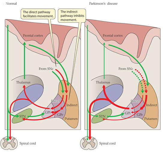
We have previously discussed Lrrk2 (and you can find that post here). It is a gene that is particularly interesting with regards to Parkinson’s disease because mutations in that gene are associated with susceptibility to the condition.

The Lrrk2 gene gives rise to an enzyme that has different functions in our cells. The researchers in the current study extracted the lrrk2 enzyme from the solid waste of urine and started analysing the “phosphorylation status of the enzyme”.

Ok, um,…what is Phosphorylation?

Phosphorylation is the process by which a phosphoryl group is added to a molecule.

And what is a phosphoryl group?!?

Oh, never you mind. Just remember that phosphorylation is basically the way in which many enzymes – like Lrrk2 – are turned on (and off when they are dephosphorylated). Through phosphorylation the function/activity of an enzyme is changed. They can go from dormant to active through this process. And this addition of the phosphoryl group to the molecule can occur at different places on that molecule, affecting the resulting activity in different ways.

So what did the researchers find?

The scientists found that people with Parkinson’s disease who also have a particular mutation in the Lrrk2 gene (that mutation is called p.G2019S) had almost 5 times more phosphorylation at a particular part of the Lrrk2 enzyme than normal healthy control subjects. Interestingly, those levels were also 4.5 times higher than those of people with PD, but who did not have the Lrrk2 mutation.

This means that the researchers have found a potential biomarker of the Lrrk2 mutation (independent of Parkinson’s disease itself). This finding could offers us a means of determining people with the Lrrk2 mutation – who may be susceptible to Parkinson’s disease – with a simple urine test.

But the researchers also noticed that among all of the study participants who have the Lrrk2 mutation,those who also had Parkinson’s disease had levels of phosphorylation twice as high as those who did not have Parkinson’s disease. Thus the overall results suggest that regardless of mutation status, higher levels of Lrrk2 phosphorylation are associated with a greater risk or the presence of Parkinson’s disease.

Lrrk-Urine1

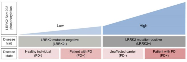
A diagram graph illustrating the findings of the Lrrk2 study. Source: Neurology

What does this mean?

Firstly, we need to point out that the study was conducted on a small population of men (two studies actually – the first had 14 subjects, and the second had 62 subjects). The results need to be independently replicated in larger groups (ideally also containing some female participants).

The results are very exciting, however, as they may point towards potential therapeutic pathways. It could also provide a means of monitoring clinical trials – a feature that the University of Alabama researchers are currently testing in another clinical trial. They are investigating if a LRRK2 inhibitor drug, called Sunitinib, results in lower leaves of phosphorylated Lrrk2 in the urine.

The research is also encouraging with regards to the search for biomarkers in Parkinson’s disease – a quest that has struggled somewhat until recently. Novel biomarkers provide useful tools in our fight against this terrible disease.

Brain (not Heart) warming research

The great Isaac Asimov once said:

“The most exciting phrase to hear in science, the one that heralds new discoveries, is not ‘Eureka!’ but ‘huh, that’s funny’…”

Here at the Science of Parkinson’s disease we suspect that this was the situation when some Italian scientists made a curious discovery in some early-onset Parkinson’s disease subjects.

Brain-scan

An image of a brain scan. Source: DailyMail

Last week they published their observation in the journal Movement Disorders:

temp-title

Title: Abnormal Brain Temperature in Early-Onset Parkinson’s Disease
Authors: Rango M, Piatti M, Di Fonzo A, Ardolino G, Airaghi L, Biondetti P, & Bresolin N.
Journal: Movement Disorders, 2016 Mar;31(3):425-6.
PMID: 26873586

The researchers were conducting brain scans of 5 people with early onset Parkinson’s disease (3 men and 2 women, with an average age of 41±6 years) and 10 control/normal subjects (6 men and 4 women with an average age of 43±7 years). The study was a follow on from a smaller previous study conducted by the same researchers. There was absolutely no difference in the average body temperature of all the subjects (36.7±0.48°C) and healthy subjects (36.5±0.84 °C).

But when the researchers began looking at different brains regions, they found there were substantial increases in temperature in the early-onset Parkinson’s patients when compared to the control subjects.

The areas of the brain where significant temperature differences were observed included:

  • the hypothalamus (38.50±0.20 vs. 37.01±0.60 °C; PD subjects vs controls)
  • the posterior cingulate gyrus (37.60±0.20 vs. 36.70±0.40 °C)
  • the centrum semiovale (38.00±0.60 vs. 36.60±0.60 °C)
  • the lenticulate nucleus (38.80±0.80 vs. 37.40±0.60 °C)

There was also a slight difference in the visual cortex in the patient group, but this was not statistically significant (37.20±0.20 vs. 36.80±0.40 °C).

Dysfunction in the hypothalamus is known to occur in Parkinson’s disease (click here for more information on this). Changes in the posterior cingulate gyrus (an area involved with emotion) have also been reported (click here for more information on this). But our knowledge of the role of the centrum semiovale and lenticulate nucleus in Parkinson’s disease requires further investigation.

Please remember that all things being equal, there should be no difference whatsoever in brain temperatures. The brain is an extremely sensitive organ and its temperature is rigidly controlled.

So why is there a difference?

Basically the researchers have no idea and, to their credit, they admit as much.

They also point out to the reader that any temperature change in the hypothalamus – an area of the brain that regulates temperature in the body – should result in a corrective response to restore proper temperature in the brain. But apparently in the early-onset Parkinsonian brain it doesn’t. They also note that dopamine-based Parkinson’s treatments (such as levodopa) should decrease overall brain temperature because they increase cerebral blood flow (a natural cooling system for the brain). But again, this doesn’t appear to be happening.

They speculate that maybe these temperature differences are the result of ongoing disease-related activities in the brain, and they offer some well considered ideas as to why this maybe. But there are many other areas of the brain that are affected by Parkinson’s disease – why is there no change in temperature in those regions?

The researchers also ask whether cooling the brain may be considered as a therapeutic option. An interesting idea but this still needs to be tested. And the results of the current study also need to be replicated – independently validated by other groups.

In those replication studies it would be interesting to conduct the same experiment on people with Parkinson’s disease at different stages of the disease to see the effect is consistent or changing over time.

A curious result. Opening up new areas of research. And further evidence that it’s the ‘huh, that’s funny’ results that ultimately lead to the  ‘Eureka!’ moments.

The science of focused ultrasound therapy

Novel therapeutic approaches for Parkinson’s disease are popping up all the time.

Recently there has been quite a bit of noise in the media regarding something called ‘focused ultrasound-based therapies‘ for Parkinson’s disease. We’re talking about reports such as this and this.

The initial results look very exciting and the Michael J Fox foundation has helped to fund a phase one clinical trial of the technology, but what exactly is focused ultrasound?

Let’s start at the beginning: Ultrasound

Ultrasound is defined as sounds at frequencies greater than 20 kHz. These are at high frequencies and short wavelengths.

Ultrasound_range_diagram.svg

Approximate frequency ranges corresponding to ultrasound. Source: Wikipedia

The human ear is not designed to register ultrasound, but it is still useful to us. Since ultrasound was first used by Paul Langevin to detect submarines in 1917, we have found many uses for it, most of them in the field of medicine (notably the imaging of unborn babies and breaking up kidney stones).

The use of ultrasound in procedures involving the brain has previously been very limited because of that natural protective helmet we call the skull. Normal ultrasound does not penetrate the skull very well. High intensity ultrasound, however, does.

So what is focused ultrasound?

High intensity focused ultrasound, also known as magnetic resonance guided focused ultrasound, is a procedure that uses very intense ultrasound generated from multiple points but focused on one specific area. The waves from those different points of emission are all in phase – that is to say their waves match. All that ultrasound concentrated in one location generates a lot of heat at that focal point. That heat allows for the destruction of diseased or damaged tissue at that particular point of focus.


07_14_13632_02b_cmyk

A schematic demonstrating the focused ultrasound technique. Source: Ghanouni et al (2015)

The procedure is relatively quick, non-invasive (no surgery). The subject is placed on a bed and their head is covered by a cooling unit. Around the cooling unit is the ultrasound transducer array – multiple generators of the ultrasound pointing in toward the skull.

U3

Images demonstrating the focused ultrasound technique: Source: Ghanouni et al (2015)

The subject, the cooling unit, and the transducer array are then placed inside an MRI brain scanning machine to allow for extremely accurate focusing of the ultrasound waves. Once everything is in place, the procedure will begin. According to the scientists conducting the research, the procedure – from start to finish – takes approximately 2 hours.

07_14_13632_03a_cmyk A brain scan image of the area being targeted (red cross). The skull is in green, and the cooled water unit is is red.  Source: Ghanouni et al (2015)

So what has been done research wise?

The research about using focused ultrasound in Parkinson’s disease is still very new. The first papers investigating the utility of the technology with regards to PD were only published a couple of years ago.

These papers include:

Magara_title

Title: First experience with MR-guided focused ultrasound in the treatment of Parkinson’s disease.
Authors: Magara A, Bühler R, Moser D, Kowalski M, Pourtehrani P, Jeanmonod D.
Journal: J Ther Ultrasound. 2014 May 31;2:11.
PMID: 25512869  (This article is OPEN SOURCE if you would like to read it)

This study was the first report of focused ultrasound being employed in humans with Parkinson’s disease. The researchers performed a pallidothalamic tractotomy (or destruction of the fibres connecting the basal ganglia to the thalamus – see our previous post about these structures). This removed the inhibitory signal being sent to the thalamus, allowing it to function more freely.

In this paper, the procedure was performed on 13 patients, demonstrating the safety and efficacy of the approach. The investigators reported a 60% improvement in Parkinson’s motor assessment (using the UPDRS) at 3 months post procedure.


Schlesinger_title

Title: MRI Guided Focused Ultrasound Thalamotomy for Moderate-to-Severe Tremor in Parkinson’s Disease.
Authors: Schlesinger I, Eran A, Sinai A, Erikh I, Nassar M, Goldsher D, Zaaroor M.
Journal: Parkinsons Dis. 2015;2015:219149.
PMID: 26421209  (This article is also OPEN SOURCE if you would like to read it)

This study involved 7 subjects with Parkinson’s disease who received a thalamotomy (the destruction of part of the thalamus) using focused ultrasound. The subjects demonstrated a better than 50% improvement in their motors scores and the effects were sustained for at least 7 months post procedure.


Na_title
Title: Unilateral magnetic resonance-guided focused ultrasound pallidotomy for Parkinson disease.
Authors: Na YC, Chang WS, Jung HH, Kweon EJ, Chang JW.
Journal: Neurology. 2015 Aug 11;85(6):549-51.
PMID: 26180137

In this case study report, the researchers conducted a pallidotomy (the destruction of the source of the fibres projecting to the thalamus) using focused ultrasound in a patient with Parkinson’s disease who had extremely severe dyskinesias (uncontrollable movements associated with long term use of PD medications). They reported 70-80% reductions in many of the motor assessments at 6 months post-procedure. The researchers noted many of the complications of deep brain stimulation that can be avoided with this technique (those complications include infection & hemorrhage due to surgery, and hardware-related complications with the expensive devices).

Importantly all of these study illustrated that the focused ultrasound technique was safe and had beneficial effects.

In Summary:

Focused ultrasound therapy is a new experimental treatment that is being tested in people with Parkinson’s disease. It is currently being oriented as a treatment option for people with Parkinson’s who have severe dyskinesias.

The pros of the focused ultrasound technology/approach:

  • No surgery – the procedure is non-invasive
  • No risk of infection
  • Little collateral damage (e.g. due to instruments passing through the cortex to get to the target region)
  • The effect is relatively immediate

The cons:

  • It does not cure the disease, merely  alleviates motor features (tremors, etc)
  • It is very important to note that the focused ultrasound procedure is irreversible.
  • Once performed, the effect is unadjustable – there is no ‘volume control’ on the resulting effect.
  • The technique is still experimental and the researchers do not know how long the effects will last.


We here at the Science of Parkinson’s have presented this post for information purposes. We are not affiliated with any of the groups/companies currently promoting this treatment. We have tried to remain unbiased in our explanations and assessment.

We would like to see some long-term data on the focused ultrasound approach before passing judgement on this treatment approach. Importantly, we need to know how long the effect can last – this will most likely vary from person to person. Given that the disease will still be progressing in individuals who have this treatment, it would be interesting to see the longer term consequences.

We would also like to see a comparative study between focused ultrasound and deep brain stimulation (the current best option for people with severe dyskinesias). Deep brain stimulation is an invasive, surgical procedure in which a ‘pacemaker’ like probe is inserted into the brain. Importantly deep brain stimulation is adjustable (the electrical signal being sent into the brain can be fine tuned) while focused ultrasound is not adjustable once the procedure is completed. Given this last detail, we believe that the focused ultrasound should remain a last choice option in any treatment approach to Parkinson’s disease (at least for the time being).

New Research – on how movement is controlled


A couple of very interesting studies were published a week ago that help us to better understand how we move. They are particularly important with respects to Parkinson’s disease.


The parts of the brain involved in movement

Movement is largely controlled by the activity in a specific collection of brain regions, collectively known as the ‘Basal ganglia‘.

B9780702040627000115_f11-01-9780702040627

The location of the basal ganglia structures (blue) in the human brain. Source: iKnowledge

The basal ganglia receives signals from the overlying cortex, processes that information before sending the signal on down the spinal cord to the muscles that are going to perform the movement.

There is also another important participant in the regulation of movement: the thalamus.

Brain_chrischan_thalamus

A brainscan illustrating the location of the thalamus in the human brain. Source: Wikipedia

The thalamus is a structure deep inside the brain that acts like the central control unit of the brain. Everything coming into the brain from the spinal cord, passes through the thalamus. And everything leaving the brain, passes through the thalamus. It is aware of most everything that is going on and it plays an important role in the regulation of movement.

The direct/indirect pathways

The processing of movement in the basal ganglia involves a direct pathway and an indirect pathway. In simple terms, the direct pathway encourages movement, while the indirect pathway does the opposite (inhibits it). The two pathways work together like a carefully choreographed symphony.

The motor features of Parkinson’s disease (slowness of movement and resting tremor) are associated with a breakdown in the processing of those two pathways, which results in a stronger signal coming from the indirect pathway – thus inhibiting/slowing movement.

Pathways

Excitatory signals (green) and inhibitory signals (red) in the basal ganglia, in both a normal brain and one with Parkinson’s disease. Source: Animal Physiology 3rd Edition

Both the direct and indirect pathways finish in the thalamus, but their effects on the thalamus are very different. The direct pathway leaves the thalamus excited and active, while the indirect pathway causes the thalamus to be inhibited.

The thalamus will receive signals from the two pathways and then decide – based on those signals – whether to send an excitatory or inhibitory message to the cortex, telling it what to do (‘get excited and movement’ or ‘don’t get excited and don’t move’, respectively).

Where does dopamine come into the picture?

In Parkinson’s disease, the cells in the brain that produce the chemical dopamine are lost. These cells reside in a structure called the substantia nigra (or SNc in the figure above). What effect does this cell loss have on the direct and indirect pathways? Under normal circumstances the dopamine neurons excite the direct pathway and inhibit the indirect pathway.

In Parkinson’s disease the loss of dopamine neurons results in increased activity in the indirect pathway. As a result, the thalamus is kept inhibited. With the thalamus subdued, the overlying motor cortex has trouble getting excited, and thus the motor system is unable to work properly.

So what was published last week?

Two papers.

Both from the same lab (Well done!)

One in the prestigious scientific journal, Cell and the other in her sister journal, Neuron:

Roseberry-title

Title: Cell-Type-Specific Control of Brainstem Locomotor Circuits by Basal Ganglia.
Authors: Roseberry TK, Lee AM, Lalive AL, Wilbrecht L, Bonci A, Kreitzer AC.
Journal: Cell, 2016 Jan 28;164(3):526-537.
PMID: 26824660

The researchers in this study discovered signal sent from the basal ganglia that selectively activates a group of neurons an area of the brainstem called the ‘mesencephalic locomotor region’. Some of the neurons in this area release a chemical called glutamate. Glutamate is a neurotransmitter that excites the cells it comes into contact with. The researchers who conducted this study found that these glutamate-releasing cells in the mesencephalic locomotor region are responsible for initiating movement.

Print

The researchers used a new technique called ‘optogenetics’ that allows light to activate or inhibit specific cells in the brain. By using this technique on the cells in the direct (dMSN in the figure above) or indirect pathways (iMSN) of the basal ganglia, the researchers were able to control the glutamate-releasing neurons in the mesencephalic locomotor region of mice -initiating or inhibiting their movement, respectively.

The researchers then took the study one step further and used the optogenetics approach directly on the glutamate-releasing neurons in the mesencephalic locomotor region, and they were able to control the initiation of movement in the mice irrespective of the signal being generated by the direct or indirect pathways. That is to say, when the glutamate-releasing neurons in the mesencephalic locomotor region were activated, the mouse would move even when the basal ganglia was sending an inhibitory signal.

So what does it all mean?

While some of the findings of the study were already known, the researchers here have elegantly linked the workings of the basal ganglia and the mesencephalic locomotor region, helping us to better understand the neurological functioning of movement. Deep brain stimulation of the mesencephalic locomotor region has already been attempted and it has demonstrated mixed results in people with Parkinson’s disease (it does appear to help with regards to reducing falls – click here and here for more on this).

It will be interesting to follow the research resulting from this current study.


Parker-title
Title: Pathway-specific remodeling of thalamostriatal synapses in Parkinsonian mice
Authors: Parker PRL, Lalive AL, Kreitzer AC.
Journal: Neuron, 2016
PMID: 26833136

In the second study, the researchers (the same folks who gave us the first paper!) found that the basal ganglia is biased towards the direct pathway. The signal coming from the neurons involved in the direct pathway are stronger than those in the indirect pathway. When dopamine is removed however (as in the case of Parkinson’s disease), the system swings in the opposite direction and becomes biased toward the indirect pathway – the neurons in the direct pathway begin to produce a weaker signal than their counters in the indirect pathway which increase the strength of their signal.

Given that both pathways influence the activity of the thalamus, the researchers next turned their attention to that structure. Again using the ‘optogenics‘ (light-activation) technique, the investigators reduced the inhibitory signal coming from the thalamus and were able to reversibly correct the motor impairs observed in the mice with Parkinson’s-like features.

What does this mean for Parkinson’s disease?
This study turns our attention away from what is happening in the basal ganglia and focuses it on the thalamus, which has not receive the same amount of attention with regards to Parkinson’s disease.

There is a lot already known about changes in the thalamus in Parkinson’s disease (click here for more on this), and deep brain stimulation of structures in neighbouring regions is a regular therapy for Parkinson’s disease (targeting the subthalamic nuclei). But this new paper further breaks down the circuitry of movement for us and offers novel directions for future therapeutic approaches for Parkinson’s disease.

We can be sure that a lot of Parkinson’s disease research is now going to focus on the thalamus.


Improving diagnosis

An inconvenient truth:

The diagnosis of Parkinson’s disease can only be definitively achieved at the postmortem stage.

There is currently no diagnostic test for this task and we are reliant on the training and skills of the neurologists making the diagnosis. Brain imaging techniques (such as DAT-scans) are great, but they can only aid physicians in their final decision.

And those decisions are not always right.

In 1992, a study looking at the brains of 100 subjects who had died with Parkinson’s disease, found that 24% of the cases did not fulfill the pathological requirements for the diagnosis of Parkinson’s disease. That study was:

Accuracy

Title: Accuracy of clinical diagnosis of idiopathic Parkinson’s disease: a clinico-pathological study of 100 cases.
Authors: Hughes AJ, Daniel SE, Kilford L, Lees AJ.
Journal: Journal of Neurol Neurosurg Psychiatry. 1992 Mar;55(3):181-4.
PMID: 1564476

Unfortunately, despite years of research, it would appear that there is still a large degree of error in the clinical diagnosis of Parkinson’s disease. A study published in 2014 in the journal Neurology that suggested that there is currently a 15% rate of misdiagnosis. That study was:


Adler-title

Title: Low clinical diagnostic accuracy of early vs advanced Parkinson disease: clinicopathologic study.
Authors: Adler CH, Beach TG, Hentz JG, Shill HA, Caviness JN, Driver-Dunckley E, Sabbagh MN, Sue LI, Jacobson SA, Belden CM, Dugger BN.
Journal: Neurology. 2014 Jul 29;83(5):406-12.
PMID: 24975862

It has to be said that clinicians face a very difficult task in diagnosing Parkinson’s disease. The variety of features (symptoms) that patients present with in the clinic, and the lack of diagnostic tools, leave neurologists making a judgement based largely on clinical observations.

But this degree of error ultimately has a huge impact on clinical studies and trials: if 10-20% of the participants are not Parkinsonian, are we really going to observe an accurate result?

Better diagnostic tests/tools are critically required.


In November last year, a study was published in the journal Immunology Letters which may help in this regard:
Blood1

Title: Potential utility of autoantibodies as blood-based biomarkers for early detection and diagnosis of Parkinson’s disease.
Authors: DeMarshall CA, Han M, Nagele EP, Sarkar A, Acharya NK, Godsey G, Goldwaser EL, Kosciuk M, Thayasivam U, Belinka B, Nagele RG; Parkinson’s Study Group Investigators.
Journal: Immunol Letters, 168(1), 80-8.
PMID: 26386375  (this article is OPEN access if you would like to read it)

The researchers took 398 subjects, including 103 early-stage Parkinson’s disease subjects and they collected blood samples from them. They then screened the blood for 9,486 different autoantibodies that could be useful as biomarkers for Parkinson’s disease.

Antibodies are produced by our immune system to determine what is ‘self’ and not ‘self’. They are the foundation of our defenses against the big, bad germ/bacteria world. Autoantibodies are antibodies produced by our immune system that are directed against our own tissues. They target ‘self’.

And yeah, that is bad. Autoantibodies are associated with autoimmune diseases such as Lupus.

We are not sure why we produce autoantibodies. The causes of their production vary greatly and are not well understood. In Parkinson’s disease, however, autoantibodies may be produced as a result of the cell death in the brain. Some of the debris resulting from the dying cells will make its way into the bloodstream, to be removed from the body. Whilst in the blood, some of that debris could trigger the immune system, thus resulting in the production of autoantibodies.

De Marshall et al (the researchers who conducted this study) were hoping to take advantage of this autoantibody production and use them as biomarkers to not only differentiate between people with and without Parkinson’s disease, but also to differentiate between different stages of Parkinson’s disease (see the figure below).

1-s2.0-S0165247815300341-gr3

Attempting to differentiate between different stages of Parkinson’s disease. Source: Immuno Letters

The researchers found that using the top 50 autoantibodies that they associated with Parkinson’s disease, they could successfully differentiate between people with and without Parkinson’s disease with 90% prediction accuracy in a blind analysis (they actually found that just the top 4 autoantibodies were enough).

Interestingly, the researchers then compared the early Parkinson’s group with a mild-moderate Parkinson’s group and they found that they could differentiate between the two groups with an overall accuracy of 97.5%!


These are very exciting results and we will be following this work with interest – not only from the standpoint of biomarkers, but also the role of autoantibodies in Parkinson’s disease.

New research – the disorder of Alpha Synuclein

A couple of interesting scientific papers were published this week dealing with the Parkinson’s disease-related protein, Alpha Synuclein. If you are not familiar with it, we suggest that you check out our primer page on Alpha Synuclein before reading any further.

So, what’s new in the world of Alpha Synuclein?

Two studies.

One in the prestigious journal Nature and the other in her sister Nature Communications. Both studies came from the same lab (good job guys!)

The first study :

Theillet-title

Title: Structural disorder of monomeric α-synuclein persists in mammalian cells.
Authors: Theillet FX, Binolfi A, Bekei B, Martorana A, Rose HM, Stuiver M, Verzini S, Lorenz D, van Rossum M, Goldfarb D, Selenko P.
Journal: Nature. 2016 Jan 25.
PMID: 26808899

This first study presented a very detailed analysis of the structure of alpha synuclein – at the atomic level – inside living cells.

Interestingly, when the researchers injected alpha synuclein (at concentrations that have been observed in normal neurons) into 5 different types of cells (both neuron and others types), they found that the protein remains extremely disordered – it changed shape rapidly. They determined this by using nuclear magnetic resonance spectroscopy (try saying that 3 times really fast!), which provides a shallow peak readout for stable proteins and a sharp peak for disordered proteins (see image below).

nature16871-f1

The researchers found a lot of sharp peaks in cells that they injected Alpha Synuclein into. Source: Nature

Rather remarkably, despite the fact that disordered proteins are usually removed from cells by enzymatic degradation, the alpha synuclein that was injected by these researchers appears to have remained intact in the cells for several days (50+ hours). And the cells did not seem to be adversely affected by this.

The second Alpha Synuclein study published this week illustrated an equally interesting result:

Binolfi-title

Title: Intracellular repair of oxidation-damaged α-synuclein fails to target C-terminal modification sites.
Authors: Binolfi A, Limatola A, Verzini S, Kosten J, Theillet FX, May Rose H, Bekei B, Stuiver M, van Rossum M, Selenko P.
Journal: Nature Communications, 2016 Jan 25;7:10251.
PMID: 26807843

In this study, the researchers injected damaged alpha synuclein into cells and then watched the cells try to repair that damaged protein. There are specific enzymes that help to maintain/repair proteins like Alpha Synuclein inside each cell. This is a normal recycling process for cells, but something interesting happened with this damaged version of alpha synuclein: only one end of the protein was repaired. The other end (called the C-terminus) was left damaged and this end failed to function correctly.

fnins-09-00059-g001

The structure of Alpha Synuclein. The c-terminus is the area in red. Source: Frontiers in Neuroscience

This led the authors to conclude that damage can cause the accumulation of chemically and functionally altered Alpha Synuclein in cells.

What does this mean for Parkinson’s disease?

The results are very interesting and the researchers should be congratulated on the complexity of their work. The findings add to our understanding of Alpha Synuclein, but both of these results need to be replicated and expanded on before we can fully appreciate their impact.

One possible implications of the results is that designing drugs to target Alpha Synuclein may be more complicated than originally thought. If the protein remains as disordered as the first study suggests, it could be difficult to target. Further investigations, however, focused on the c-terminus end of Alpha synuclein may offer novel targets for therapies looking to clear damaged proteins from cells.

If Alpha Synuclein is the big, bad enemy in Parkinson’s disease, we now know a lot more about him and we can focus on his weaknesses.

New Research -Shared genetic features

There was an interesting new study published yesterday:

Sanchez-Mut-Title

Title: Human DNA methylomes of neurodegenerative diseases show common epigenomic patterns.
Author: Sanchez-Mut JV, Heyn H, Vidal E, Moran S, Sayols S, Delgado-Morales R, Schultz MD, Ansoleaga B, Garcia-Esparcia P, Pons-Espinal M, de Lagran MM, Dopazo J, Rabano A, Avila J, Dierssen M, Lott I, Ferrer I, Ecker JR, Esteller M.
Journal: Transl Psychiatry. 2016 Jan 19;6:e718. doi: 10.1038/tp.2015.214.
PMID: 26784972 – this article is OPEN ACCESS if you would like to read it.

The researchers were curious to look for common genetic markers between the major neurodegenerative disease. It is often forgotten that the different neurodegenerative conditions, such as Alzheimer’s disease and Parkinson’s disease, share some common pathological features (the characteristic signs of the diseases in the brain).

For example, when you look at the brains of people with Alzheimer’s disease, approximately 50% of them will also have the alpha-synuclein-containing ‘Lewy bodies’ in their brains, which are more commonly associated with Parkinson’s disease. Likewise, Beta-amyloid plaques and neurotangles, which are characteristic features of Alzheimer’s disease are commonly found in Parkinson’s disease brains (click here and click here for more on this topic).

To find these shared genetic markers, the researcher extracted DNA from the prefrontal cortex (Brodmann area 9) of the brains of people with Alzheimer’s disease, dementia with Lewy bodies, Parkinson’s disease and Alzheimer-like neurodegenerative profile associated with Down’s syndrome samples (more than 75 percent of people with Down Syndrome aged 65 and older develop Alzheimer’s disease – click here for more on this).

Importantly, the researchers were looking at DNA methylation, which is a commonly used tool that allows a cell to fix genes in the “off” position. That is to say, the gene can not be activated. Thus the researchers were looking for regions of DNA that have to closed down.

They found that a very defined set of genes are turned off in these neurodegenerative disorders, suggesting that these condition might have similar underlying mechanisms or processes that subsequently develop into different clinical entities. These newly identified regions of DNA methylation will be further investigated with the goal that one day they may be used as biomarkers in diagnosis and also as potential new targets for the regenerative therapies.

A call to arms

While our primary goal here at the Science of Parkinson’s is to highlight and explain new research dealing with Parkinson’s disease, we are also keen to encourage the general public to get involved with efforts to cure this debilitating condition.

To this end, we would like to bring your attention to the fact that 2017 represents the 200th anniversary of the first report of Parkinson’s disease by one Dr James Parkinson:

320px-Parkinson,_An_Essay_on_the_Shaking_Palsy_(first_page)

Although there were several earlier descriptions of individuals suffering from rigidity and a resting tremor, Dr Parkinson’s 66 page publication of six cases of ‘Shaking Palsy’, is considered the seminal report that gave rise to what we now call Parkinson’s disease. The report was published in 1817.

The 200th anniversary represents a fantastic opportunity to raise awareness about the disease and a rallying point for a concerted research effort to deal with the condition once and for all. It is still a year away, but now is the time to start planning events and building awareness. We would encourage you to mark 2017 as the year of Parkinson’s disease, share this with everyone you know, and endeavour to make some small effort to help in the fight against this condition.

Beginnings

Welcome to the Science of Parkinson’s Disease – a blog that has been set up by scientists to provide information and understanding about the neurodegenerative condition known as Parkinson’s disease.

Over the last 10 years, the advocacy for Parkinson’s disease has been tremendous and real awareness has been created by groups such as the Michael J Fox foundation, the Cure PD Trust, and Parkinson’s UK. They have generated enormous amounts of funding for scientific research and provided hope for disease halting therapies, while supporting and improving the general welfare of people suffering from this condition.

The media regularly announces new breakthroughs in the medical and scientific world, but there are few forums available for the general public to ask questions related to the science being conducted.  The Science of Parkinson’s disease has been set up for this purpose.

The Science of Parkinson’s disease is run by research scientists working in the field, and it was begun with several goals in mind:

  • Try to answer any questions you may have about Parkinson’s disease.
  • Report each week on interesting/exciting research in the world of Parkinson’s disease.
  • Interview Parkinson’s disease researchers, providing a face to the people doing the work.
  • Help you to understand this disease better.

We look forward to hearing from you.

The Team