Cell transplantation – Replacing what has been lost

Parkinson’s disease is a neurodegenerative condition. That means that cells in the brain (neurons) are dying. By the time the motor features of Parkinson’s disease (rigidity, slowness of movement, and a resting tremor or shaking of a limb) become apparent to an individual, they will have lost 60-70% of the dopamine neurons in a region of the brain called the midbrain.

CD-Pathology-Parkinson'sDisease Chptr19-Fig 19-3 Parkinson DiseaseGrossMidbrain copy

Above are slices of human brain, taken from the midbrain of a healthy control subject (left) and an individual who died with Parkinson’s disease. The dopamine cells in the control subject can be seen on both sides of the brain with the eye because they produce a chemical (neuromelanin) that makes them black. These cells are noticeably absent in the Parkinsonian brain. Source: Springer

While there is a lot of research investigating how to stop or slow down the disease, at present, the only realistic way to deal with what has already been lost is to replace it. This could be done with cell transplantation.

Cell transplantation has had a long and colourful history of trial and error with regards to Parkinson’s disease. Importantly, given that there is currently no clinically approved cell transplantation procedure for Parkinson’s disease, the approach must be considered experimental, at best. This has not, however, stop numerous unscrupulous practitioners from advertising their services and preying on desperate individuals. They offer expensive operations, that have little if any peer-reviewed scientific evidence backing them.

Let us repeat: there is currently NO clinically approved cell transplantation procedure for Parkinson’s disease.


The history of cell transplantation in Parkinson’s disease

There has been an enormous amount of cell transplant work conducted in rodent models of Parkinson’s disease – in which the dopamine system is lesioned unilaterally. Many different types of cells have been used, but by far the most successful have been immature dopamine neurons (collected from embryos). The success of that work resulted in numerous clinical trials in the 1990s. Those trials began with a group in Lund (Sweden) who, in 1991, transplanted fetal midbrain tissue into six patients: four with advanced idiopathic Parkinson’s disease and two from the ‘Frozen addicts’ cohort (see the book “The case of the frozen addicts” for an explanation of these two individuals). Similar programmes had been initiated in England, Spain, Mexico, Cuba, France, and Belgium. These were all considered to be relatively successful, except for the fact that they were all open-label/not blinded studies, meaning that everyone involved in the study knew who was getting transplanted.

In the USA, these developments took place amid a major debates about the ban on federal funding for fetal tissue research that had been introduced by the Reagan administration in 1988. The Clinton administration lifted this ban in January, 1993, and this reversal opened the way for the National Institutes of Health (NIH) to provide funding for the two placebo-controlled studies. Those two trials were:

Trial no. 1: The Colorado/Columbia Trial:

A double blind trial in which 40 subjects (with advanced PD) received transplants of fetal midbrain tissue and 34 additional subjects (with advanced PD) had a sham surgery and were considered controls. Critically, neither the subjects nor the practitioners knew who was in which group. No patients in either arm of the study received immunosuppression, meaning that their immune systems were free to attack the injected cells (which would have been considered foreign by the body). The patients were followed up for 1 year after surgery and the success of the trial was judged on the basis of a self-report rating of clinical improvement or deterioration, scored by patients in their own homes and then sent to the investigator. The report of the study was published in the New England Journal of Medicine:

Title: Transplantation of embryonic dopamine neurons for severe Parkinson’s disease.
Authors: Freed CR, Greene PE, Breeze RE, Tsai WY, DuMouchel W, Kao R, Dillon S, Winfield H, Culver S, Trojanowski JQ, Eidelberg D, Fahn S.
Journal: New England Journal of Medicine 2001 Mar 8;344(10):710-9.
PMID: 11236774

Trial no. 2: The Tampa Bay Trial:

34 patients were randomly assigned either to receive a transplant of fetal midbrain tissue or to undergo a sham surgery. All patients received 6 months of immunosuppression after surgery. The primary endpoint for this study was a significant difference between the groups at 24 months after surgery. The report of this study was published in the journal, Annals of Neurology:

Title: A double-blind controlled trial of bilateral fetal nigral transplantation in Parkinson’s disease.
Authors: Olanow CW, Goetz CG, Kordower JH, Stoessl AJ, Sossi V, Brin MF, Shannon KM, Nauert GM, Perl DP, Godbold J, Freeman TB.
Journal: Ann Neurol. 2003 Sep;54(3):403-14.
PMID: 12953276

Both of these clinical studies failed to show any significant improvement at their endpoints. In addition, 15% – 50% of transplanted subjects developed what are called ‘graft induced dyskinesias’ (or GID’s). This is where the subjects display uncontrollable/erratic movement. Interestingly, patients under 60 years of age did show signs of improvement on when assessed both clinically (using the UPDRS-III) and when assessed using brain imaging techniques (increased F-dopa uptake on PET). But the overall negative results left a shadow over the technique for the better part of a decade.

So what is currently happening?

There are new clinical trials currently underway for cell transplantation in Parkinson’s disease. Primary amongst these is the Transeuro being conducted in Europe.

Transeuro_Logo_100

The Transeuro trial. Source: Transeuro

The Transeuro trial is an open label study, involving 40 subjects, transplanted in sites across Europe. They will receive immunosuppression for at least 12 months post surgery, and the end point of the study will be 3 years post surgery, and based on brain imaging of dopamine release from the transplanted cells (PET scans). Based on the previous double blind studies discussed above, only subject under 65 years of age have been enrolled in the study.

transeuro

The European consortium behind the Transeuro trial. Source: Transeuro

Do the transplants slow down the disease?

The evidence thus far is not clear, but some of the original patients from the 1991 Sweden trial were able to stop/cut back on their L-dopa treatment. Recently, some of the patients who received transplants have pass away and their brains have been examined post-mortem. One very interesting finding is that some of the cells in the transplants (1-5%) have lewy bodies in them. This suggests that the disease is passed on to the healthy transplanted cells in some way.

gr3

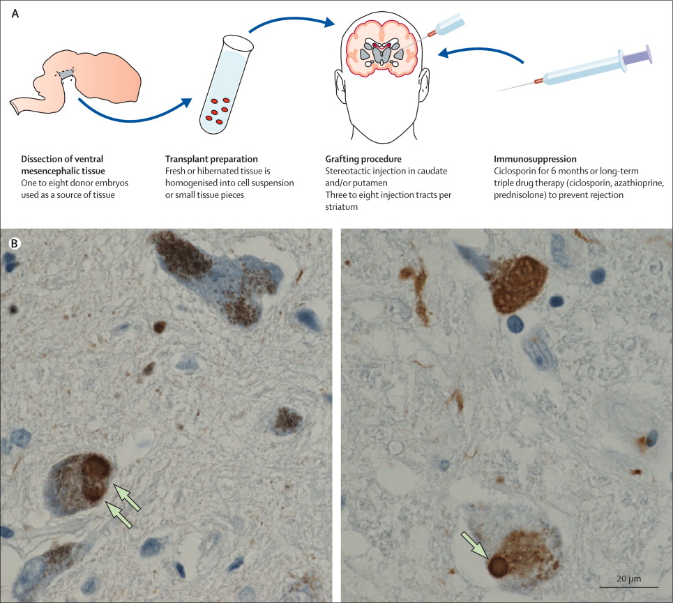
Above are photos of neurons from the post-mortem brains of people with Parkinson’s that received transplants. White arrows in the images above indicate lewy bodies inside transplanted cells. Source: The Lancet

For more information on this, see these articles:

Title: Lewy bodies in grafted neurons in subjects with Parkinson’s disease suggest host-to-graft disease propagation.
Authors: Li JY, Englund E, Holton JL, Soulet D, Hagell P, Lees AJ, Lashley T, Quinn NP, Rehncrona S, Björklund A, Widner H, Revesz T, Lindvall O, Brundin P.
Journal: Nature Medicine. 2008 May;14(5):501-3.
PMID: 18391963

Title: Lewy body pathology in long-term fetal nigral transplants: is Parkinson’s disease transmitted from one neural system to another?
Authors: Kordower JH, Brundin P.
Journal: Neuropsychopharmacology. 2009;3 (1):254.
PMID: 19079079 (this review article is freely available)

Thus it would appear that cell transplantation will not stop the disease. At best it will simply allow us to replace the lost cells and reverse some of the motor related features of the condition. Ideally, this approach would be conducted in concert with additional therapies that slow or halt the disease (such as a vaccine possibly).

What next for cell transplantation?

Given the moral and ethical issues surrounding the use of fetal tissue, research has shifted towards using embryonic stem (ES) cells and turning them into midbrain dopamine neurons. And the results here have been very promising, with recent reports suggesting that ES cells can be converted into dopamine neurons and transplanted into rodent models of Parkinson’s disease with equal efficiency to that of fetal midbrain tissue.

Grelish-title

Title: Human ESC-derived dopamine neurons show similar preclinical efficacy and potency to fetal neurons when grafted in a rat model of Parkinson’s disease.
Authors: Grealish S, Diguet E, Kirkeby A, Mattsson B, Heuer A, Bramoulle Y, Van Camp N, Perrier AL, Hantraye P, Björklund A, Parmar M.
Journal: Cell Stem Cell. 2014 Nov 6;15(5):653-65.
PMID: 25517469

The take home message regarding cell transplantation is that at present it represents one of the only ways to replace what has been lost, but as of yet there is not an approved protocol for this approach in humans. As we said above, at best this should be considered experimental, and individuals selling expensive procedures should be avoided. The future looks very bright for this field, however, and we will keep you updated as more information comes to hand.


EDITORIAL NOTE: Happy new year everyone! We look forward to bringing you all the exciting news related to the science of Parkinson’s disease in 2016.

A call to arms

While our primary goal here at the Science of Parkinson’s is to highlight and explain new research dealing with Parkinson’s disease, we are also keen to encourage the general public to get involved with efforts to cure this debilitating condition.

To this end, we would like to bring your attention to the fact that 2017 represents the 200th anniversary of the first report of Parkinson’s disease by one Dr James Parkinson:

320px-Parkinson,_An_Essay_on_the_Shaking_Palsy_(first_page)

Although there were several earlier descriptions of individuals suffering from rigidity and a resting tremor, Dr Parkinson’s 66 page publication of six cases of ‘Shaking Palsy’, is considered the seminal report that gave rise to what we now call Parkinson’s disease. The report was published in 1817.

The 200th anniversary represents a fantastic opportunity to raise awareness about the disease and a rallying point for a concerted research effort to deal with the condition once and for all. It is still a year away, but now is the time to start planning events and building awareness. We would encourage you to mark 2017 as the year of Parkinson’s disease, share this with everyone you know, and endeavour to make some small effort to help in the fight against this condition.

Parkinson’s disease and the cancer drug

In October, 40,000 neuroscientists from all over the world gathered in Chicago for the annual Society for Neuroscience conference. It is one of the premier events on the ‘brain science’ calendar each year and only a few cities in the USA have the facilities to handle such a huge event.


agu20141212-16

Science conference. Source: JPL

During the five day neuroscience marathon, hundreds of lecture presentations were made and thousands of research poster were exhibited. Many new and exciting findings  were presented to the world for the first time, including the results of an interesting pilot study that has left everyone in the Parkinson’s research community very excited, but also scratching their heads.

The study (see the abstract here) was a small clinical trial (12 subjects; 6 month study) that was aiming to determine the safety and efficacy of a cancer drug, Nilotinib (Tasigna® by Novartis), in advanced Parkinson’s Disease and Lewy body dementia patients. In addition to checking the safety of the drug, the researchers also tested cognition, motor skills and non-motor function in these patients and found 10 of the 12 patients reported meaningful clinical improvements.

The study investigators reported that one individual who had been confined to a wheelchair was able to walk again; while three others who could not talk before the study began were able to hold conversations. They suggested that participants who were still in the early stages of the disease responded best, as did those who had been diagnosed with Lewy body dementia.

So what is Nilotinib?

Nilotinib (pronounced ‘nil-ot-in-ib’ and also known by its brand name Tasigna) is a small-molecule tyrosine kinase inhibitor, that has been approved for the treatment of imatinib-resistant chronic myelogenous leukemia (CML). That is to say, it is a drug that can be used to treat a type of leukemia when the other drugs have failed. It was approved for this treating cancer by the FDA in 2007.

The researchers behind the study suggest that Nilotinib works by turning on autophagy – the “garbage disposal machinery” inside each neuron. Autophagy is a process that clears waste and toxic proteins from inside cells, preventing them from accumulating and possibly causing the death of the cell.

Print

The process of autophagy – Source: Wormbook

Waste material inside a cell is collected in membranes that form sacs (called vesicles). These vesicles then bind to another sac (called a lysosome) which contains enzymes that will breakdown and degrade the waste material.


Some details about the study:

  • The study was run at the Georgetown University Medical Center
  • The patients were given increasing doses of Nilotinib (150mg to 300mg/day) that were are significantly lower than the doses of Nilotinib used for CML treatment (800-1200mg/day).
  • The researchers took cerebrospinal fluid (CSF; the liquid surrounding the brain) and blood samples at the start of the study, 2 and 6 months into the study.
  • Nilotinib was detected in the CSF, indicating that it had no problem crossing the protective blood-brain-barrier – the membrane covering the brain that blocks many drugs from entering.
  • Participants exhibited positive changes in various cerebrospinal fluid biomarkers with statistically significant changes in an important protein called, Tau, which have been shown to increase with the onset of dementia.
  • The researchers found a significant reduction (>60%) in levels of α-Synuclein detected in the blood, but no change in CSF levels of α-Synuclein. 
  • The investigators report that one individual confined to a wheelchair was able to walk again; three others who could not talk were able to hold conversations.

If the outcomes of this study are reproducible, then we here at the Science of Parkinson’s are assuming that Nilotinib is working by turning on the garbage disposal system of the remaining cells in the brain and allowing them to function better. This would suggest that there is a certain level of dysfunction in those remaining cells, which would be expected as this is a progressive disease. The study researchers reported that the small, daily dose of nilotinib turns on autophagy for about four to eight hours, and if that is enough to have such remarkable effects, then this treatment deserves more research.

The results of the study are intriguing and the participants of the study will continue to be treated and followed to see if the improvements continue.

BUT before we go getting too excited:

While these results sound extremely positive, there are several issues with this study that need to be considered before we celebrate the end of Parkinson’s disease.

Firstly, this study was an open-label trial – that means that everyone involved in the study (both researchers and subjects) knew what drug they were taking. There was also no control group or control treatment for comparative analysis in the study. Given these conditions there is always the possibility that what some of the subjects were experiencing was simply a placebo effect. Indeed the lead scientist on the project, Dr Fernando Pagan, pointed out that “It is critical to conduct larger and more comprehensive studies before determining the drug’s true impact.”

In addition, according to Novartis (the producer of the drug), the current cost of Nilotinib is about $10,360 (£6,900) per month for the daily 800mg dose used for cancer treatment. Even if the dose used in this study was only 150 to 300 mg/daily, it would still make this treatment extremely expensive. 

Thirdly, Nilotinib has a number of adverse side-effects when used as an anti-cancer drug (at 800mg/day). These include headache, fatigue, nausea, vomiting, diarrhea, constipation, muscle/joint pain, skin issues, flu-like symptoms, and reduced blood cell count. It may not be the nicest of treatments to tolerate.

There are important reasons for optimism, however, with the results of this study:

In 2010, a group of researchers published a paper demonstrating the neuroprotective effects of another cancer drug very similar to Nilotinib. That drug was ‘Gleevec’

Gleevec-PD1

Title: Phosphorylation by the c-Abl protein tyrosine kinase inhibits parkin’s ubiquitination and protective function.
Authors: Ko HS, Lee Y, Shin JH, Karuppagounder SS, Gadad BS, Koleske AJ, Pletnikova O, Troncoso JC,Dawson VL, Dawson TM.
Journal: Proc Natl Acad Sci U S A. 2010 Sep 21;107(38):16691-6.
PMID: 20823226

And that Gleevec publication was followed up a couple of years ago with a second study demonstrating the neuroprotective effects of another Abl-inhibitor: Nilotinib!

Gleevec-PD2

Title: The c-Abl inhibitor, nilotinib, protects dopaminergic neurons in a preclinical animal model of Parkinson’s disease.
Authors: Karuppagounder SS, Brahmachari S, Lee Y, Dawson VL, Dawson TM, Ko HS
Journal: Sci Rep. 2014 May 2;4:4874.
PMID: 24786396

These studies provided a strong rationale for testing brain permeable c-Abl inhibitors as potential therapeutic agents for the treatment of PD. The phase 2 trial at Georgetown will be starting in early 2016 and we will be watching this trial very closely.

“Red hair, sir, in my opinion, is dangerous”

Henna-Hair-Red-Hair-Ginger-Hair-Benefits-Beauty

The quote entitling this post is from a PG Wodehouse book ‘Very Good, Jeeves!’.


We have previously discussed the curious connection between melanoma and Parkinson’s disease. There is also a well known connection between melanoma and red hair. And believe it or not, there is another really strange relationship between Parkinson’s disease and red hair.

Redhair

Title: Genetic determinants of hair color and Parkinson’s disease risk.
Authors: Gao X, Simon KC, Han J, Schwarzschild MA, Ascherio A.
Journal: Ann Neurol. 2009 Jan;65(1):76-82.
PMID: 19194882

In 2009, researchers from Harvard University found a relationship between hair color and risk of Parkinson’s disease, when they examined the records of 131,821 US men and women who participated in the two large longitudinal studies, the Health Professionals Follow-up Study (HPFS) and the Nurses’ Health Study (NHS).


The HPFS, which started in 1986, sends questionnaires to US health professionals (dentists, optometrists, etc) – aged 40-75.  Every couple of years, members of the study receive questionnaires dealing with diseases and health-related issues (e.g. smoking, physical activity, etc). The questionnaire is supplemented by another questionnaires which is sent every four years, that deals with dietary information.

The NHS study – which was established in 1976 and then expanded in 1989 – has also collected questionnaire-based information from 238,000 registered nurses. Similar to the HPFS, every two years the study participants receive a questionnaire dealing with diseases and health-related topics.


In their study, the investigators found 264 of the male and 275 of the female responders to the HPFS and NHS questionnaires had been diagnosed with Parkinson’s disease. Of these individuals, 33 were black haired, 418 had brown hair, 62 were blond and 26 were redheads. Given that redheads make up just 1% of the general population but 5% of the people who were diagnosed with Parkinson’s disease in their study, the authors suggested that red haired people have a higher risk of developing Parkinson’s disease. Interestingly, they found a stronger association between hair color and Parkinson’s disease in younger-onset of PD (that is being diagnosed before 70 years of age) than those with age of onset greater than 70 years. When they took health and age related matters into account, the authors concluded that people with red hair are almost four times more likely to develop Parkinson’s disease than people with black hair.

NOTE: This result does not mean that people with red hair are definitely going to develop Parkinson’s disease, it simply suggests that they may be more vulnerable to the condition. And we should add that this result have never been replicated and we are not sure if anyone has ever attempted to reproduce it with a different database.

So how does (or could) this work?

The short answer is: we really don’t know.

The long answer involves explaining where there are no connections:

Red hair results from a genetic mutation. 80% of people with red hair have a mutation in a gene called MC1R – full name: melanocortin-1 receptor. Another gene associated with red hair is called HCL2 – ‘Hair colour 2’. We know that the connection between red hair and Parkinson’s disease is not genetic, as there is no association between MC1R mutations and Parkinson’s disease (for more on this, click here). We are not sure about HCL2, but this gene has never been associated with any disease.

What we do know is that redheads:

  • are more sensitive to cold (for more on this, click here)
  • are less responsive to subcutaneously (under the skin) administered anaesthetics (for more on this, click here)
  • suffer more from toothaches (for more on on this, click here)
  • are more sensitive to painkillers (for more on this, click here)
  • require more anesthetic for surgery (for more on this, click here)

Common myths associated with red hair include:

  • redheads bled more than others (this is not true – click here)…but they do bruise easier!
  • redheads are at greater risk of developing endometriosis (this is not true – click here)
  • redheads are more frequently left-handed (I can find no evidence for this, so I’ll put it in the myth basket until corrected).

There is also a strange link between red hair and multiple sclerosis, but it is too complicated to understand at the moment (women with red hair are more vulnerable to multiple sclerosis than men with red hair, for more on this, click here).

How any of these findings relates to Parkinson’s disease is unclear – we provide them here for those who are interested in following up this curious relationship.

One important caveat regarding this study is that incidence rates of Parkinson’s disease in countries with very high levels of red hair do not support the relationship (PD & red hair). In Scotland, approx. 10% of the population have red hair (source), and yet the England has a higher incidence of Parkinson’s disease (28.0/10,000 in England vs 23.9/10,000 in Scotland – source).

It may well be, however, that there is no direct connection between red hair and Parkinson’s disease. And until the results of the 2009 study mentioned above are replicated or supported by further findings, we here at the ‘Science of Parkinson’s disease’ shall consider this simply as a curious correlation.


The difference between men and women

At the bottom of our previous post, we mentioned that Japan is the only country where women have a higher incidence of Parkinson’s disease than men.

JapanPanorama_top

We also suggested that we have no idea why this difference exists. Well, a study presented at the Cardiovascular, Renal and Metabolic Diseases conference in Annapolis City (Maryland) last week may now be able to explain why this is.


The prevalence of Alzheimer’s disease is significantly higher in women compared to men. One recent estimate suggested that almost two-thirds of individuals diagnosed with Alzheimer’s disease are women (More information here). One possible reason for this is that Alzheimer’s disease is a condition of the elderly and women live longer.

So why is it then is the exact opposite true in Parkinson’s disease???


elderly-cake_2165089b

Source: The Telegraph Newspaper

Men are approximately twice as likely to develop Parkinson’s disease as females (More information here)

In addition, women are on average diagnosed 2 years later than men (More information here)

This gender difference has long puzzled the Parkinson’s research community. But now a group from the University of North Texas Health Science Center think that they may have the answer.

UNTHSC-copy

The researchers – lead by Shaletha Holmes from Dr Rebecca Cunningham’s lab – observed that when they stressed dopamine neurons, adding the male hormone testosterone made the damage worse. Interestingly, they found that testosterone was doing this by acting on a protein called cyclooxygenase 2 (or COX2). When they blocked the actions of COX2 while stressing dopamine neurons, they found that they also blocked the damaging effect of testosterone. The researchers concluded that testosterone may exacerbate the damage (and death) in dopamine neurons that occurs in Parkinson’s disease, thus possibly explaining the sex differences described above.

Now, there are several interesting aspects to this finding:

Firstly, the use of Ibuprofen, the nonsteroidal anti-inflammatory drug used for relieving pain, has long been associated with reducing the risk of Parkinson’s disease (More information here).

Ibuprofen is a COX2 inhibitor.

But more importantly, several years ago it was shown that Japanese men have lower levels of testosterone than their Western equivalents. Here is the study:

Japan1

Title: Evidence for geographical and racial variation in serum sex steroid levels in older men.
Authors: Orwoll ES, Nielson CM, Labrie F, Barrett-Connor E, Cauley JA, Cummings SR, Ensrud K, Karlsson M, Lau E, Leung PC, Lunggren O, Mellström D, Patrick AL, Stefanick ML, Nakamura K, Yoshimura N, Zmuda J, Vandenput L, Ohlsson C; Osteoporotic Fractures in Men (MrOS) Research Group.
Journal: Journal of Clinical Endocrinol. Metab. 2010 Oct;95(10):E151-60.
PMID: 20668046

The study suggested that total testosterone levels (while similar in men from Sweden, Tobago and the US) were 16 per cent higher in men from Hong Kong and Japan. BUT – and here’s the catch – Japanese men also had higher levels of a testosterone-binding hormone (Sex hormone-binding globulin or SHBG), so there is less of the testosterone floating around free to act. As a result, Japanese men had the lowest levels of active testosterone in the study.

Intriguingly, the researchers found that Japanese men who emigrated to the US had similar testosterone levels to men of European descent, suggesting that environmental influences may be having an effect of testosterone levels. Diet perhaps?

If testosterone is found to play a role in the gender difference found in Parkinson’s disease, the lower levels of free testosterone observed in Japanese men may explain why women in Japan have a higher risk of Parkinson’s disease than men.


EDITOR’S NOTE: WHILE WE HAVE NO DOUBTS REGARDING THE RESEARCH OF DR CUNNINGHAM AND HER GROUP, WE ARE TAKING A LEAP IN THIS POST BY APPLYING THE TESTOSTERONE RESULTS TO THE GENDER DIFFERENCE IN JAPAN. THIS IS PURE SPECULATION ON OUR PART. WE HAVE SIMPLY SAT DOWN AND TRIED TO NUT OUT POSSIBLE REASONS AS TO WHY THERE IS A REVERSED GENDER DIFFERENCE FOR PARKINSON’S DISEASE IN JAPAN. OUR THEORY IS YET TO BE TESTED, AND MAY BE COMPLETELY BONKERS. WE PRESENT IT HERE PURELY FOR DISCUSSION SAKE AND WELCOME YOUR THOUGHTS.

The Honolulu Heart Study

In 1950, Dr Tavia Gordon noticed that while the overall mortality rates for men in the USA and Japan were very similar, the incidence of heart disease was significantly lower in Japan. This observation resulted in three longitudinal studies – one of which became known as the Honolulu Heart Study.

Dr Travis Gordon. Source: JSTOR


The original purpose of the study was to determine whether there was a difference in heart disease incidence between Japanese people living in Japan and individuals of Japanese ancestry living in Hawaii.

The subjects recruited for the study were “non-institutionalized men of Japanese ancestry, born 1900-1919, resident on the island of Oahu.” In all, 12,417 men were identified as meeting the criteria. Of those contacted, 1,269 questionnaires were ‘return to sender’, 2,962 men declined to participate in the study, and 180 died before the study commenced. That left 8,006 participants who would be studied and followed for the rest of their lives.

From October 1965 onwards, the participants were interviewed and given physical examinations every few years. The interview processed asked for:

  • Family and personal history of illness
  • Sociological history
  • Smoking status
  • Physical activity level

The physical examination was very thorough, looking at:

  • ECG (Electrocardiography – electrical activity of the heart)
  • Urine analysis
  • Measurements of weight, height, skinfold thickness, etc.
  • Blood pressure and serum cholesterol

As a result, the study built up a HUGE amount of epidemiological information regarding these 8,006 individuals.

So, what does this have to do with Parkinson’s disease????

Given the enormous number of individuals involved in the study and the length of time that they were followed, it was inevitable that a certain percentage of them would develop Parkinson’s disease as the study progressed. As a result, the Honolulu Heart Study represents one of the largest epidemiological study of Parkinson’s to date. In 1994, a group of research involved in the study, published some very interesting findings relating to Parkinson’s disease. That published article was:

Morens


Title: Epidemiologic observations on Parkinson’s disease: incidence and mortality in a prospective study of middle-aged men.
Authors: Morens DM, Davis JW, Grandinetti A, Ross GW, Popper JS, White LR.
Journal: Neurology, 1996 Apr;46(4):1044-50.
PMID: 8780088

In total, 92 of the 8006 individuals enrolled in the study developed Parkinson’s disease. The incidence of Parkinson’s cases was registered between 1965 and November 30th 1994. The majority of the cases were diagnosed between 55 and 79 years of age (n=80). Diagnosis after the age of 80 was very rare. It is interesting to note that when the researchers divided the group into those ‘born before 1910’ and those ‘born after 1910’, the older group (born before 1910) had a lower risk of Parkinson’s disease.

In another study, the same group of investigators noted


Smoking


Title: Prospective study of cigarette smoking and the risk of developing idiopathic Parkinson’s disease.
Authors:  Grandinetti A, Morens DM, Reed D, MacEachern D.
Journal: American Journal of Epidemiology 1994 Jun 15;139(12):1129-38.
PMID: 8209872

In this study the authors found that men who had smoked cigarettes at any time prior to their enrollment in the study in 1965, had a reduced risk of developing idiopathic Parkinson’s disease (relative risk = 0.39). That is to say, smoking reduced the chance of developing Parkinson’s disease. And a few years later the authors published a follow up paper which rejected the possibility that smoking was killing people before they could develop Parkinson’s disease (selective mortality representing a false positive). That follow up report can be found here.

EDITOR’S NOTE: THIS DOES NOT MEAN THAT EVERYONE SHOULD RUSH OUT AND START SMOKING. THERE DOES, HOWEVER, APPEAR TO BE SOME INGREDIENT IN CIGARETTES THAT REDUCES THE INCIDENCE OF PARKINSON’S DISEASE. A LOT OF RESEARCH IS CURRENTLY TRYING TO IDENTIFY THAT INGREDIENT.

This finding was made alongside other interesting correlations (Note: coffee and alcohol reduce the risk of Parkinson’s disease):

Smoking-table

From Grandinetti et al (1994).

It should be noted that many of these associations (smoking in particular) had been reported before, but the Honolulu Heart Study was the first epidemiological study providing definitive proof. And it should be noted that subsequent epidemiological studies have found similar results.

INTERESTING FACTS ABOUT THE JAPANESE:

  1. The Japanese as a population have a lower incidence of Parkinson’s disease (much like most of the Asian nations) than their western equivalents, despite living longer.
  2. Japan is the only country in the world where females have a higher incidence of Parkinson’s disease than men (and we have no idea why!). Look here for more on this.

Beginnings

Welcome to the Science of Parkinson’s Disease – a blog that has been set up by scientists to provide information and understanding about the neurodegenerative condition known as Parkinson’s disease.

Over the last 10 years, the advocacy for Parkinson’s disease has been tremendous and real awareness has been created by groups such as the Michael J Fox foundation, the Cure PD Trust, and Parkinson’s UK. They have generated enormous amounts of funding for scientific research and provided hope for disease halting therapies, while supporting and improving the general welfare of people suffering from this condition.

The media regularly announces new breakthroughs in the medical and scientific world, but there are few forums available for the general public to ask questions related to the science being conducted.  The Science of Parkinson’s disease has been set up for this purpose.

The Science of Parkinson’s disease is run by research scientists working in the field, and it was begun with several goals in mind:

  • Try to answer any questions you may have about Parkinson’s disease.
  • Report each week on interesting/exciting research in the world of Parkinson’s disease.
  • Interview Parkinson’s disease researchers, providing a face to the people doing the work.
  • Help you to understand this disease better.

We look forward to hearing from you.

The Team