New Research – on how movement is controlled


A couple of very interesting studies were published a week ago that help us to better understand how we move. They are particularly important with respects to Parkinson’s disease.


The parts of the brain involved in movement

Movement is largely controlled by the activity in a specific collection of brain regions, collectively known as the ‘Basal ganglia‘.

B9780702040627000115_f11-01-9780702040627

The location of the basal ganglia structures (blue) in the human brain. Source: iKnowledge

The basal ganglia receives signals from the overlying cortex, processes that information before sending the signal on down the spinal cord to the muscles that are going to perform the movement.

There is also another important participant in the regulation of movement: the thalamus.

Brain_chrischan_thalamus

A brainscan illustrating the location of the thalamus in the human brain. Source: Wikipedia

The thalamus is a structure deep inside the brain that acts like the central control unit of the brain. Everything coming into the brain from the spinal cord, passes through the thalamus. And everything leaving the brain, passes through the thalamus. It is aware of most everything that is going on and it plays an important role in the regulation of movement.

The direct/indirect pathways

The processing of movement in the basal ganglia involves a direct pathway and an indirect pathway. In simple terms, the direct pathway encourages movement, while the indirect pathway does the opposite (inhibits it). The two pathways work together like a carefully choreographed symphony.

The motor features of Parkinson’s disease (slowness of movement and resting tremor) are associated with a breakdown in the processing of those two pathways, which results in a stronger signal coming from the indirect pathway – thus inhibiting/slowing movement.

Pathways

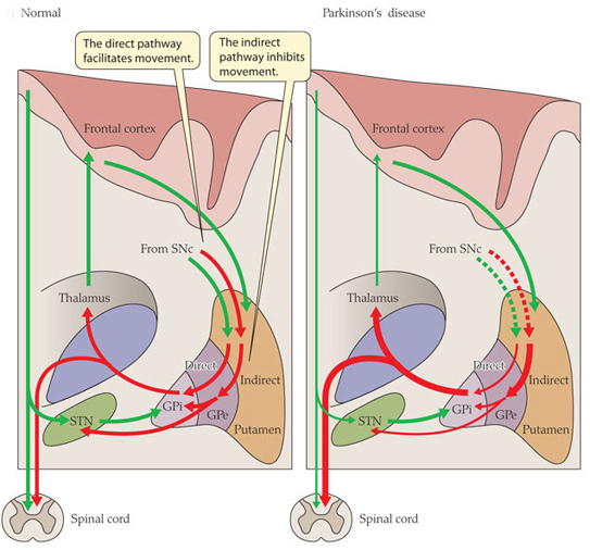
Excitatory signals (green) and inhibitory signals (red) in the basal ganglia, in both a normal brain and one with Parkinson’s disease. Source: Animal Physiology 3rd Edition

Both the direct and indirect pathways finish in the thalamus, but their effects on the thalamus are very different. The direct pathway leaves the thalamus excited and active, while the indirect pathway causes the thalamus to be inhibited.

The thalamus will receive signals from the two pathways and then decide – based on those signals – whether to send an excitatory or inhibitory message to the cortex, telling it what to do (‘get excited and movement’ or ‘don’t get excited and don’t move’, respectively).

Where does dopamine come into the picture?

In Parkinson’s disease, the cells in the brain that produce the chemical dopamine are lost. These cells reside in a structure called the substantia nigra (or SNc in the figure above). What effect does this cell loss have on the direct and indirect pathways? Under normal circumstances the dopamine neurons excite the direct pathway and inhibit the indirect pathway.

In Parkinson’s disease the loss of dopamine neurons results in increased activity in the indirect pathway. As a result, the thalamus is kept inhibited. With the thalamus subdued, the overlying motor cortex has trouble getting excited, and thus the motor system is unable to work properly.

So what was published last week?

Two papers.

Both from the same lab (Well done!)

One in the prestigious scientific journal, Cell and the other in her sister journal, Neuron:

Roseberry-title

Title: Cell-Type-Specific Control of Brainstem Locomotor Circuits by Basal Ganglia.
Authors: Roseberry TK, Lee AM, Lalive AL, Wilbrecht L, Bonci A, Kreitzer AC.
Journal: Cell, 2016 Jan 28;164(3):526-537.
PMID: 26824660

The researchers in this study discovered signal sent from the basal ganglia that selectively activates a group of neurons an area of the brainstem called the ‘mesencephalic locomotor region’. Some of the neurons in this area release a chemical called glutamate. Glutamate is a neurotransmitter that excites the cells it comes into contact with. The researchers who conducted this study found that these glutamate-releasing cells in the mesencephalic locomotor region are responsible for initiating movement.

Print

The researchers used a new technique called ‘optogenetics’ that allows light to activate or inhibit specific cells in the brain. By using this technique on the cells in the direct (dMSN in the figure above) or indirect pathways (iMSN) of the basal ganglia, the researchers were able to control the glutamate-releasing neurons in the mesencephalic locomotor region of mice -initiating or inhibiting their movement, respectively.

The researchers then took the study one step further and used the optogenetics approach directly on the glutamate-releasing neurons in the mesencephalic locomotor region, and they were able to control the initiation of movement in the mice irrespective of the signal being generated by the direct or indirect pathways. That is to say, when the glutamate-releasing neurons in the mesencephalic locomotor region were activated, the mouse would move even when the basal ganglia was sending an inhibitory signal.

So what does it all mean?

While some of the findings of the study were already known, the researchers here have elegantly linked the workings of the basal ganglia and the mesencephalic locomotor region, helping us to better understand the neurological functioning of movement. Deep brain stimulation of the mesencephalic locomotor region has already been attempted and it has demonstrated mixed results in people with Parkinson’s disease (it does appear to help with regards to reducing falls – click here and here for more on this).

It will be interesting to follow the research resulting from this current study.


Parker-title
Title: Pathway-specific remodeling of thalamostriatal synapses in Parkinsonian mice
Authors: Parker PRL, Lalive AL, Kreitzer AC.
Journal: Neuron, 2016
PMID: 26833136

In the second study, the researchers (the same folks who gave us the first paper!) found that the basal ganglia is biased towards the direct pathway. The signal coming from the neurons involved in the direct pathway are stronger than those in the indirect pathway. When dopamine is removed however (as in the case of Parkinson’s disease), the system swings in the opposite direction and becomes biased toward the indirect pathway – the neurons in the direct pathway begin to produce a weaker signal than their counters in the indirect pathway which increase the strength of their signal.

Given that both pathways influence the activity of the thalamus, the researchers next turned their attention to that structure. Again using the ‘optogenics‘ (light-activation) technique, the investigators reduced the inhibitory signal coming from the thalamus and were able to reversibly correct the motor impairs observed in the mice with Parkinson’s-like features.

What does this mean for Parkinson’s disease?
This study turns our attention away from what is happening in the basal ganglia and focuses it on the thalamus, which has not receive the same amount of attention with regards to Parkinson’s disease.

There is a lot already known about changes in the thalamus in Parkinson’s disease (click here for more on this), and deep brain stimulation of structures in neighbouring regions is a regular therapy for Parkinson’s disease (targeting the subthalamic nuclei). But this new paper further breaks down the circuitry of movement for us and offers novel directions for future therapeutic approaches for Parkinson’s disease.

We can be sure that a lot of Parkinson’s disease research is now going to focus on the thalamus.


Improving diagnosis

An inconvenient truth:

The diagnosis of Parkinson’s disease can only be definitively achieved at the postmortem stage.

There is currently no diagnostic test for this task and we are reliant on the training and skills of the neurologists making the diagnosis. Brain imaging techniques (such as DAT-scans) are great, but they can only aid physicians in their final decision.

And those decisions are not always right.

In 1992, a study looking at the brains of 100 subjects who had died with Parkinson’s disease, found that 24% of the cases did not fulfill the pathological requirements for the diagnosis of Parkinson’s disease. That study was:

Accuracy

Title: Accuracy of clinical diagnosis of idiopathic Parkinson’s disease: a clinico-pathological study of 100 cases.
Authors: Hughes AJ, Daniel SE, Kilford L, Lees AJ.
Journal: Journal of Neurol Neurosurg Psychiatry. 1992 Mar;55(3):181-4.
PMID: 1564476

Unfortunately, despite years of research, it would appear that there is still a large degree of error in the clinical diagnosis of Parkinson’s disease. A study published in 2014 in the journal Neurology that suggested that there is currently a 15% rate of misdiagnosis. That study was:


Adler-title

Title: Low clinical diagnostic accuracy of early vs advanced Parkinson disease: clinicopathologic study.
Authors: Adler CH, Beach TG, Hentz JG, Shill HA, Caviness JN, Driver-Dunckley E, Sabbagh MN, Sue LI, Jacobson SA, Belden CM, Dugger BN.
Journal: Neurology. 2014 Jul 29;83(5):406-12.
PMID: 24975862

It has to be said that clinicians face a very difficult task in diagnosing Parkinson’s disease. The variety of features (symptoms) that patients present with in the clinic, and the lack of diagnostic tools, leave neurologists making a judgement based largely on clinical observations.

But this degree of error ultimately has a huge impact on clinical studies and trials: if 10-20% of the participants are not Parkinsonian, are we really going to observe an accurate result?

Better diagnostic tests/tools are critically required.


In November last year, a study was published in the journal Immunology Letters which may help in this regard:
Blood1

Title: Potential utility of autoantibodies as blood-based biomarkers for early detection and diagnosis of Parkinson’s disease.
Authors: DeMarshall CA, Han M, Nagele EP, Sarkar A, Acharya NK, Godsey G, Goldwaser EL, Kosciuk M, Thayasivam U, Belinka B, Nagele RG; Parkinson’s Study Group Investigators.
Journal: Immunol Letters, 168(1), 80-8.
PMID: 26386375  (this article is OPEN access if you would like to read it)

The researchers took 398 subjects, including 103 early-stage Parkinson’s disease subjects and they collected blood samples from them. They then screened the blood for 9,486 different autoantibodies that could be useful as biomarkers for Parkinson’s disease.

Antibodies are produced by our immune system to determine what is ‘self’ and not ‘self’. They are the foundation of our defenses against the big, bad germ/bacteria world. Autoantibodies are antibodies produced by our immune system that are directed against our own tissues. They target ‘self’.

And yeah, that is bad. Autoantibodies are associated with autoimmune diseases such as Lupus.

We are not sure why we produce autoantibodies. The causes of their production vary greatly and are not well understood. In Parkinson’s disease, however, autoantibodies may be produced as a result of the cell death in the brain. Some of the debris resulting from the dying cells will make its way into the bloodstream, to be removed from the body. Whilst in the blood, some of that debris could trigger the immune system, thus resulting in the production of autoantibodies.

De Marshall et al (the researchers who conducted this study) were hoping to take advantage of this autoantibody production and use them as biomarkers to not only differentiate between people with and without Parkinson’s disease, but also to differentiate between different stages of Parkinson’s disease (see the figure below).

1-s2.0-S0165247815300341-gr3

Attempting to differentiate between different stages of Parkinson’s disease. Source: Immuno Letters

The researchers found that using the top 50 autoantibodies that they associated with Parkinson’s disease, they could successfully differentiate between people with and without Parkinson’s disease with 90% prediction accuracy in a blind analysis (they actually found that just the top 4 autoantibodies were enough).

Interestingly, the researchers then compared the early Parkinson’s group with a mild-moderate Parkinson’s group and they found that they could differentiate between the two groups with an overall accuracy of 97.5%!


These are very exciting results and we will be following this work with interest – not only from the standpoint of biomarkers, but also the role of autoantibodies in Parkinson’s disease.

New research – new targets of Lrrk2

This is Sergey Brin.

sergey_brin

He’s a dude.

Having brought us ‘Google’, he is now turning his attention to other projects.

One of these other projects is close to our hearts: Parkinson’s disease.


In 1996, Sergey’s mother started experiencing numbness in her hands. Initially it was believed to be RSI, but then her left leg started to drag. In 1999, following a series of tests, Sergey’s mother was diagnosed with Parkinson’s disease. It was not the first time the family had been affected by the condition: Sergey’s late aunt had also had Parkinson’s disease.

Both Sergey and his mother have had their genome scanned for mutations that increase the risk of Parkinson’s disease. And both of them discovered that they were carrying a mutation on the 12th chromosome, in a gene called Leucine-rich repeat kinase 2 or Lrrk2.

Not everyone with this particular mutation will go on to develop Parkinson’s disease, but Sergey has decided that his chances are 50:50. Being one of the founders of a large company like Google, however, has left Sergey with resources at his disposal. And he has chosen to focus some of those resources on Lrrk2 research (call it an insurance  policy).

Today, the fruits of some of that research has been published and the results are really interesting:

Elife


Title: Phosphoproteomics reveals that Parkinson’s disease kinase LRRK2 regulates a subset of Rab GTPases
Authors: Martin Steger, Francesca Tonelli, Genta Ito, Paul Davies, Matthias Trost, Melanie Vetter, Stefanie Wachter, Esben Lorentzen, Graham Duddy, Stephen Wilson, Marco AS Baptista, Brian K Fiske, Matthew J Fell, John A Morrow, Alastair D Reith, Dario R Alessi, Matthias Mann
Journal: Elife 2016;10.7554/eLife.12813
PMID: 26824392  (This report is openly available for reading on the Elife website)

So what is Lrrk2?

Also known as dardarin (Basque for ‘trembling‘), Lrrk2 is a gene in our DNA that is responsible for making an enzyme. That Lrrk2 enzyme is involved in many different aspects of cell biology. From cellular remodeling and moving (‘trafficking’) various proteins around in the cell, to protein degradation and stabilization, Lrrk2 has numerous roles.

Discovered in 2004, Lrrk2 was quickly associated with Parkinson’s disease because mutations in this gene are amongst the most common in ‘familial Parkinson’s‘ (where an inherited genetic mutation is present in the sufferer; accounting for about 10-20% of all cases of Parkinson’s disease). The most common mutation of LRRK2 gene is G2019S, which is present in 5–6% of all familial cases of Parkinson’s disease, and is also present in 1–2% of all sporadic cases.

Curiously, mutations in Lrrk2 are also associated with increased risk of Crohn’s disease and cancer.

image1

The structure of Lrrk2 and where various mutations lie. Source: Intech

Given the association with Parkinson’s disease, there have been attempts to develop inhibitors of Lrrk2 as a means of treating the condition. These efforts, however, have been hampered by a poor agreement as to which proteins are interacting with Lrrk2.

The goal of the current study was to identify the key proteins that Lrrk2 acts upon.

What did they discover?

Using various techniques to accomplish their task, the scientists began with 30,000 possible targets and gradually whittled that number down to a small group of Lrrk2 targets.

Most importantly, they found that Lrrk2 is deactivating certain proteins that are called ‘Rabs’. The Rab family are heavily involved with trafficking (and that’s not the mafia drug variety!). Trafficking in cells in moving proteins around within the cell itself. And Lrrk2 was found to deactivate 4 Rab family members (3, 8, 10 and 12).

This is a very important result as not only does it provide us with novel Lrrk2 targets, but it also offers us an excellent tool with which we can determine if Lrrk2 inhibitors are actually working  – a functioning Lrrk2 inhibitor will lower the activity of Rab 3, 8 10 & 12 and this can be measured.

The results represent a major leap forward in our understanding of Lrrk2 and a significant return on investment for one Mr Sergey Brin.


New research – the disorder of Alpha Synuclein

A couple of interesting scientific papers were published this week dealing with the Parkinson’s disease-related protein, Alpha Synuclein. If you are not familiar with it, we suggest that you check out our primer page on Alpha Synuclein before reading any further.

So, what’s new in the world of Alpha Synuclein?

Two studies.

One in the prestigious journal Nature and the other in her sister Nature Communications. Both studies came from the same lab (good job guys!)

The first study :

Theillet-title

Title: Structural disorder of monomeric α-synuclein persists in mammalian cells.
Authors: Theillet FX, Binolfi A, Bekei B, Martorana A, Rose HM, Stuiver M, Verzini S, Lorenz D, van Rossum M, Goldfarb D, Selenko P.
Journal: Nature. 2016 Jan 25.
PMID: 26808899

This first study presented a very detailed analysis of the structure of alpha synuclein – at the atomic level – inside living cells.

Interestingly, when the researchers injected alpha synuclein (at concentrations that have been observed in normal neurons) into 5 different types of cells (both neuron and others types), they found that the protein remains extremely disordered – it changed shape rapidly. They determined this by using nuclear magnetic resonance spectroscopy (try saying that 3 times really fast!), which provides a shallow peak readout for stable proteins and a sharp peak for disordered proteins (see image below).

nature16871-f1

The researchers found a lot of sharp peaks in cells that they injected Alpha Synuclein into. Source: Nature

Rather remarkably, despite the fact that disordered proteins are usually removed from cells by enzymatic degradation, the alpha synuclein that was injected by these researchers appears to have remained intact in the cells for several days (50+ hours). And the cells did not seem to be adversely affected by this.

The second Alpha Synuclein study published this week illustrated an equally interesting result:

Binolfi-title

Title: Intracellular repair of oxidation-damaged α-synuclein fails to target C-terminal modification sites.
Authors: Binolfi A, Limatola A, Verzini S, Kosten J, Theillet FX, May Rose H, Bekei B, Stuiver M, van Rossum M, Selenko P.
Journal: Nature Communications, 2016 Jan 25;7:10251.
PMID: 26807843

In this study, the researchers injected damaged alpha synuclein into cells and then watched the cells try to repair that damaged protein. There are specific enzymes that help to maintain/repair proteins like Alpha Synuclein inside each cell. This is a normal recycling process for cells, but something interesting happened with this damaged version of alpha synuclein: only one end of the protein was repaired. The other end (called the C-terminus) was left damaged and this end failed to function correctly.

fnins-09-00059-g001

The structure of Alpha Synuclein. The c-terminus is the area in red. Source: Frontiers in Neuroscience

This led the authors to conclude that damage can cause the accumulation of chemically and functionally altered Alpha Synuclein in cells.

What does this mean for Parkinson’s disease?

The results are very interesting and the researchers should be congratulated on the complexity of their work. The findings add to our understanding of Alpha Synuclein, but both of these results need to be replicated and expanded on before we can fully appreciate their impact.

One possible implications of the results is that designing drugs to target Alpha Synuclein may be more complicated than originally thought. If the protein remains as disordered as the first study suggests, it could be difficult to target. Further investigations, however, focused on the c-terminus end of Alpha synuclein may offer novel targets for therapies looking to clear damaged proteins from cells.

If Alpha Synuclein is the big, bad enemy in Parkinson’s disease, we now know a lot more about him and we can focus on his weaknesses.

New research – Urate and Parkinson’s

New research this week lends further support to ongoing clinical trials focused on urate in Parkinson’s disease:

urate

Title: Prospective study of plasma urate and risk of Parkinson disease in men and women.
Authors: Gao X, O’Reilly ÉJ, Schwarzschild MA, Ascherio A.
Journal: Neurology. 2016 Jan 13.
PMID: 26764029

The researchers in this study looked at 90,214 participants who are involved in three ongoing US-based longitudinal studies (the Nurses’ Health Study (NHS), the Cancer Prevention Study II Nutrition (CPS-IIN), and the Health Professionals Follow-up Study (HPFS)). They identified 388 people in these cohorts who had developed Parkinson’s disease (202 men and 186 women) since their respective longitudinal studies began, and they matched them to 1,267 randomly selected control subjects.

Blood samples that had been taken from the Parkinson’s and control subjects were analysed, and the level of urate was measured. Normal levels of urate range from 3.5-7.2 milligrams per deciliter (mg/dL). The researchers found that there was no difference between in spectrum of urate levels in the women (with or without Parkinson’s).

In men, however, things were very different. The men with the lowest levels of urate had less than 4.9 mg/dL, while those with the highest levels had 6.3-9.0 mg/dL. Among the men with Parkinson’s disease, 45 had the highest level of urate and 58 had the lowest – if no difference existed, this number should be 50:50, but instead there is more than 30% difference. Men with high levels of urate had a lower chance of developing Parkinson’s disease.

The researchers then combined their results with the results from three previous studies on the same topic and found a very similar result. This led the researchers to conclude that men, but not women, with higher urate concentrations had a lower future risk of developing Parkinson’s, suggesting that urate could be protective against Parkinson’s risk or could slow disease progression during the preclinical stage of disease.

So, what is urate?

During the breaking down of dietary proteins, the liver produces large amounts of a chemical called ‘ammonia’. Ammonia is toxic for the body, so the liver breaks it down further, and one of the products of that process is urate. If the body does not get rid of it, urate can build up and form crystals within the joints. High blood concentrations of urate can lead to gout and is also associated with other medical conditions, such as diabetes and the formation of kidney stones.

Paradoxically, urate is also an ‘antioxidant’ – a chemical that prevents tissue from being damaged by the negative effects of oxygen (yes, we need oxygen but not too much). Other antioxidants include vitamin C, and vitamin E. It is this antioxidant function of urate that researchers believe has a positive effect in Parkinson’s disease.

What are the clinical trials we mentioned?

rfi-util-logo

In September 2015, a Phase III trial of Inosine was initiated. The study will involve 270 people with early-stage Parkinson’s. Inosine is a chemical precursor to urate and Phase III is the ‘acid test’ – a double blind test of treatment efficacy. A Michael J Fox Foundation-funded Phase II study showed that Inosine is safe and tolerable, and it also raised levels of urate in people with early-stage Parkinson’s disease. Now it is time to see if this raising of urate levels has a positive outcome. Enrollment for this trial is currently underway and -given the results of the study published this week – it will be interesting to see if there is a stronger effect in men in this phase III trial.


IMPORTANT EDITOR’S NOTE HERE: Inosine is commercially available as a dietary supplement, but we must stress that patients should act with caution. Inosine has not yet been proven as a therapy for Parkinson’s disease, and, as we indicated above, it can cause serious conditions such as gout and kidney stones. Please do not initiate usage of this chemical without first discussing it with your physician.

New Research -Shared genetic features

There was an interesting new study published yesterday:

Sanchez-Mut-Title

Title: Human DNA methylomes of neurodegenerative diseases show common epigenomic patterns.
Author: Sanchez-Mut JV, Heyn H, Vidal E, Moran S, Sayols S, Delgado-Morales R, Schultz MD, Ansoleaga B, Garcia-Esparcia P, Pons-Espinal M, de Lagran MM, Dopazo J, Rabano A, Avila J, Dierssen M, Lott I, Ferrer I, Ecker JR, Esteller M.
Journal: Transl Psychiatry. 2016 Jan 19;6:e718. doi: 10.1038/tp.2015.214.
PMID: 26784972 – this article is OPEN ACCESS if you would like to read it.

The researchers were curious to look for common genetic markers between the major neurodegenerative disease. It is often forgotten that the different neurodegenerative conditions, such as Alzheimer’s disease and Parkinson’s disease, share some common pathological features (the characteristic signs of the diseases in the brain).

For example, when you look at the brains of people with Alzheimer’s disease, approximately 50% of them will also have the alpha-synuclein-containing ‘Lewy bodies’ in their brains, which are more commonly associated with Parkinson’s disease. Likewise, Beta-amyloid plaques and neurotangles, which are characteristic features of Alzheimer’s disease are commonly found in Parkinson’s disease brains (click here and click here for more on this topic).

To find these shared genetic markers, the researcher extracted DNA from the prefrontal cortex (Brodmann area 9) of the brains of people with Alzheimer’s disease, dementia with Lewy bodies, Parkinson’s disease and Alzheimer-like neurodegenerative profile associated with Down’s syndrome samples (more than 75 percent of people with Down Syndrome aged 65 and older develop Alzheimer’s disease – click here for more on this).

Importantly, the researchers were looking at DNA methylation, which is a commonly used tool that allows a cell to fix genes in the “off” position. That is to say, the gene can not be activated. Thus the researchers were looking for regions of DNA that have to closed down.

They found that a very defined set of genes are turned off in these neurodegenerative disorders, suggesting that these condition might have similar underlying mechanisms or processes that subsequently develop into different clinical entities. These newly identified regions of DNA methylation will be further investigated with the goal that one day they may be used as biomarkers in diagnosis and also as potential new targets for the regenerative therapies.

Viruses and Parkinson’s – a hit and run story?

SupportGroup

I was recently presenting a talk at a Parkinson’s support group meeting. Afterwards I sat with some of the attendees and we chatted over tea and cookies. At one point the lady sitting beside me tapped me on the arm and said:

“The other day we were discussing some of the commonalities that we [people with Parkinson’s] share. I wonder if they would be of interest to you?”

“Absolutely”‘ I replied, “Let’s hear them”

“Well, firstly, most of us have little or any sense of smell” she said

And I nodded, “this is a common feature amongst people with Parkinson’s disease” (Click here for more on this)

“Ok. Number two, we all have trouble doing ‘number twos'”

I nodded again, and explained that constipation and gastrointestinal problems are also common features of Parkinson’s disease. (Click here for more on this)

“Interesting”, she said, before aiding: “Thirdly, none of us have ever had chickenpox”

I confess I looked at her a long time.

I was speechless.

I had never heard of anything like that.

In science, we are always looking for the presence of possible causal agents – not their absence. I was so intrigued that I took her contact details and told her that I would go away and do some homework on the matter.

I’d like to share my findings here as part of a larger discussion on viruses and Parkinson’s disease.


Given the random and indiscriminate way in which Parkinson’s disease attacks people, scientists have looked for a virus that may be causing the condition.

283615-virus

Throughout our lives, our immune system is constantly under attack from viruses. They are small infectious agent that thrive by replicating themselves inside the living cells of other organisms. Technically speaking they are not alive as they lack most of the machinery which characterizes ‘life’ (most importantly the components that is necessary for reproduction). We currently know of approx. 3000 viruses, but we can only guess at the total number of viruses (it may be in the millions!).

There are several different ways that Parkinson’s disease could theoretically be caused in some way by a virus:

1. There may be a specific virus that we are unaware of that infects the body at some point in one’s life causing the slow progressive disease. This could be consider the ‘lightning bolt’ theory – a single unlucky event with terrible consequences. Such a theory has weight as it would explain why some clusters of Parkinson’s disease is sometimes observed. People often use the example of Michael J Fox and his TV work colleagues in this theory.


leadership-fox-m-img_2

Actor Michael J Fox and three other people who worked on the Canadian TV show ‘Leo & me’ went on to develop Parkinson’s disease. Image source: Michael J Fox Foundation

2. A virus attacking the body coincides with a secondary event (e.g. a bacterial infection) that may result in the slow progressive events that result in Parkinson’s disease. The secondary event may be a genetic mutation or exposure to an environmental toxin. The virus attack in itself may not be enough in itself.

The two theories outlined above are just theories. We do not know if Parkinson’s disease is caused by a viral infection.

There is, however, some lines of evidence supporting the idea:

Influenza and Parkinson’s disease

Between January 1918 and December 1920 there were two outbreaks of an influenza virus during an event that became known as the 1918 flu pandemic. Approximately 500 million people across the globe were infected, and this resulted in 50 to 100 million deaths (basically 3-5% of the world’s population). Given that is occurred during World War 1, censors limited the media coverage of the pandemic in many countries in order to maintain morale. The Spanish media were not censored, however, and this is why the 1918 pandemic is often referred to as the ‘Spanish flu’.

Influenza is the virus that causes ‘the flu’. Most commonly in a mild form (runny nose, sore throat, coughing, and fatigue), the symptom will arise two days after exposure and last for about a week. There are three types of influenza viruses, called Type A, Type B, and Type C. Type A are the most virulent in humans. The influenza virus behind both of the outbreaks in the 1918 pandemic was a Type A. It was called H1N1.

NOTE: The “H” (hemagglutinin) and the “N” (neuraminidases) are both proteins that are found on the outer surface of the virus. Different viruses have different hemagglutinin and neuraminidase proteins, hence the numbering.

At the same time that H1N1 was causing havoc, a Romanian born neurologist named Constantin von Economo reported a number of unusual symptoms which were referred to as encephalitis lethargica (EL). This disease left many of the victims in a statue-like condition, both motionless and speechless. You may be familiar with the Oliver Sacks book ‘Awakenings’ which was turned into a film starring Robin Williams and Robert De Niro – the patients in that book were victims of EL.

robin_williams_con_robert_de_niro_en_1990

Robin Williams and Robert De Niro in Awakenings

Historically, it was believed that EL was caused by the influenza virus from the 1918 Spanish influenza pandemic. This was largely due to a temporal association and the finding of influenza antigens in some of the suffers of EL. More recent evidence rejects this hypothesis (e.g. an absence of viral RNA recovered from the brains of postencephalitic PD patients – click here for more on this). We genuinely don’t know what caused EL.

But there has recently been some evidence suggesting a link between Parkinson’s disease and influenza:

Jang-title

Title: Highly pathogenic H5N1 influenza virus can enter the central nervous system and induce neuroinflammation and neurodegeneration.
Author: Jang H, Boltz D, Sturm-Ramirez K, Shepherd KR, Jiao Y, Webster R, Smeyne RJ.
Journal: Proc Natl Acad Sci U S A. 2009 Aug 18;106(33):14063-8.
PMID: 19667183

The researchers in this study found that when they injected the highly infectious H5N1 influenza virus into mice, the virus progressed from the periphery into the brain, where it induced Parkinson’s disease-like symptoms. The virus also caused a significant increase in the aggregation of the protein Alpha Synuclein. Importantly, they witnessed the loss of dopamine neurons in the midbrain of the mice 60 days after resolution of the infection.

This study supports the theory we discussed above (theory 1.) of a virus possibly causing Parkinson’s disease. These same researchers have also looked at other influenza viruses and found additional results:

Sadasivan-title

Title: Induction of microglia activation after infection with the non-neurotropic A/CA/04/2009 H1N1 influenza virus.
Author: Sadasivan S, Zanin M, O’Brien K, Schultz-Cherry S, Smeyne RJ.
Journal: PLoS One. 2015 Apr 10;10(4):e0124047.
PMID: 25861024

In this second study, however, the different type of influenza (H1N1) did not infect the brain, but did cause the immune system to flare up. This is an interesting example of the second theory we discussed above (theory 2.), the double hit theory of Parkinson’s disease, in which the virus doesn’t necessarily cause Parkinson’s disease but plays a supportive role to some other toxic agent in attacking the body.

In a follow up study to their 2009 report on H5N1, these same researchers found that the Parkinson’s disease-like symptoms that they observed were actually only temporary:

Jang-title2

Title: Inflammatory effects of highly pathogenic H5N1 influenza virus infection in the CNS of mice.
Authors: Jang H, Boltz D, McClaren J, Pani AK, Smeyne M, Korff A, Webster R, Smeyne RJ.
Journal: Journal for Neuroscience, 2012 Feb 1;32(5):1545-59.
PMID: 22302798

This third study may give further support to the double hit theory (theory 2.), but also indicates how complicated a viral component to Parkinson’s disease can be.

And influenza is not the only virus to be associated with Parkinson’s disease.

Hepatitis C and Parkinson’s disease

Hepatitis C is a contagious liver disease, which is caused by the hepatitis C virus (HCV). The virus has been found in the brains of infected people, and it has also been shown to kill dopamine neurons in cell culture. Only in the last few months, however, has a more direct association with Parkinson’s disease been proposed:

HepC-Title

Title: Hepatitis C virus infection as a risk factor for Parkinson disease: A nationwide cohort study.
Authors: Tsai HH, Liou HH, Muo CH, Lee CZ, Yen RF, Kao CH.
Journal: Neurology, 2015 Dec 23. Published early online.
PMID: 26701382

The researchers in this study wanted to investigate whether hepatitis C could be a risk factor for Parkinson’s disease. They did this by analyzing data from 2000-2010 drawn from the Taiwan National Health Insurance Research Database.

The database included 49,967 people with either hepatitis B, hepatitis C or both, in addition to 199,868 people without hepatitis. During the 12 year period, 270 participants who had a history of hepatitis developed Parkinson’s disease (120 still had hepatitis C). This compared with 1,060 participants who were free of hepatitis, but went on to develop Parkinson’s disease.

When the researchers controlled for potentially confounding factors (such as age, sex, etc), the researchers found participants with hepatitis C had a 30% greater risk of developing Parkinson’s disease than the controls.

Summary

It is tempting to consider a viral theory for Parkinson’s disease, especially as the condition seems to strike indiscriminately from out of the blue. Maybe a virus works in cahoots with another factor (unable to do the job alone). The evidence of this, however, has not been apparent to allow for a definitive conclusion.


Finally, regarding my homework… 

Never having Chickenpox could mean two different things – never being exposed to it OR being exposed to it and not getting infected (missing a particular protein required for infection). The first would suggest that exposure to the virus would given some kind of resistance to Parkinson’s disease. The latter would suggest that a protein which makes you vulnerable to Chickenpox gives you resistance to Parkinson’s disease.

As to the scientific literature, there have been two studies published regarding Chickenpox and Parkinson’s disease. The first:

Semchuk-title

Title: Parkinson’s disease: a test of the multifactorial etiologic hypothesis.
Authors: Semchuk KM, Love EJ, Lee RG.
Journal: Neurology, 1993 Jun;43(6):1173-80.
PMID: 8170564

In this study, the researchers collected life-time information (family history, occupational and medical records, etc) from 130 people with Parkinson’s disease. When the looked at all of the variables, they noted that a family history of Parkinson’s had the strongest association with Parkinson’s disease. This was followed by head trauma and occupational herbicide use. The subjects with Parkinson’s disease did not differ from control subjects with regards to:

  • exposure to smoking or ionizing radiation
  • family history of essential tremor
  • work-related contact with aluminum, carbon monoxide, cyanide, manganese, mercury, or mineral oils
  • history of arteriosclerosis, chicken pox, encephalitis, hypertension, hypotension, measles, mumps, rubella, or Spanish flu.

They proposed that the results supported the idea of a multifaceted cause of Parkinson’s disease, “probably involving genetic, environmental, trauma, and possibly other factors”.

And the second published study was:

Title: Infections as a risk factor for Parkinson’s disease: a case-control study.
Authors: Vlajinac H, Dzoljic E, Maksimovic J, Marinkovic J, Sipetic S, Kostic V.
Journal: Int J Neurosci. 2013 May;123(5):329-32. doi: 10.3109/00207454.2012.760560. Epub 2013 Feb 4.
PMID: 23270425

In this study the researchers found that Parkinson’s Disease was significantly associated to mumps, scarlet fever, influenza, whooping cough and herpes simplex infections. But they found no association between Parkinson’s disease and Tuberculosis, measles or chickenpox

So it would appear that chickenpox is not associated with Parkinson’s disease. And at a subsequent Parkinson’s support group meeting I asked the audience for a raise of hands as to who has had chickenpox and there was a sea of hands.

Back to the drawing board I guess.


The Placebo effect and Parkinson’s disease

According to our friends at Wikipedia:

A placebo (/pləˈsiboʊ/ plə-see-boh; Latin placēbō, “I shall please” from placeō, “I please”) is a simulated or otherwise medically ineffectual treatment for a disease or other medical condition intended to deceive the recipient. Sometimes patients given a placebo treatment will have a perceived or actual improvement in a medical condition, a phenomenon commonly called the placebo effect or placebo response.


power-of-placebo-effect

In our previous post we wrote about cell transplantation and we cited the two double-blind clinical studies that found little positive effect resulting from the procedure.

In both of those studies, half the participants were given a sham surgery – that is, they were put into the surgery room, anesthetized, an incision was made in their scalps, but nothing was injected into their brains. They (and their assessing investigators) were not told if they were in the transplant group or the sham/control group and they were left in this ‘blind’ state for 12-18 months.

Time is a funny thing.

After a couple of weeks of wondering which group they were in and self assessing their abilities since the surgery, some of the individuals in those studies may have started to think that you are in one group or the other. This is a very human thing to do.

The effect is VERY strong. And it can mess with a clinical study in terrible ways.

In one of the double-blind clinical studies discussed in the last post (Freed et at, 2001), one of the patients had described herself as ‘not being physically active for several years’ before her surgery. Shortly after her surgery, she found that she was able to hike and ice skate again. A miraculous change in situations.

Twelve months after the surgery, however, she found out that she’d had been in the sham/control surgery group. Nothing had been injected into her brain. She had received NO treatment.

Her response was solely due to the placebo effect.

The Placebo effect in Parkinson’s disease

Early last year there was an interesting study conducted that looked at the placebo effect and Parkinson’s disease.

Placebo1

Title: Placebo effect of medication cost in Parkinson disease: a randomized double-blind study.
Author: Espay AJ, Norris MM, Eliassen JC, Dwivedi A, Smith MS, Banks C, Allendorfer JB, Lang AE, Fleck DE, Linke MJ, Szaflarski JP.
Journal: Neurology. 2015 Feb 24;84(8):794-802.
PMID: 25632091

The investigators conducted a double-blind study involving 12 patients with moderate-severe Parkinson disease (average age of the subjects was 62.4 ± 7.9 years; and their average time since diagnosis was 11 ± 6 years). The study involved two visits to the clinic – the first visit involved a clinical assessment while the subjects were both ‘off’ and ‘on’ their standard medication. The assessment also involved a brain scan (fMRI). This was done to determine the magnitude of the dopaminergic benefit of their standard medication.
During the second visit, the subjects were told that they would be given two formulations – a “cheap” and “expensive” – of a “novel injectable dopamine agonist”. Both of these solutions were simply the same saline (medical salt water) solution. Four hours after being give the first injection, the subjects were given the other solution. In this manner, the subjects were exposed to both the ‘cheap’ and ‘expensive’ solutions. During visit 2, the subjects were clinically assessed and brain scanned 3 times, once before the first solution was injected, once after the first solution, and once after the second solution was given.

Below is a flowchart illustrating the structure study:

NEUROLOGY2014583914FF1

Source: Neurology

The results were interesting:

  • Both of the placebos improved motor function when compared with the baseline (no medication state)
  • The expensive placebo had more effect than the cheap placebo (remember: they were the same solution!)
  • The benefits were greater when patients were randomized first to expensive placebo followed by the cheap.
  • There was a significant difference in the level of improvement between the cheap and expensive placebos (UPDRS-III), with the expensive placebo giving better benefits
  • Brain imaging demonstrated that activation was greater when the cheap placebo was given first.

The authors concluded that the “expensive placebo significantly improved motor function and decreased brain activation in a direction and magnitude comparable to, albeit less than, levodopa. Perceptions of cost are capable of altering the placebo response in clinical studies“.

The authors also wrote a summary of the debriefing that followed the study, where the subjects were informed about the true nature of the study. They told the subjects that rather than being injected with a novel dopamine agonist, they were simply given a saline solution – the same solution for both ‘cheap’ and ‘expensive’. They reported that “responses ranged from disbelief to amazement regarding changes experienced“. It must have been rather bewildering to have been told that the positive benefits you experienced were ‘all in your head’ and not based on any pharmacological effect.

While extremely unethical, we here at the SoPD can’t help but wonder about long this placebo effect could last. Would the difference between the cheap and expensive solutions still exist in 12 months time if the subjects were left blinded and continued to take them?

So how might this work?
We know that the placebo effect in Parkinson’s disease is controlled by the release of dopamine – one of the chemicals in the brain that is affected by Parkinson’s disease. Importantly, we know that it the endogenous dopamine that is causing the effect – that is the dopamine our brains are producing naturally as opposed to the L-dopa treatment.

The dopamine that helps to control our motor movements is also involved with positive anticipation, motivation, and response to novelty. Thus when the placebo solution was given to subjects in this study, they believed that they were receiving an active drug and demonstrated an “expectation of reward” response. And the more expensive solution simply heightened the expectation and positive anticipation, therefore increasing the amount of dopamine produced/released.

Given that dopamine is involved with both the features of Parkinson’s disease AND with the mechanisms of anticipation/expectation, you can begin to understand why the placebo effect is such an enormous problem for clinicians undertaking clinical trials.

It would be nice, however, to have a better understanding of the placebo effect and try to harness its positive benefits while also treating Parkinson’s with diseasing slowing/halting therapies.

A call to arms

While our primary goal here at the Science of Parkinson’s is to highlight and explain new research dealing with Parkinson’s disease, we are also keen to encourage the general public to get involved with efforts to cure this debilitating condition.

To this end, we would like to bring your attention to the fact that 2017 represents the 200th anniversary of the first report of Parkinson’s disease by one Dr James Parkinson:

320px-Parkinson,_An_Essay_on_the_Shaking_Palsy_(first_page)

Although there were several earlier descriptions of individuals suffering from rigidity and a resting tremor, Dr Parkinson’s 66 page publication of six cases of ‘Shaking Palsy’, is considered the seminal report that gave rise to what we now call Parkinson’s disease. The report was published in 1817.

The 200th anniversary represents a fantastic opportunity to raise awareness about the disease and a rallying point for a concerted research effort to deal with the condition once and for all. It is still a year away, but now is the time to start planning events and building awareness. We would encourage you to mark 2017 as the year of Parkinson’s disease, share this with everyone you know, and endeavour to make some small effort to help in the fight against this condition.

Parkinson’s disease and the cancer drug

In October, 40,000 neuroscientists from all over the world gathered in Chicago for the annual Society for Neuroscience conference. It is one of the premier events on the ‘brain science’ calendar each year and only a few cities in the USA have the facilities to handle such a huge event.


agu20141212-16

Science conference. Source: JPL

During the five day neuroscience marathon, hundreds of lecture presentations were made and thousands of research poster were exhibited. Many new and exciting findings  were presented to the world for the first time, including the results of an interesting pilot study that has left everyone in the Parkinson’s research community very excited, but also scratching their heads.

The study (see the abstract here) was a small clinical trial (12 subjects; 6 month study) that was aiming to determine the safety and efficacy of a cancer drug, Nilotinib (Tasigna® by Novartis), in advanced Parkinson’s Disease and Lewy body dementia patients. In addition to checking the safety of the drug, the researchers also tested cognition, motor skills and non-motor function in these patients and found 10 of the 12 patients reported meaningful clinical improvements.

The study investigators reported that one individual who had been confined to a wheelchair was able to walk again; while three others who could not talk before the study began were able to hold conversations. They suggested that participants who were still in the early stages of the disease responded best, as did those who had been diagnosed with Lewy body dementia.

So what is Nilotinib?

Nilotinib (pronounced ‘nil-ot-in-ib’ and also known by its brand name Tasigna) is a small-molecule tyrosine kinase inhibitor, that has been approved for the treatment of imatinib-resistant chronic myelogenous leukemia (CML). That is to say, it is a drug that can be used to treat a type of leukemia when the other drugs have failed. It was approved for this treating cancer by the FDA in 2007.

The researchers behind the study suggest that Nilotinib works by turning on autophagy – the “garbage disposal machinery” inside each neuron. Autophagy is a process that clears waste and toxic proteins from inside cells, preventing them from accumulating and possibly causing the death of the cell.

Print

The process of autophagy – Source: Wormbook

Waste material inside a cell is collected in membranes that form sacs (called vesicles). These vesicles then bind to another sac (called a lysosome) which contains enzymes that will breakdown and degrade the waste material.


Some details about the study:

  • The study was run at the Georgetown University Medical Center
  • The patients were given increasing doses of Nilotinib (150mg to 300mg/day) that were are significantly lower than the doses of Nilotinib used for CML treatment (800-1200mg/day).
  • The researchers took cerebrospinal fluid (CSF; the liquid surrounding the brain) and blood samples at the start of the study, 2 and 6 months into the study.
  • Nilotinib was detected in the CSF, indicating that it had no problem crossing the protective blood-brain-barrier – the membrane covering the brain that blocks many drugs from entering.
  • Participants exhibited positive changes in various cerebrospinal fluid biomarkers with statistically significant changes in an important protein called, Tau, which have been shown to increase with the onset of dementia.
  • The researchers found a significant reduction (>60%) in levels of α-Synuclein detected in the blood, but no change in CSF levels of α-Synuclein. 
  • The investigators report that one individual confined to a wheelchair was able to walk again; three others who could not talk were able to hold conversations.

If the outcomes of this study are reproducible, then we here at the Science of Parkinson’s are assuming that Nilotinib is working by turning on the garbage disposal system of the remaining cells in the brain and allowing them to function better. This would suggest that there is a certain level of dysfunction in those remaining cells, which would be expected as this is a progressive disease. The study researchers reported that the small, daily dose of nilotinib turns on autophagy for about four to eight hours, and if that is enough to have such remarkable effects, then this treatment deserves more research.

The results of the study are intriguing and the participants of the study will continue to be treated and followed to see if the improvements continue.

BUT before we go getting too excited:

While these results sound extremely positive, there are several issues with this study that need to be considered before we celebrate the end of Parkinson’s disease.

Firstly, this study was an open-label trial – that means that everyone involved in the study (both researchers and subjects) knew what drug they were taking. There was also no control group or control treatment for comparative analysis in the study. Given these conditions there is always the possibility that what some of the subjects were experiencing was simply a placebo effect. Indeed the lead scientist on the project, Dr Fernando Pagan, pointed out that “It is critical to conduct larger and more comprehensive studies before determining the drug’s true impact.”

In addition, according to Novartis (the producer of the drug), the current cost of Nilotinib is about $10,360 (£6,900) per month for the daily 800mg dose used for cancer treatment. Even if the dose used in this study was only 150 to 300 mg/daily, it would still make this treatment extremely expensive. 

Thirdly, Nilotinib has a number of adverse side-effects when used as an anti-cancer drug (at 800mg/day). These include headache, fatigue, nausea, vomiting, diarrhea, constipation, muscle/joint pain, skin issues, flu-like symptoms, and reduced blood cell count. It may not be the nicest of treatments to tolerate.

There are important reasons for optimism, however, with the results of this study:

In 2010, a group of researchers published a paper demonstrating the neuroprotective effects of another cancer drug very similar to Nilotinib. That drug was ‘Gleevec’

Gleevec-PD1

Title: Phosphorylation by the c-Abl protein tyrosine kinase inhibits parkin’s ubiquitination and protective function.
Authors: Ko HS, Lee Y, Shin JH, Karuppagounder SS, Gadad BS, Koleske AJ, Pletnikova O, Troncoso JC,Dawson VL, Dawson TM.
Journal: Proc Natl Acad Sci U S A. 2010 Sep 21;107(38):16691-6.
PMID: 20823226

And that Gleevec publication was followed up a couple of years ago with a second study demonstrating the neuroprotective effects of another Abl-inhibitor: Nilotinib!

Gleevec-PD2

Title: The c-Abl inhibitor, nilotinib, protects dopaminergic neurons in a preclinical animal model of Parkinson’s disease.
Authors: Karuppagounder SS, Brahmachari S, Lee Y, Dawson VL, Dawson TM, Ko HS
Journal: Sci Rep. 2014 May 2;4:4874.
PMID: 24786396

These studies provided a strong rationale for testing brain permeable c-Abl inhibitors as potential therapeutic agents for the treatment of PD. The phase 2 trial at Georgetown will be starting in early 2016 and we will be watching this trial very closely.