We have talked a lot about a protein called Alpha Synuclein on this blog (see our primer page here and our previous post).
It is very closely associated with Parkinson’s disease, given that people with genetic mutations in the alpha synuclein gene are more vulnerable to the condition, AND the protein is a key component in the disease-related circular aggregations (called ‘Lewy bodies’) in the brain. Recently researchers have identified proteins that may be involved with the transfer of Alpha Synuclein between cells – the method by which the disease is believed to be spreading. By blocking or removing these proteins, the researchers have been able to block the transfer of alpha synuclein.
In this post, we will review the research and discuss what this could mean for Parkinson’s disease.
At the recent annual Society for Neuroscience conference in sunny San Diego, Dr Ravindran Kumaran, a neuroscientist in the laboratory of Professor Mark Cookson (at the National Institute on Aging in Bethesda, Maryland) stood up and presented data about an interesting protein that few people in the audience had ever heard of.
Title: High-content siRNA screen identifies cellular modifiers of pre-formed alpha-synuclein fibril uptake Authors: Kumarani R, Fernandez D, Werner-Allen JW, Buehler E, Bax A, Lai-Nag M, Cookson MR. Source: Click here to see the full abstract
Dr Kumaran and his colleagues had systematically removed the function of each gene – one by one – in cell cultures of human cancer cells, and then measured the efficiency of the cells to absorb (or ‘take up’) the Parkinson’s related protein, alpha synuclein. An absolutely laborious task (remember there are over 20,000+ genes), but when they turned off a gene called TM9SF2, something amazing happened:
The cells absorbed 75% less of the free floating alpha synuclein than normal health cells.
This caused a bit of excitement in the Parkinson’s research community. Here was a potential method of blocking the spreading of alpha synuclein.
The funny thing is: few people had ever heard of TM9SF2, and yet Dr Kumaran then showed that TM9SF2 is in the top 3% of all proteins present in the brain. In fact, the highest concentrations of TM9SF2 are in the substantia nigra and other brain regions that are most affected by Parkinson’s disease.
So you can hopefully understand why some people in the Parkinson’s research community thought that this was a wee bit exciting.
Plus, this data presentation came on the back of another study that was published in September which presented another protein (called Lag3) that exhibited a similar ability to reduce the absorption of alpha synuclein:
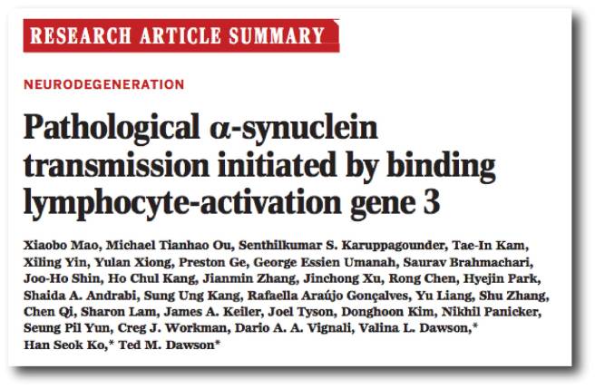
Title: Pathological α-synuclein transmission initiated by binding lymphocyte-activation gene 3. Authors: Mao X, Ou MT, Karuppagounder SS, Kam TI, Yin X, Xiong Y, Ge P, Umanah GE, Brahmachari S, Shin JH, Kang HC, Zhang J, Xu J, Chen R, Park H, Andrabi SA, Kang SU, Gonçalves RA, Liang Y, Zhang S, Qi C, Lam S, Keiler JA, Tyson J, Kim D, Panicker N, Yun SP, Workman CJ, Vignali DA, Dawson VL, Ko HS, Dawson TM. Journal: Science. 2016 Sep 30;353(6307). PMID:27708076
In this study, the researchers conducted a screen of 352 proteins that sit on the membrane of cells. They were measuring the level of alpha synuclein binding. They identified three interesting candidates for further investigation, include lymphocyte-activation gene 3 (LAG3), neurexin 1β, and amyloid β precursor-like protein 1 (APLP1).
When the researchers compared the three, they found that by removing LAG3 less alpha synuclein was taken into the cell (by endocytosis) than the other two proteins. In addition, when they increased the amount of LAG3 that a cell produces, they observed a similar increase in the amount of alpha synuclein absorbed by cells.
Next the researchers investigated the transmission of alpha synuclein between brain cells in both normal cells and cells that had no LAG3, and they found not only that LAG3 is required for the transmission, but the absence of LAG3 reduces the damage caused by the transmission.
Finally the researchers used small proteins (antibodies) to bind to and block LAG3, and they observed less transmission and damage caused by alpha synuclein. In their conclusions, the authors pointed out that LAG3 is not the only protein involved with the transmission of alpha synclein – there will be others – but it represents a potential future target for therapeutic intervention in Parkinson’s disease.
So what does this mean?
If the theory of alpha synuclein – that this protein is passed between cells, causing the spread of the disease – is correct, then any agent that can block that transmission should slow down or halt Parkinson’s disease. We have previously talked about vacines and antibodies against alpha synuclein being tested in the clinic (Click here, here and here for more on this), but blocking TM9SF2 and LAG3 represent a new method of preventing the transmission of alpha synuclein. This is very exciting. The more angles of attack that we have for designing a treatment the better our options.
Schematic of how LAG3 may be working. Source: Science
We will be watching the field very closely and will keep you posted as new information comes to hand.
We have previously written about the benefits of drinking coffee in reducing one’s chances of developing Parkinson’s disease (Click here for that post). Today, however, we shift our attention to another popular beverage: Tea.
Green tea in particular. Why? Because of a secret ingredient called Epigallocatechin Gallate (or EGCG).
Today’s post will discuss why EGCG may be of great importance to Parkinson’s disease.
INTERESTING FACT: after water, tea is the most widely consumed drink in the world.
In the United Kingdom only, over 165 million cups of tea were drunk per day in 2014 – that’s a staggering 62 billion cups per year. Globally 70 per cent of the world’s population (over the age of 10) drank a cup of tea yesterday.
Tea is derived from cured leaves of the Camellia sinensis, an evergreen shrub native to Asia.
The leaves of Camellia sinensis. Source: Wikipedia
There are two major varieties of Camellia sinensis: sinensis (which is used for Chinese teas) and assamica (used in Indian Assam teas). All versions of tea (White tea, yellow tea, green tea, etc) can be made from either variety, the difference is in the processing of the leaves.
The processing of different teas. Source: Wikipedia
There are at least six different types of tea based on the way the leaves are processed:
White: wilted and unoxidized;
Yellow: unwilted and unoxidized but allowed to yellow;
Green: unwilted and unoxidized;
Oolong: wilted, bruised, and partially oxidized;
Black: wilted, sometimes crushed, and fully oxidized; (called “red tea” in Chinese culture);
Post-fermented: green tea that has been allowed to ferment/compost (“black tea” in Chinese culture).
(Source: Wikipedia)
More than 75% of all tea produced in this world is considered black tea, 20% is green tea, and the rest is made up of white, Oolong and yellow tea.
What is the difference between Green tea and Black tea?
Green tea is made from Camellia sinensis leaves that are largely unwilted and heated through steaming (Japanese style) or pan-firing (Chinese style), which halts oxidation so the leaves retain their color and fresh flavor. Black tea leaves, on the other hand, are harvested, wilted and allowed to oxidize before being dried. The oxidation process causes the leaves to turn progressively darker.
So what does green tea have to do with Parkinson’s disease?
In 2006,this research paper was published:
Title: Small molecule inhibitors of alpha-synuclein filament assembly Authors: Masuda M, Suzuki N, Taniguchi S, Oikawa T, Nonaka T, Iwatsubo T, Hisanaga S, Goedert M, Hasegawa M. Journal: Biochemistry. 2006 May 16;45(19):6085-94. PMID:16681381
In this study, the researchers tested 79 different chemical compounds for their ability to inhibit the assembly of alpha-synuclein into fibrils. They found several compounds of interest, but one of them in particular stood out: Epigallocatechin Gallate or EGCG
The chemical structure of EGCG. Source: GooglePatents
Now, before we delve into what exactly EGCG is, let’s take a step back and look at what is meant by the “assembly of alpha-synuclein into fibrils” (???).
Alpha Synuclein
We have previously written a lot about alpha synuclein (click here for our primer page). It is a protein that has been closely associated with Parkinson’s disease for some time now. People with mutations in the alpha synuclein gene are more vulnerable to developing Parkinson’s disease, and the alpha synuclein protein is found in the dense circular clumps called Lewy bodies that are found in the brains of people with Parkinson’s disease.
A lewy body (brown with a black arrow) inside a cell. Source: Cure Dementia
What role alpha synuclein plays in Parkinson’s disease and how it ends up in Lewy bodies is the subject of much research and debate. Many researchers, however, believe that it all depends on how alpha synuclein ‘folds’.
The misfolding of alpha synuclein
When a protein is produced (by stringing together amino acids in a specific order set out by RNA), it will then be folded into a functional shape that do a particular job.
Alpha synuclein is slightly different in this respect. It is normally referred as a ‘natively unfolded protein’, in that is does not have a defined structure. Alone, it will look like this:
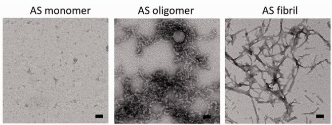
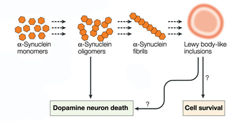
By itself, alpha synuclein is considered a monomer, or a single molecule that will bind to other molecules to form an oligomer (a collection of a certain number of monomers in a specific structure). In Parkinson’s disease, alpha-synuclein also aggregates to form what are called ‘fibrils’.
Microscopic images of Monomers, oligomers and fibrils. Source: Brain
Oligomer versions of alpha-synuclein are emerging as having a key role in Parkinson’s disease. They lead to the generation of fibrils and may cause damage by themselves.
It is believed that the oligomer versions of alpha-synuclein is being passed between cells – and this is how the disease may be progressing – and forming Lewy bodies in each cells as the condition spreads.
For this reason, researchers have been looking for agents that can block the production of alpha synuclein fibrils and stabilize monomers of alpha synuclein.
And now we can return to EGCG.
What is EGCG?
Epigallocatechin Gallate is a powerful antioxidant. It has been associated with positive effects in the treatment of cancers (Click here for more on that).
And as the study mentioned near the top of this blog suggested, EGCG is also remarkably good at blocking the production of alpha synuclein fibrils and stabilizing monomers of alpha synuclein. If the alpha synuclein theory of Parkinson’s disease is correct, then EGCG could be the perfect treatment.
And there have been many studies replicating this effect:
Title: EGCG remodels mature alpha-synuclein and amyloid-beta fibrils and reduces cellular toxicity Authors: Bieschke J, Russ J, Friedrich RP, Ehrnhoefer DE, Wobst H, Neugebauer K, Wanker EE. Journal: Proc Natl Acad Sci U S A. 2010 Apr 27;107(17):7710-5. doi: 10.1073/pnas.0910723107. PMID:20385841 (This article is OPEN ACCESS if you would like to read it)
In this particular study, the researchers found that EGCG has the ability to not only block the formation of of alpha synuclein fibrils and stabilize monomers of alpha synuclein, but it can also bind to alpha synuclein fibrils and restructure them into the safe form of aggregated monomers.
And again, what has Green tea got to do with Parkinson’s disease?
Green tea is FULL of EGCG.
In the production of Green tea, the picked leaves are not fermented, and as a result they do not go through the process of oxidation that black tea undergoes. This leaves green tea extremely rich in the EGCG, and black tea almost completely void of EGCG. Green tea is also superior to black tea in the quality and quantity of other antioxidants.
What clinical studies have been done on EGCG and Parkinson’s disease?
Two large studies have looked at whether tea drinking can lower the risk of Parkinson’s disease. Both studies found that black tea is associated with a reduced risk of Parkinson’s disease, but one of the studies found that drinking green tea had no effect (Click here and here for more on this). Now the positive effect of black tea is believed to be associated with the high level of caffeine, which is a confounding variable in these studies. Even Green tea has some caffeine in it – approximately half the levels of caffeine compared to black tea.
The levels of EGCG in these studies were not determined and we are yet to see a proper clinical trial of EGCG in Parkinson’s disease. EGCG has been clinically tested in humans (Click here for more on that), so it seems to be safe. And there is an uncompleted clinical trial of EGCG in Huntington’s disease (Click here for more) which we will be curious to see the results of.
So what does it all mean?
Number 1.
It means that if the alpha-synuclein theory of Parkinson’s disease is correct, then more research should be done on EGCG. Specifically a double-blind clinical trial looking at the efficacy of this antioxidant in slowing down the condition.
Number 2.
It means that I now drink a lot of green tea.
Usually mint flavoured (either Teapigs or Twinnings – please note: SoPD is not a paid sponsor of these products, though some free samples would be appreciated!).
Here at SoPD we try to remain politically neutral.
That said, we do have a vested interest when it comes to political events and their impact on government research funding for Parkinson’s disease (or simply medical research in general).
In the wake of the recent BREXIT vote in the UK and the poll-defining victory of Mr Donald Trump in the US presidential elections, there have been many in the research community who are expressing concerns about the future of research funding.
In this post we thought it would be interesting to have a look at US and UK Government research funding and where things may be heading after the election of Mr Trump and the BREXIT vote.
What is the current situation for federal research funding in the USA?
According to the American Association for the Advancement of Science (AAAS), the US federal government appropriates almost $140 billion per year to research and development. That is a remarkably big number (it is more than the entire GDP of Hungary!).
The grandeur of this number, however, hides a disturbing fact. That $140 billion is down from a 2010 peak of about $160 billion (in constant dollars – inflation adjusted). And this reduction in funding has had trickle down effects.
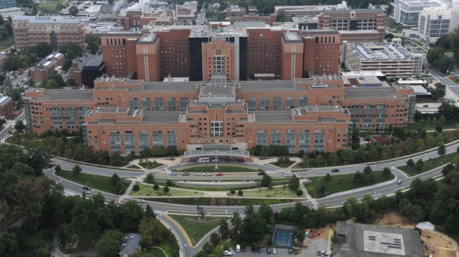
The NIH headquarters in Maryland, USA. Source: NPR
The National Institute for Health (NIH) is one of the largest funders of medical research in the world. In 2015 it had a budget of $31,381 million. More than 83 percent of their budget goes to more than 300,000 research personnel at over 3,000 universities, medical schools, and other research institutions in the USA and around the world (Source: NIH). Few other research funding institutions wield the kind of power that the NIH has.
Again, however, the impressive numbers hides a secret.
As displayed in the graph above, from 2003 to 2015, NIH funding from the US government dropped by 22% of its capacity to fund research due to budget cuts, sequestration, and inflationary losses.
In very real terms, medical research funding from the US federal Government has been falling – and this started long before the global financial crisis.
What is the current situation for Government research funding in the UK?
The UK spends approximately £25bn per year on research.While not as impressive as our cousins across the pond, that number is still a large chunk of change. Approximately 1/3 (£7.98bn) comes from the UK Government. And again that sounds like a lot of money, but here is the terrible truth of the matter:
Science research funding as a % of GDP. Source: Scienceogram
At a time where the population is ageing and requiring more assistance due to conditions like Parkinson’s disease, we are spending less (based on GDP) on research than most of our neighbours. Yes, we are still recovering for the global economic crisis (9 years and counting, dear bankers), but the trend for the UK in the graph above is of some concern. Especially when you consider that back in the 1980s the UK was spending over 2% of GDP on research:
The difference in % of GDP spent on research between 1985 and 2007. Source: Keith’s Blog
For academic research, there are seven Research Councils that receive funding from the Government’s Science Budget. Each year, they invest around £3 billion in research, covering the full spectrum of academic disciplines. This arrangement may change shortly, with all of the seven councils coming together under one umbrella: Research UK (but that is an entirely different controversy – click here for more on this).
A total investment of £26.3 billion has been planned by the Government between 2016/17 to 2020/21 (Source: Gov.uk), but this may well change in the wake of BREXIT. All eyes in the UK are focused on the Autumn budget statement on Wednesday 23 November. This will be the first confirmation from Theresa May’s government as to their stance on research funding.
In addition to Government funding of research, the UK research community has benefitted considerably from belonging to the EU. Between 2007 to 2013, the UK contributed nearly £4.3bn towards EU research projects, BUT it received nearly £7bn back over the same period. That £2.7bn excess was equivalent to more than £400m in research funds a year. By leaving the EU, this enormous stream of funding is now in jeopardy.
The UK is the leading country in terms of number of projects won from Horizon 2020. Source: LSE
We remain fully paid-up members of Horizon 2020, the EU’s eighth Framework Programme for funding research and innovation, and as the graph above shows we are one of the most successful countries in the EU with regards to projects being awarded funding. The Horizon 2020 scheme has a total budget of just over €70 billion for funding research until 2020. But beyond that…
Critically for researchers, the lack of clarity in the UK position with the EU leaves the potential for international collaborations up in the air.
So what is the outlook for the US?
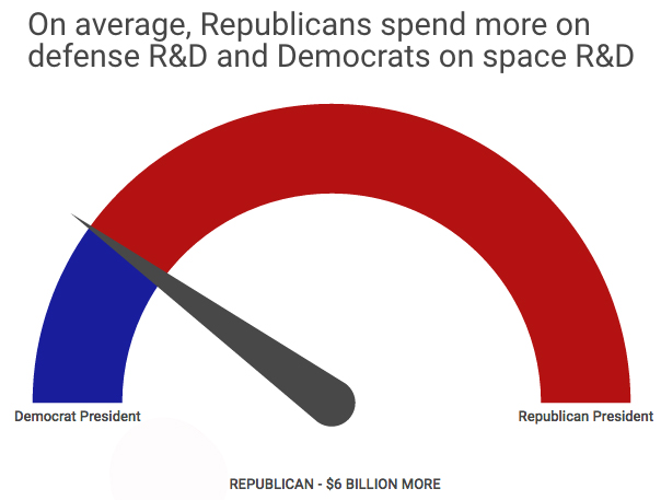
The good news is that historically new Republicans presidents generally spend more on research than democrats:
The bad news is that much of that increase is predominantly on the defence research side of things (Click here to read more on this – the original study).
Mr Trump has given little indication regarding his thoughts on research funding. And it is difficult to get any real sense of where things may be going based on the mass media news outlets, which seem to be more interested in scandal rather than in depth investigative journalism.
Mr Trump has been quoted as saying:
“Though there are increasing demands to curtail spending and to balance the federal budget, we must make the commitment to invest in science, engineering, healthcare, and other areas that will make the lives of Americans better, safer and more prosperous. We must have programs such as a viable space program and institutional research that serve as incubators to innovation and the advancement of science and engineering in a number of fields.”
Adding, however:
“In a time of limited resources, one must ensure that the nation is getting the greatest bang for the buck. We cannot simply throw money at these institutions and assume that the nation will be well served.”
Mr Trump appears to be intent on bringing the US federal deficit under control. But he has also indicated plans for cutting taxes (for all incomes), eliminating the estate tax, and providing a significant child care credit. He believes that the increased economic activity resulting from these cuts would counteract that drop in tax income. Such policies do not bode well for research funding (an easy section of the budget to reduce).
With regards to neurodegeneration research, during the election campaign Mr Trump told a New Hampshire audience that Alzheimer’s was a “total top priority” for him. So there may be some hope there for closely associated Parkinson’s disease (we can hope).
We will simply have to wait and see.
And what is the outlook for the UK?
The winning team in the BREXIT vote. Source: Telegraph
The UK’s public finances have worsened by approximately £25 billion since the March Budget (source: Independent), with the impact of the BREXIT vote apparently being a major contributing factor. This hole in the finances is going to require the Government to borrow more and spend less, which may well impact research funding in the up coming Autumn budget statement. And the Autumn statement is causing very real concerns for many in the research community (Click here for a recent editorial in the journal Nature).
To counter any reduction in the levels of Government research funding, incentives could be put in place for commercial/industrial resources to step in. The pharmaceuticals industry accounts for 48% of all corporate research funding in the UK, and much of this funding is at the University research institute level.
With regards to the huge pot of EU funding that could be lost, the UK could ‘buy-back’ into the EU research programmes as an ‘Associated Member’. But this approach would have several major drawbacks:
No political say into the formation and direction of future research funding programmes.
A 12% contribution of funds requirement for just a 16% gain of competitive funds.
Any changes to UK immigration policies at any stage would cause major disruption to future programmes.
Obviously clarity is required. We will wait to see what the Autumn statement brings.
EDITORIAL NOTE: I have tried to remain unbiased here, ignoring much of the negative comments in the media regarding Mr Trump’s proposed policies and the BREXIT related scaremongering in the UK. It is however difficult to sort through the mess and differentiate fact from opinion. This post was never intended to be a post, just a personal investigation of the state of play in research funding for Parkinson’s disease. But I decided to share it here for general interest (and I hope it was of interest). It is a very serious matter.
This is one of the first immuno-therapies being tested in Parkinson’s disease, and the results indicate that the treatment was active and well tolerated.
In this post we will review the press release and what it tells us regarding this clinical trial.
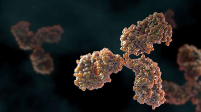
When your body is infected by a foreign agent, it begins to produce some things called antibodies. In most cases, these are Y-shaped proteins that binds to the un-wanted invader and act as a beacon for the immune system. It is a very effective system, allowing us to go about our daily business without getting sick on a regular basis. Antibodies allow us to build up immunity, or resistance of an organism to infection or disease.
Scientist have harnessed the power of this natural process, and they have use it to develop methods of helping our bodies fight off disease.
The first approach is called Acquired Immunity (or adaptive immunity), and it is based on the idea that exposure of the immune system to a pathogen (disease/damage causing agent) creates an ‘immunological memory’ within our immune system, and this leads to an enhanced response to subsequent future encounters with that same pathogen.
Scientists have used the idea of acquired immunity to develop what we call vaccines – which are simply small, neutral fragments of specific pathogen that help the immune system to build up immunity (or resistance) before the body is attacked by the disease-causing pathogen itself.
Passive immunisation is simply the sharing of antibodies. And that might sound a bit disturbing, but it is actually a naturally occurring process. For example, a mother’s antibodies are transferred to her baby in the womb via the placenta.
And again, scientists have devised ways of producing passive immunisation artificially. And recently researchers have been using this approach to attack many medical conditions (particularly cancer), in an area of medicine called immunotherapy.
Think of it as simply boosting the immune system by supplementing the supply of antibodies. Scientists can produce high levels of antibodies that specifically target a particular pathogen and then transfer those antibodies to affected people via an intravenous injection.
How is this being used for Parkinson’s disease?
Well, we have previously discussed the idea of a vaccine for Parkinson’s disease (click here to read that post), and we have been closely following the progress of an Austrian company, AffiRis, who are leading the vaccination approach (Click here for that post).
The vaccine approach is targeting the Parkinson’s disease associated protein, Alpha synuclein. It is believed that a bad kind of alpha synuclein is causing the spread of the condition, by being passed from cell to cell. The goal of the vaccine is to capture and remove all of the alpha synuclein being passed between cells and thus (hopefully) halt the progress of – or at least slow down – the disease.
And this week, another company – Prothena – has reported the results of their phase 1 trial for a passive immunity approach to Parkinson’s disease. They have been injecting subjects in the trial with a treatment called PRX002.
(Remember that a phase 1 trial simply tests the safety of a treatment in humans, it is not required to test efficacy of the treatment. Efficacy comes with phases 2 & 3 trials)
What is PRX002?
PRX002 is a monoclonal antibody. The scientists at the biotech company Prothena have artificially produced large amounts of antibodies to alpha synuclein and these have been injected into people with Parkinson’s disease.
Prothena provide a short video explaining this concept (click here to view the video).
So what were the results of the Prothena study?
The study was conducted in collaboration the pharmaceutical company Roche. It was a double-blind (so both the researchers and subjects did not know what they were receiving until the conclusion of the study), placebo-controlled study involving 80 people with Parkinson’s disease. The subjects were randomly assigned to one of six groups, which received either PRX002 or a placebo. There were six doses of PRX002 tested in the study (0.3, 1, 3, 10, 30 or 60 mg/kg).
The study was conducted over six-month, during which patients received three once-a-month injections of either PRX002 or placebo. The subjects were then followed for an observational period of three months.
According to the press release, no serious treatment-related adverse events were reported in PRX002 treated patients. Mild treatment-related adverse events (greater than anything experienced within the placebo group) were noted in 4 of the 12 subjects in the highest dosage group of PRX002, including constipation and diarrhoea.
Importantly, the investigators reported that thePRX002 antibodies were crossing the blood brain barrier and entering the brain. This resulted in a rapid reduction of alpha-synuclein levels (in some cases by up to 97 percent after a single dose!).
The follow-on Phase 2 clinical study is expected to begin in 2017.
What is the difference between the vaccine and the passive immunity approaches?
Basically, it comes down to levels of control. With a vaccination, once you have injected the vaccine and the immune system is activated, there isn’t much you can do to control the response of the body. And that immune memory is going to last a long time. The passive immunity response, on the other hand, requires regular injections of antibodies which can be stopped if adverse effects are noted.
Plus – and forgive me if I sound a little bit cynical here – drug companies prefer a regular treatment approach (which they can charge for each visit) compared to a one-shot cure. It’s simply a better business model.
What happens next?
In both cases – the vaccine and the passive immunity approaches – phase 2 trials are being set up by the respective companies and we will wait to see have affective these treatments are at slowing down Parkinson’s disease.
If they are affective, expect big headlines in the media and plans for adults everywhere to start being vaccinated. If they fail,…. well, we will have to re-address our understanding of the role of alpha synuclein in Parkinson’s disease.
Interesting times lie ahead.
The banner for todays post was sourced from Prothena
It seems everyday we read stories in the media about the benefits of these things called antioxidants. We are repeatedly told that we ‘need more antioxidants in our diet’, because they will help to stave off debilitating conditions like Parkinson’s disease.
Last week, however, a study was published which indicates that this may not be the case.
In todays post we look at antioxidants and their impact on Parkinson’s disease.
Berries are a wonderful source of antioxidants. Source: Steroidal
Antioxidants are one of those subjects that is often discussed, but not well understood. So before we review the study that was published last week, let’s first have a look at what we mean when we talk about antioxidants.
What is an antioxidant?
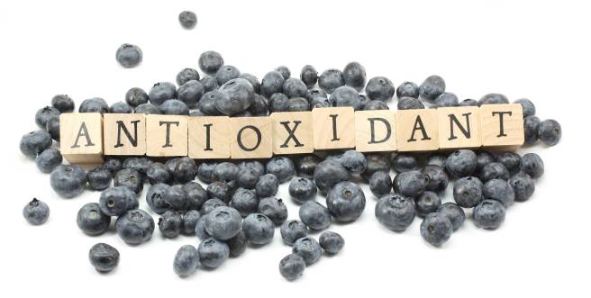
An antioxidant is simply a molecule that prevents the oxidation of other molecules.
OK, but what does that mean?
Well, the cells in your body are made of molecules. Molecules are combinations atoms of one or more elements joined by chemical bonds. Atoms consist of a nucleus, neutrons, protons and electrons.
Oxidation is the loss of electrons from a molecule, which in turn destabilises the molecule. Think of iron rusting. Rust is the oxidation of iron – in the presence of oxygen and water, iron molecules will lose electrons over time. Given enough time, this results in the complete break down of objects made of iron.
The exact same thing happens in biology. Molecules in your body go through a similar process of oxidation – losing electrons and becoming unstable. This chemical reaction leads to the production of what we call free radicals, which can then go on to damage cells.
What is a free radical?
A free radical is an unstable molecule – unstable because they are missing electrons. They react quickly with other molecules, trying to capture the needed electron to re-gain stability. Free radicals will literally attack the nearest stable molecule, stealing an electron. This leads to the “attacked” molecule becoming a free radical itself, and thus a chain reaction is started. Inside a living cell this can cause terrible damage, ultimately killing the cell.
Antioxidants are thus the good guys in this situation. They are molecules that neutralize free radicals by donating one of their own electrons. The antioxidant don’t become free radicals by donating an electron because by their very nature they are stable with or without that extra electron.
How free radicals and antioxidants work. Source: h2miraclewater
What are good sources of antioxidants?
While human being are pretty poor at producing antioxidants, plants produce LOTS! Thus vegetables and fruits are a fantastic source of antioxidants.
Sources of antioxidants (no. 3 is our favourite). Source: DrAxe
The Oxygen radical absorbance capacity (ORAC) score mentioned in the figure above is a method of measuring the antioxidant capacity of various substances. For comparative sake, a piece of tofu has an ORAC score of approximately 90, a beef steak has an ORAC score of approximately 10, and a ‘Redbull’ energy drink has an ORAC score of 0 (as they all have very few antioxidants – Source:Superfoodly).
A source of major antioxidants are vitamins (such as beta-carotene, vitamin C, and vitamin E). Vitamins are essential nutrients that our bodies needs (in small amounts) to function properly. Many of them are also potent antioxidants.
Vitamin C (or ascorbic acid), in particular, is a powerful antioxidant and it is found in both animals and plants. Unfortunately for humans, however, one of the enzymes needed to make ascorbic acid was lost by a genetic mutation during primate evolution, and so we must obtain it from our diet (eat lots of oranges folks).
How could antioxidants work for Parkinson’s disease?
Postmortem analysis of the brains of people who had Parkinson’s disease has revealed numerous signs of oxidative damage, and this has lead to many researchers hypothesising that oxidation is a key component of the disease.
So what research was published last week?
The results of this study:
Title: Intake of antioxidant vitamins and risk of parkinson’s disease. Authors: Hughes KC, Gao X, Kim IY, Rimm EB, Wang M, Weisskopf MG, Schwarzschild MA, Ascherio A. Journal: Movement Disorders. 2016 Oct 27. doi: 10.1002/mds.26819. PMID:27787934
In this study, the investigators wanted to look at the consumption of antioxidant vitamins and the risk of developing Parkinson’s disease. In order to do this, they needed large pools of medical data that they could analyse. They used the databases from the Nurses’ Health Study (NHS) and the Health Professionals Follow-Up Study (HPFS) in the USA.
NHS study was started in 1976 when 121,700 female registered nurses (aged 30 to 55 years) completed a mailed questionnaire. They provided an overview of their medical histories and health-related behaviours. The HPFS study was established in 1986, when 51,529 male health professionals (40 to 75 years) responded to a similar questionnaire. Both the NHS and the HPFS send out follow-up questionnaires every 2 years.
The investigators in the current study, removed the data from people who reported ‘implausible total energy intake at baseline (<660 or >3,500 kcal/day for women and <800 or >4,200 kcal/day for men)’, missed reporting for any survey, or had a diagnosis of Parkinson’s disease at the start of the study. This left them with the survey results of 80,750 women and 48,672 men to analyse.
From these pools of subjects, they found a total of 1036 people with Parkinson’s disease (554 in HPFS and 482 in NHS). When the investigators looked at antioxidant vitamin consumption, they found that vitamin E was not associated with an increased or decreased risk of Parkinson’s disease. Vitamin C intake showed indications of reducing the risk of developing Parkinson’s, but this not significant.
The investigators concluded that their results do not support the hypothesis that consumption of antioxidant vitamins reduces the risk of Parkinson’s disease.
What about other Parkinson’s disease research on antioxidants?
There have been several clinical trials for antioxidants and Parkinson’s disease. Of particular interest has been the research surrounding Coenzyme Q10 (also known as ubiquinone and ubidecarenone).Coenzyme Q10 is an antioxidant that exhibited positive preclinical results for Parkinson’s disease, and this led to several large clinical trials:
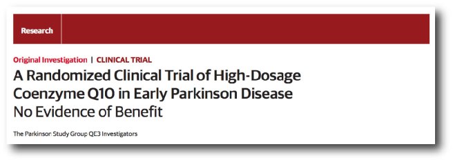
Title: A randomized clinical trial of high-dosage coenzyme Q10 in early Parkinson disease: no evidence of benefit. Authors: Parkinson Study Group QE3 Investigators., Beal MF, et al. Journal: JAMA Neurol. 2014 May;71(5):543-52. PMID:24664227
This article reported the results of a phase III randomized, placebo-controlled, double-blind clinical trial at 67 North American sites, consisting of 600 participants. While Coenzyme Q10 was safe and well tolerated by the subjects in the study, it demonstrated no evidence of clinical benefit.
One justified critique of this study, however, was the variety of subjects with Parkinson’s disease involved in the study. It has been suggested that a clinical trial should be performed with coenzyme Q10 in people with Parkinson’s disease who have a proven mutation in the PINK1 gene as these are the subjects who are most likely to benefit from this approach. That would be an interesting trial.
So what does it all mean?
Well, the study published last week needs to be replicated with another large database before any serious conclusions can be made. For all the hype around antioxidants, however, there is a worrying lack of supporting evidence that they actually have any effect (in the case of lung cancer there are even suggestions that some vitamin antioxidants could exacerbate the situation – click here for more on this).
The results of the study reviewed above do suggest that our view of oxidation in Parkinson’s disease needs to be re-addressed. It may be that oxidation may simply be an end step in the condition, and trying to block it with antioxidants is fruitless.
It should be noted that we are not suggesting here that people should stop taking antioxidants – they are an important part of any balanced diet, necessary for normal biological functioning. We are simply presenting the evidence that some of the hype surrounding their potential is unfounded.
As usual, as more information comes to hand, we shall present it here. Watch this space.
The banner for todays post was sourced from Pinkhope
There is a very interesting article in this week’s issue of Nature – one of the most eminent scientific journals.
With the 200 year anniversary of Parkinson’s disease coming up next year, the editorial team at Nature are keen to explore what is happening in the field.
There are numerous interesting articles about Parkinson’s disease available on their outlook site, but we thought this one is particularly interesting as it deals with the most controversial topic in Parkinson’s disease research.
Enjoy.
The banner for this brief post was sourced from the HuffingtonPost
For more than 50 years, L-dopa (a critical ingredient used by the brain to produce the chemical dopamine) has been one of the primary therapies used in the treatment of Parkinson’s disease. Over those years, there have been several different versions of L-dopa, providing advantages over previous forms. Last week, the results of clinical trials involving a new inhalable version of L-dopa were published.
In this post we will review the results of those studies.
The motor features (a resting tremor in one of the limbs, slowness of movement, and rigidity in the limbs) of Parkinson’s disease begin to appear when most of the dopamine producing neurons in the brain have been lost (specifically, >60% of the midbrain dopamine neurons). Thus for the last 50 years the primary means of treating Parkinson’s disease has been via dopamine replacement therapies.
Why don’t we just inject people with dopamine?
The chemical dopamine has a very difficult time crossing the blood-brain barrier, which is a thick membrane surrounding the brain. This barrier protects the brain from unwanted undesirables (think toxic chemicals), but it also blocks the transfer of some chemicals that exert a positive impact (such as dopamine).
When dopamine is blocked from entering the brain, other enzymes can convert it into another chemical called ‘norepinephrine’ (or epinephrine) and this conversion can cause serious side effects in blood pressure and glucose metabolism.
In addition, any dopamine that does find its way into the brain is very quickly broken down by enzymes. Thus, the amount of time that dopamine has to act is reduced, resulting in a very limited outcome. And these reasons are why doctors turned to L-dopa instead of dopamine in the treatment of Parkinson’s disease.
What is L-dopa?
Basically, Levodopa (L-dopa) is a chemical intermediary in the production of dopamine. That is to say, you need L-dopa to make dopamine. L-dopa is very stable inside the body and crosses the blood-brain-barrier very easily.
In the UK, a commonly used version is known as ‘Sinemet®‘(produced by Merck).
The chemical structure of L-dopa. Source: Wikipedia
The best way to understand what L-dopa is probably be to explain the history of this remarkable chemical.
The history of L-dopa
Until the 1950s there were few treatment options for Parkinson’s disease, but a young scientist in Sweden was about to change that.
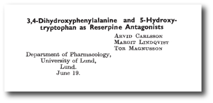
In 1957, he discovered that when he injected the brains of rabbits with a neurotoxin (reserpine) it killed the dopamine neurons (and the animals exhibited reduced movement). He also discovered that by injecting the dopamine precursor –L-dopa – into those same animals, he was able to rescue their motor ability. Importantly, he found that the serotonin precursor (called 5-hydroxytryptophan) was not capable of reversing the reduction in motor ability, indicating that the effect was specific to L-dopa.
Here is the 1957 report:
Title: 3,4-Dihydroxyphenylalanine and 5-hydroxytryptophan as reserpine antagonists.
Authors: Carlsson A, Lindqvist M, Magnusson T.
Journal: Nature. 1957 Nov 30;180(4596):1200. No abstract available.
PMID: 13483658 (the article on the Nature website – access required)
At the time, we did not know that dopamine was depleted in Parkinson’s disease. And people with Parkinson’s continued to suffer.
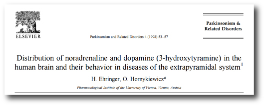
It was not until 1960 that the critical discovery of Parkinson’s disease was made by another young European scientist. Carlsson’s research (and that of others) inspired the Austrian researcher, Oleh Hornykiewicz to look at dopamine levels in people with Parkinson’s disease.
In his study, Hornykiewicz found very high levels of dopamine in the basal ganglia of normal postmortem adult brains, but a marked and consistent reduction (approx. 10-fold) in six postmortem cases of Parkinsonisms. The basal ganglia is one of the main regions of the brain that dopamine neurons communicate with (releasing dopamine there).
Title: Distribution of noradrenaline and dopamine (3-hydroxytyramine) in the human brain and their behavior in diseases of the extrapyramidal system Authors: Ehringer H, Hornykiewicz O. Journal: Parkinsonism Relat Disord. 1998 Aug;4(2):53-7. No abstract available. PMID:18591088
Importantly, Hornykiewicz did not stop there.
In November 1960, Hornykiewicz approached Walther Birkmayer, a doctor at a home for the aged in Vienna, and together they began some clinical trials of L-dopa in July 1961. Birkmayer injected 50 to 150 mg intravenously in saline into 20 volunteers with Parkinsonism. In their report, Birkmayer and Hornykiewicz wrote this regarding the results:
“The effect of a single intravenous injection of l-dopa was, in short, a complete abolition or substantial relief of akinesia. Bedridden patients who were unable to sit up, patients who could not stand up when seated, and patients who when standing could not start walking performed after l-dopa all of these activities with ease. They walked around with normal associated movements, and they could even run and jump. The voiceless, aphonic speech, blurred by palilalia and unclear articulation, became forceful and clear as in a normal person. For short periods of time the people were able to perform motor activities, which could not be prompted to any comparable degree by any other known drug”
Despite their initial excitement, Birkmayer and Hornykiewicz found that the response to L-dopa was very limited in its duration. In addition, subsequent trials by others were not able to achieve similar results, with many failing to see any benefit at all.
And that was when George stepped into the picture.
Dr George Cotzias…and yes, he is holding a brain. Source: New Scientist
Dr George Cotzias was a physician working in New York who became very interested in the use of L-dopa for Parkinson’s disease. And he discovered that by starting with very small doses of L-dopa, given orally every two hours and gradually increasing the dose gradually he was able to stabilize patients on large enough doses to cause a dramatic changes in their symptoms. His studies led ultimately to the Food and Drug Administration (FDA) approving the use of L-dopa for use in PD in 1970. Cotzias and his colleagues were also the first to describe L-dopa–induced dyskinesias.
How does L-dopa work?
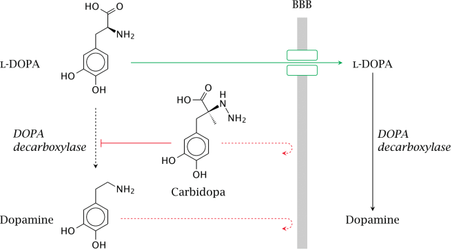
When you take an L-dopa tablet, the chemical will enter your blood. Via your bloodstream, it arrives in the brain where it will be absorbed by cells. Inside the cells, another chemical (called DOPA decarboxylase) then changes it into dopamine. And that dopamine is released, and that helps to alleviate the motor features of Parkinson’s disease.
The production of dopamine, using L-dopa. Source: Watcut
Outside the brain, there is a lot of DOPA decarboxylase in other organs of the body, and if this is not blocked then the effect of L-dopa is reduced in the brain, as less L-dopa reaches the brain. To this end, people with Parkinson’s disease are also given Carbidopa (Lodosyn) which inhibits DOPA decarboxylase outside of the brain (Carbidopa does not cross the blood-brain-barrier).
How does the L-dopa inhaler work?
The company behind this new product, Acorda Therapeutics, spent many years developing a powdered version of levodopa that could be delivered to the lungs. Early on in this developmental process the scientists realised a problem: while normal asthma inhalers only need to release micrograms of their medicine to the lungs, a L-dopa inhaler would need to deliver 1,000 times more than that to have any effect. The huge amounts were needed to ensure that enough L-dopa would get from the lungs into the brain to be effective. Thus, the ARCUS inhaler delivers 25 to 50 milligrams in two breaths.
The inhaler contains capsules of L-dopa, which are designed to break open so that the powder can escape. By sucking on the inhaler (see image below), the open capsule starts spinning, releasing the levodopa into the air and subsequently into the lungs.
Pretty straightforward, right? Nice idea, cool design, easy to use.
But does it work?
What were the results of the clinical trials?
Title: Preclinical and clinical assessment of inhaled levodopa for OFF episodes in Parkinson’s disease. Authors: Lipp MM, Batycky R, Moore J, Leinonen M, Freed MI. Journal: Sci Transl Med. 2016 Oct 12;8(360):360ra136. PMID:27733560 (This article is OPEN ACCESS if you would like to read it)
In their research report, the scientists provided data from three studies: preclinical, phase one clinical, and phase two clinical. In the preclinical work, they measured the levels of L-dopa in dogs who had inhaled levodopa powder. When they looked at blood samples, they found that levodopa levels peaked in all of the animals 2.5 min after administration. This represented a very quick route to the blood system, as dogs that were given levodopa plus carbidopa orally did not exhibit peak blood levodopa levels until 30 min after administration.
In the phase one (safety) clinical trial, 18 healthy persons were enrolled, and again comparisons were made between inhaled CVT-301 and orally administered carbidopa/levodopa. This study demonstrated that CVT-301 was safe and had a similar rapidity of action as in the preclinical dog study.
Next, the researchers conducted a phase two (efficacy) clinical study. This involve 24 people with Parkinson’s disease inhaling CVT-301 as a single 50mg dose during an OFF episode (periods of no prescribed medication). 77% of the CVT-301 treated subjects showed an increase in plasma levodopa within 10 min. By comparison, only 27% of a group of subjects taking oral doses of carbidopa/levodopa at a 25-mg/100-mg dose achieved the same levels within that time. Improvements in timed finger tapping and overall motor function (as measured by the Unified Parkinson’s Disease Rating Scale) were observed between 5 and 15 minutes after administration.
The most common adverse event was cough, but all of the coughing events were considered mild to moderate, generally occurring at the time of inhalation. In most cases, they were resolved rapidly and became less frequent after initial dosing.
So what does it all mean?
Inhalation of L-dopa may represent a novel means of treating people with Parkinson’s disease, especially those who struggle with swallowing pills. The most obvious benefit is the speed with which the subjects see results.
The amount of L-dopa being used is very high, however, and we will be interested to see the results of more long term studies before passing judgement on the inhaler approach. We’ll keep you informed as more information comes to hand.
The banner for today’s post is sourced from the BBC
In today’s post we are going to review the results of a phase 1 trial for new kind of drug being oriented at Parkinson’s disease. The results were announced in late September, and they indicate that the drug was well tolerated by subjects taking part in the study.
Here at the Science of Parkinson’s disease we are always on the look out for novel drug therapies. Many of the treatments currently being tested in the clinic are simply different versions of L-dopa or a dopamine agonist.
So when Prexton Therapeutics recently announced the results of their phase 1 clinical trial for their lead drug, PXT002331, we sat up and took notes. PXT002331 (formerly called DT1687) is the first drug of its kind to be tested in Parkinson’s disease.
It is a mGluR4 positive allosteric modulator.
What on earth is mGluR4 positive allosteric modulator?
The metabotropic glutamate receptors (mGluR) are an abundant family of receptors in the brain. Proteins bind to these receptors and activate (or block) an associated function. There are many different types of these receptors and mGluR4 is simply a small subset. The mGluR4s, however, are present in the areas affected by Parkinson’s disease, and this is why this particular family of receptors has been the focus of much research attention.
But what about the positive allosteric modulator part of ‘mGluR4 positive allosteric modulator’
Yes, good question.
This is the key part of this new approach. Allosteric modulators are a new class of orally available small molecule therapeutic agents. Traditionally, most marketed drugs bind directly to the same part of receptors that the body’s own natural occurring proteins attach to. This means that those drugs are competing with those endogenous proteins, thus limiting the potential effect of the drug.
Allosteric modulators get around this problem by binding different parts of the receptor. And instead of simply turning on or off the receptor, allosteric modulators can either turn up the volume of the signal being sent by the receptor or decrease the signals. This means that when the body’s naturally occurring protein binds in the receptor, allosteric modulators can either amplify the effect or reduce it depending on which type of allosteric modulators is being administered.
There are two different types of allosteric modulators: positive and negative. And as the label suggests, positive allosteric modulators (or PAMs) increase the signal from the receptor while negative allosteric modulators (or NAMs) reduce the signal. Thus, mGluR4 PAMS are amplifying the signal of the mGluR4 receptors.
Why do we want an amplification of a particular signal?
That is a hard question to answer.
Here’s the short explanation:
When you are planning to make a movement with your body, the process of actually initiating that movement begins in the cortex, specifically the primary motor cortex:
A cross section of the human brain illustrating the primary motor cortex. Source: Droso4schools
The primary motor cortex receives information from other regions of the brain (such as the prefrontal cortex where you make a lot of your decisions), and it will then send a signal down into the brain and down the spinal cord telling the limbs to move. On the way down through the brain, the signal will pass through a series of check points that will filter the signal and determine the final strength of it.
A schematic of the feedback loop of check points. Source: Parkinson’s Biology
EDITOR’S NOTE: We have borrowed this image from the Parkinson’s biology blog, which we are huge fans of. We highly recommend people visit that site as well as our lovely site. They also provide easy to understand explanations of the biology of Parkinson’s disease.
These checkpoints represent a large feedback loop. The critical step in this process is the processing being conducted in the basal ganglia, which can be broken down into different subregions:
A schematic of the components of the basal ganglia. Source: Parkinson’s Biology
The globus pallidus (GPi) is the last area of the basal ganglia that the signal will pass through on it’s way to the thalamus (the ultimate decider of whether you will move or not), so if there is anything going wrong between these two structures the initiation of movement will be disrupted.
In a normal brain, the chemical dopamine is being produced in an area called the substantia nigra pars compacta (say that three times really fast). That dopamine is released in the striatum and other areas of the basal ganglia, and it has a mediating effect on the signal passing through these structures.
In Parkinson’s disease, however, the dopamine producing cells of the pars compacta are loss – 60% by the time a person starts to have the clinical motor features appearing. The loss of this dopamine leaves the whole system ‘unmediated’. The feedback loop becomes extremely inhibited, resulting in problems initiating movement.
Deep brain stimulation can un-inhibit the globus pallidus, by mediating the signal passing through that structure. But this requires surgery and the implanting of probes deep inside the brain.
A schematic of deep brain stimulation of the globus pallidus. Source: Parkinson’s Biology (great website!)
A better way of reducing the inhibition in this feedback loop is the replacement of dopamine (which we do via the taking of treatments like L-dopa). This has been the standard approach for more than 50 years.
A new method of reducing the inhibition in the feedback loop would be to chemically targeting the globus pallidus, and this is what scientists are trying to do with the mGluR4 PAMS. By amplifying the signal of mGluR4s in the globus pallidus, the scientists believe that they can reduce the level of inhibition in the feedback loop.
The hope is that this approach is a less Parkinson’s disease-affected treatment. That is to say, the globus pallidus is structurally less affected by Parkinson’s disease than the substantia nigra pars compacta, and thus any treatment of the globus pallidus should be more stable over time (as the disease progresses).
That said, it is acknowledged that mGluR4 PAMS are NOT a potential cure for Parkinson’s disease, but rather a better way of treating the condition.
What research has been done on mGluR4 PAMS and Parkinson’s disease?
In August of 2003, some researchers at the pharmaceutical company Merck published a study which indicated that activation of mGluR4 could decrease the excessive levels of inhibition in the globus pallidus.
Title: Group III metabotropic glutamate receptor-mediated modulation of the striatopallidal synapse. Authors: Valenti O, Marino MJ, Wittmann M, Lis E, DiLella AG, Kinney GG, Conn PJ. Journal: Journal of Neuroscience. 2003 Aug 6;23(18):7218-26. PMID:12904482 (This article is OPEN ACCESS if you would like to read it)
The researchers found that an mGluR4 agonist (a protein that binds to the receptor directly, encouraging the associated action) reduced inhibitory signal being produced in the globus pallidus (through a presynaptic mechanism of action). They next demonstrated that the effect did not happen in mice which do not have mGluR4s, illustrating the specificity of the effect. They finished the study by injecting the mGluR4 agonist into a rodent model of Parkinson’s disease and found beneficial effects – that were equivalent to L-dopa.
Based on this research, the scientists at Merck next turned their attention to modulating the mGluR4s in the globus pallidus using allosteric modulators:
Title: Allosteric modulation of group III metabotropic glutamate receptor 4: a potential approach to Parkinson’s disease treatment. Authors: Marino MJ, Williams DL Jr, O’Brien JA, Valenti O, McDonald TP, Clements MK, Wang R, DiLella AG, Hess JF, Kinney GG, Conn PJ. Journal: Proc Natl Acad Sci U S A. 2003 Nov 11;100(23):13668-73. PMID:14593202 (This article is OPEN ACCESS if you would like to read it)
In this article, the same researchers introduce a positive allosteric modulator called ‘PHCCC’ which has a preference for binding to mGluR4. They found that when they put PHCCC – in combination with the mGluR4 agonist used in the previous study – onto cells in petri dishes, they got an amplification of the reduction in inhibition in the cells. Administered alone, PHCCC also produced a marked reversal of the motor deficit observed in a rodent model of Parkinson’s disease.
With these results, the scientists could begin building the justification for taking mGluR4 PAMs to the clinic. They were interested, however, in what impact mGluR4 PAMs could have on the involuntary motor problems associated with long-term L-dopa use, called dyskinesias (we have previously written about these – click here to read that post). So they decided to investigate whether mGluR4 PAMs may have an impact on dyskinesias:
Title: Pharmacological stimulation of metabotropic glutamate receptor type 4 in a rat model of Parkinson’s disease and L-DOPA-induced dyskinesia: Comparison between a positive allosteric modulator and an orthosteric agonist. Authors: Iderberg H, Maslava N, Thompson AD, Bubser M, Niswender CM, Hopkins CR, Lindsley CW, Conn PJ, Jones CK, Cenci MA. Journal: Neuropharmacology. 2015 Aug;95:121-9. PMID:25749357 (This article is OPEN ACCESS if you would like to read it)
In this study, the investigators compared a mGluR4 PAM with a mGluR4 agonist (similar to that used in the previous studies) in rodent models of L-dopa induced dyskinesias. They found that the neither of the two drugs modified the development of dyskinetic behaviours, nor could they modify the behaviours when given together with L-dopa. In fact, when a low dose of L-dopa was given to the animals (resulting in only mild dyskinesias), the researchers found that by adding mGluR4 PAM the dyskinetic behaviours became more exaggerated. The investigators concluded that stimulation of mGluR4 does not have anti-dyskinetic activity. This is an important characteristic to determine before taking a drug to the clinic for Parkinson’s disease.
So what were the results of the phase 1 clinical trial?
In July of 2012, Merck spun off the research into a new company called Prexton Therapeutics. The company almost immediately started setting up a phase 1 safety clinical trial for its lead compound, the mGluR4 PAM: PXT002331. A total of 64 healthy volunteers were enrolled to evaluate the safety and tolerability of several different doses of orally taken PXT002331. The study was completed on time and demonstrated that PXT002331 is safe and well tolerated (at doses well above those that produce robust effects in Parkinson’s disease animal models).
Very positive news.
The planning of a phase 2 clinical trial in people with Parkinson’s disease is now underway. It will take place in the first half of 2017, and this study will provide the first indications as to whether this new treatment approach will be effective in human at treating the features of Parkinson’s disease. We will keep you posted on the success of that study when the results become available.
Are other biotech companies using this approach?
Yes, PAM-based therapies for Parkinson’s disease are very much in vogue at the moment.
Just this month, the biotech company Asceneuron received a grant from The Michael J. Fox Foundation for Parkinson’s Research for the development of positive allosteric modulators of the M1 muscarinic acetylcholine receptor (M1 PAMs). So we can hopefully expect more from this approach to therapies.
Interesting times. And hopefully positive results to come.
EDITOR’S NOTE: It is important to remember that any clinical trial research discussed on this blog is of an educational nature. Nothing written here can or should be mistaken as medical advice. All of these drugs are still experimental and require extensive testing before being offered to the general population. Please speak with a certified clinician before attempting any change to your current medical treatment regime.
The image used in the banner of today’s post was sourced from MedTechBoston
In 2000, a research paper investigating the incidence of Parkinson’s disease in Bulgaria was published in the journal Neuroepidemiology.
The results were rather startling.
In their study, the researchers included a subpopulation of over 6,000 gypsies. In a population of that size they had expected to find 10-30 cases of Parkinson’s disease (based on the incidence in other populations of people).
What they actually found didn’t make any sense.
In this post we will look at the incidence of Parkinson’s disease around the world and why the Bulgarian gypsies are unique in the data.
Trying to determine how frequently a particular phenomenon occurs within a given population sounds like a pretty straightforward task, right?
In practise, however, it proves to be very difficult. In some cases, almost impossible. In the western/developed world – where the medical records databases exist – the task of determining certain medical characteristics within a population of interest is slightly easier, but most experts will agree that most measures of incidence still include a pinch of error and a smidgen of guesstimating.
Beyond the developed world, determining incidence in a population is a ‘door-knocking’ job. Researchers literally have to go from house to house and asking for a survey to be filled in, or conduct doorstep evaluations of the inhabitants. A much harder task and cultural characteristics begin to play a role in the outcomes (such as lower incidence of a particular disease in communities that don’t like to ‘lose face’).
Additional problems with measuring incidence
Other problems with measuring incidence within a population include:
Unimpeded access to the population (eg. some people live in isolated locations/communities)
Accurate measures/criteria of the disease (eg. remember we don’t have an accurate diagnostic test for Parkinson’s disease)
No response bias (posted surveys receive a limited response, and many affected individuals within a community will live with a condition without alerting their doctor)
The size of the effect (if only one or two people are affected by a characteristic, the task of determining incidence becomes much harder – consider the very low incidence of juvenile onset Parkinson’s disease – Click here for more on this)
With all of that said, many efforts have been made in trying to determine the incidence of Parkinson’s disease. Some consensus has become apparent, but there are some interesting differences.
The incidence of Parkinson’s disease
The incidence of Parkinson’s disease varies around the world and there are some interesting differences.
Most studies agree, however, that the incidence of Parkinson’s disease is approximately 0.3% of the general population in industrialized countries. That is, 1 person in every 2-300. As we are all aware, Parkinson’s disease is more common in the elderly, and as such the incidence rises to about 1% (or 1 in 100) in those over 60 years of age. The incidence rate continues to rise with age to 4% of the population over 80 years of age (almost 1 in every 20 people over 80 year of age).
In 2009, Parkinson’s UK published their report on the incidence of Parkinson’s disease within the UK and their numbers are very similar to those summarised above (Click here for a PDF file of that report).
Disease burden – another way of measuring a disease
Many epidemiologists (the people who measure all of this incidence stuff) now incorporate a different kind of population-disease measurement into their analysis: ‘Disease burden’.
Below is a map of ‘hotspot’ countries (in red) around the world that have the disease burden due to Parkinson’s disease according to the World Health Organisation (WHO) (click here for their raw data – Microsoft Excel file).
A world map of Parkinson’s disease burden (red = high incidence). Source: Wikipedia
The map illustrates the disability-adjusted life year (DALY) rates from Parkinson disease by country (per 100,000 inhabitants).
Yeah I know. It sound complicated, but it isn’t really.
The DALY is simply a measure of the overall disease burden that a population experiences, and it is expressed as the number of years lost due to ill-health, disability or early death. Put another way, the DALY for any given country is calculated by taking the total number of the years of life lost due to dying early and adding it to the number of years lost due to disability. So for the map above, the Maldives (dark red dot in the Indian Ocean) exhibits the highest burden with the country loses 557 years per 100,000 inhabitants.
And importantly these measures are ‘age adjusted’, so that countries with a higher proportion of elderly people (such as Japan) do not appear to have a higher burden due to Parkinson’s disease than a country with a younger population. The WHO numbers are provided by the government health services in each country.
The highest incidence of Parkinson’s disease
Ok, so if we leave the global/macro world of Parkinson’s disease incidence and focus on particular nations/communities of people, what does the research literature tell us about the incidence of Parkinson’s disease?
Well, one of the highest incidence occurs in the Amish community of the US midwest.
The Amish communities of the American midwest. Source: DartMed
The Amish community started in Switzerland in the 17th century. In the 18th and 19th centuries, many adherents
immigrated to the USA in an attempt to flee religious persecution. They now live in communities rather culturally isolated from society – maintaining a traditional way of life, ignoring the modern conveniences, and
marrying strictly within their religion (maintaining strict endogamy). They are not completely isolated, however, as they are work/conduct business with mainstream society. From a scientific standpoint, the Amish are a wonderful cases study. They have diligently kept meticulous family records dating far back in history. In addition, they forbid consumption of alcohol or use of tobacco.
Many years ago, researchers began to notice a high incidence of Parkinson’s features within the community. Several population studies have been conducted on the Amish, including this one:
Title: A population-based study of parkinsonism in an Amish community. Authors: Racette BA, Good LM, Kissel AM, Criswell SR, Perlmutter JS. Journal: Neuroepidemiology. 2009;33(3):225-30. PMID:19641327 (This article is OPEN ACCESS if you would like to read it)
The researcher in this study tried to recruit all of the individuals over the age of 60 (total 262 people) in an Old-Order Amish community of 4,369 people. Of the 213 subjects who agreed to participate, 15 had Parkinson’s disease while a further 73 individuals had a UPDRS (Unified Parkinson’s Disease Rating Scale) motor score of >9 (indicating early stages of Parkinson’s). The researchers calculated the prevalence of Parkinson’s disease in this population of people at 5,703/100,000 or 5% of the population over 60 years of age. This was far higher than the 1% of the 60+ years population in the rest of the world.
There are over 200,000 Amish in North America, and they have played a prominent historical role in Parkinson’s disease research – the first Parkinson’s-related genetic mutations were identified in genetically isolated Amish populations (Click here for more on this). The genetics of Parkinson’s disease in the Amish is not clear, however, as a recent large population analysis demonstrated:
Title: Parkinson disease loci in the mid-western Amish. Authors: Davis MF, Cummings AC, D’Aoust LN, Jiang L, Velez Edwards DR, Laux R, Reinhart-Mercer L, Fuzzell D, Scott WK, Pericak-Vance MA, Lee SL, Haines JL. Journal: Hum Genet. 2013 Nov;132(11):1213-21. PMID:23793441 (This article is OPEN ACCESS if you would like to read it)
The scientists behind this study collected DNA samples from 798 individuals (31 with diagnosed Parkinson’s disease) who are part of a 4,998 individuals living in the Amish communities of Indiana and Ohio. Although there were a couple of areas of DNA that may confer susceptibility towards Parkinson’s disease, the researchers did not find any major/significant regions (or loci) suggesting that even within the Amish the genetics of Parkinson’s disease may be more extensive than previously appreciated.
Is there a gender bias in the incidence of Parkinson’s disease?
Yes there is.
On average women have a later onset of Parkinson’s disease than men. In addition, around the world, men are more likely to be affected by Parkinson’s disease than women by a ratio of approximately 2:1.
Curiously, there is one country that bucks this trend: Japan
There are now several studies that find the incidence of Parkinson’s disease in Japan is higher in females than males (Click here for more on this), and we have previously looked at this curious difference in a previous post (Click here to read that post)
Is there any evidence that the incidence of Parkinson’s disease is increasing?
Interesting question, and yes there is:
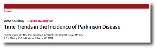
Title: Time Trends in the Incidence of Parkinson Disease Authors: Savica R, Grossardt BR, Bower JH, Ahlskog JE, Rocca WA. Journal: JAMA Neurol. 2016 Aug 1;73(8):981-9. PMID:27323276
This very recent study analysed the incidence of Parkinson’s disease by using medical records from the Rochester Epidemiology Project to identify incidence cases of Parkinson’s disease and other types of parkinsonism in Olmsted County (Minnesota) between 1976 to 2005. And the researchers made an interesting discovery: between 1976 and 2005, the incidence of Parkinson’s disease has increased, particularly in men 70 years and older. The researchers speculate as to whether this increase is associated with a dramatic decrease in the rates of smoking or other environmental/life styles changes.
We should add that there is some research that refutes this finding and we are waiting to see what follow up analysis shows us – we will report that when it is available.
So what about the Bulgarian gypsies?
Oh yeah, almost forgot.
Title: Prevalence of Parkinson’s disease in Bulgarian Gypsies. Authors: Milanov I, Kmetski TS, Lyons KE, Koller WC. Journal: Neuroepidemiology. 2000 Jul-Aug;19(4):206-9. PMID:10859500
So between January and November of 1997, the Bulgarian scientists sent out their questionnaire, and they conducted door-to-door visits, eventually collecting a pool of over 6,000 people of gypsy descent. They were trying to determine the incidence of Parkinson’s disease within this community, but what they discovered was not what they expected:
Just one case of Parkinson’s disease.
A 61 year old man.
Given the incidence in most other communities, in a population of 6,000 people one might expect to see maybe 20 cases. Not just one!
The researchers concluded that the prevalence of Parkinson’s disease in the Gypsies was found to be 16/100,000 (based on that 1 case out of 6163 people), compared to 137/100,000 for Caucasians (based on 119 cases from 87,025 people). This means that Bulgarian gypsies have the lowest incidence of Parkinson’s disease in the world.
What? How?
Our answer: ????
We really do not know. No one does.
The authors of the research paper suggest that gypsies are believed to originate from North India, and given that the inhabitants of Asia have a lower rate of Parkinson’s disease than their western counterparts, this may partly explain the low frequency in the Bulgarian gypsies. This is only applicable, however, if similar low rates of Parkinson’s disease are found in other gypsy populations. To our knowledge, these studies have not been done (please feel free to correct us on this matter).
This is the kind of post that can really get someone in quite a bit of trouble.
Both the legal kind of trouble and the social media type of trouble.
Given the online excitement surrounding a particular video that appeared on the internet last week, however, we thought that it would be useful to have a look at the research that has been done on the medicinal use of Cannabis and Parkinson’s disease.
In addition, we will assess the legal status regarding the medicinal use of Cannabis (in the UK at least).
Cannabis being grown for medicinal use. Source: BusinessWire
This week a video appeared online that caused a bit of interest (and hopefully not too many arrests) in the Parkinson’s community.
Here is the video in question:
The video was posted by Ian Frizell, a 55 year old man with early onset Parkinson’s disease. He has recently had deep brain stimulation (DBS) surgery to help control his tremors and he has also posted a video regarding that DBS surgery which people might find useful (Click here to see this).
In the video, Ian turns off his DBS stimulator and his tremors quickly become apparent. He then ‘self medicates’ with cannabis off camera and begins filming again some 20-30 minutes later to show the difference. The change with regards to his tremor are very clear and quite striking.
Here at the SoPD, we find the video very interesting, but we have two immediate questions:
How is this reduction in tremors working?
Would everyone experience the same effect?
We have previously seen many miraculous treatments online (such as coloured glasses controlling dyskinesias video from a few years ago) which have failed when tested under controlled conditions (the coloured glasses did not elicit any effect in the clinical setting – click here to read more). Some of these amazing results can simply be put down to the notorious placebo effect (we have previously discussed this in relation to Parkinson’s disease – click here to read the post), while others may vary on a person to person basis.
Thus, while we applaud Mr Frizell for sharing his finding with the Parkinson’s community, we are weary that the effect may not be applicable to everyone. For this reason, we have made a review of the scientific literature surrounding Cannabis and Parkinson’s disease.
But first:
What exactly is Cannabis?
Drawings of the Hemp plant, from Franz Eugen Köhler’s ‘Medizinal-Pflantzen’. Source: Wikipedia
Cannabis (also known as marijuana) is a family of flowering plants that can be found in three types: sativa, indica, and ruderalis. Cannabis is widely used as a recreational drug, behind only alcohol, caffeine and tobacco in its usage. It typically consumed as dried flower buds (marijuana), as a resin (hashish), or as various extracts which are collectively known as hashish oil.
While the three varieties of cannabis (sativa, indica, and ruderalis) may look very similar, pharmacologically they have very different properties. Cannabis sativa is often reported to cause a “spacey” or heady feeling, while Cannabis indica causes more of a “body high”. Cannabis ruderalis, by contrast, is less well used due to its low Tetrahydrocannabinol levels.
What is Tetrahydrocannabinol?
Tetrahydrocannabinol (or THC) is one of the principle psychoactive components in Cannabis. It a chemical that is believed to be a plant defensive mechanism against herbivores. THC is a cannabinoid, a type of chemical that attaches to the cannabinoid receptors in the body, and it is this pathway that many scientists are exploring for future neuroprotective therapies for Parkinson’s disease (For a good review on the potential cannabinoid-based therapies for Parkinson’s disease, click here).
A second type of cannabinoid is Cannabidiol (or CBD). CBD is considered to have a wider scope for potential medical applications. This is largely due to clinical reports suggesting reduced side effects compared to THC, in particular a lack of psychoactivity.
So what research has been done regarding Cannabis and Parkinson’s disease?
In 2004, a group of scientists in Prague (Czech Republic) were curious to determine cannabis use in people with Parkinson’s disease, so they conducted a study and published their results:
Title: Survey on cannabis use in Parkinson’s disease: subjective improvement of motor symptoms. Authors: Venderová K, Růzicka E, Vorísek V, Visnovský P. Journal: Mov Disord. 2004 Sep;19(9):1102-6. PMID:15372606
The researchers posted out 630 questionnaires to people with Parkinson’s disease in Prague. In total, 339 (53.8%) completed questionnaires were returned to them. Of these, 85 people reported Cannabis use (25.1% of returned questionnaires). They usually consumed it with meals (43.5%), and most of them were taking it once a day (52.9%).
After consuming cannabis, 39 responders (45.9%) described mild or substantial alleviation of their Parkinson’s symptoms in general, 26 (30.6%) improvement of rest tremor, 38 (44.7%) alleviation of rigidity (bradykinesia), 32 (37.7%) alleviation of muscle rigidity, and 12 (14.1%) improvement of L-dopa-induced dyskinesias.
Importantly, half of the people who consumed cannabis experience no effect on their Parkinson’s disease features, and four responders (4.7%) reported that cannabis actually worsened their symptoms. So while this survey suggested some positive effects of cannabis in the treatment of Parkinson’s disease, it is apparent that the effect is different between people.
Additional surveys have been conducted around the world, with similar results (Click here to read more on this).
Have there been any clinical trials?
Yes, there have.
In the 1990s, there was a very small clinical study of cannabis use as a treatment option for Parkinson’s disease, and this study failed to demonstrate any positive outcome. In the study, none of the 5 people with Parkinson’s disease experienced any effect on their Parkinson’s motor features after a week of smoking cannabis (click here for more on this).
This study was followed up by a larger study:
Title: Cannabis for dyskinesia in Parkinson disease: a randomized double-blind crossover study. Authors: Carroll CB, Bain PG, Teare L, Liu X, Joint C, Wroath C, Parkin SG, Fox P, Wright D, Hobart J, Zajicek JP. Journal: Neurology. 2004 Oct 12;63(7):1245-50. PMID:15477546
In this randomized, double-blind, placebo-controlled study, 19 people with Parkinson’s disease randomly received either oral cannabis extract or a placebo (twice daily) for 4 weeks. They then took no treatment for an intervening 2-week ‘washout’ period, before they were given the opposite treatment for 4 weeks (so if they received the cannabis extract during the first 4 weeks, they would be given the placebo during the second 4 weeks). In all cases, the participants and the researchers were ‘blind’ to (unaware of) which treatment was being given.
The results indicated that cannabis was well tolerated by all of the participants in the study, but that it had no pro- or anti-Parkinsonian actions. The researchers found no evidence for a treatment effect on levodopa-induced dyskinesia.
In addition to this study, there has been a recent double-blind clinical study of cannabidiol (CBD, mentioned above) in the treatment of Parkinson’s disease:
Title: Effects of cannabidiol in the treatment of patients with Parkinson’s disease: an exploratory double-blind trial. Authors: Chagas MH, Zuardi AW, Tumas V, Pena-Pereira MA, Sobreira ET, Bergamaschi MM, dos Santos AC, Teixeira AL, Hallak JE, Crippa JA. Journal: J Psychopharmacol. 2014 Nov;28(11):1088-98. PMID:25237116
The Brazilian researchers who conducted the study took 21 people with Parkinson’s disease and assigned them to one of three groups which were treated with placebo, small dose of CBD (75 mg/day) or high dose of CBD (300 mg/day). They found that there was no positive effects by administering CBD to people with Parkinson’s disease, except in their self-reported measures on ‘quality of life’.
So what does all of this mean?
Firstly, let us be clear that we are not trying to discredit Mr Frizell or suggest that what he is experiencing is not a real effect. The video he has uploaded suggests that he is experiencing very positive benefits by consuming cannabis to help treat his tremors.
Having said that, based on the studies we have reviewed above we (here at the SoPD) have to conclude that the clinical evidence supporting the idea of cannabis as a treatment for Parkinson’s disease is inconclusive. There does appear to be some individuals (like Mr Frizell) who may experience some positive outcomes by consuming the drug, but there are also individuals for whom cannabis has no effect.
One of the reasons that cannabis may not be having an effect on everyone with Parkinson’s disease is that many people with Parkinson’s disease actually have a reduction in the cannabis receptors in the brain (click here for more on this). This reduction is believed to be due to the course of the disease. If there are less receptors for cannabis to bind to, there will be less effect of the drug.
Ok, but how might cannabis be having a positive effect on the guy in the video?
Cannabis is known to cause the release of dopamine in the brain – the chemical classically associated with Parkinson’s disease (Click here and here for more on this). Thus the positive effects that Mr Frizell is experiencing may simply be the result of more dopamine in his brain, similar to taking an L-dopa tablet. Whether enough dopamine is being released to explain the full effect is questionable, but this is still one possible explanation.
There could be questions regarding the long term benefits of Mr Frizell’s cannabis use, as long term users of cannabis generally have reduced levels of dopamine being released in the brain (Click here for more on this). Although the drug initially causes higher levels of dopamine to be released, over time (with long term use) the levels of dopamine in the brain gradually reduce.
I live in the UK. Is it legal for me to try using Cannabis for my Parkinson’s disease?
National status on Cannabis possession for medical purposes. Source: Wikipedia
The map above is incorrect, with regards to the UK at least (and may be incorrect for other regions as well).
According to the Home Office, it is illegal for UK residents to possess cannabis in any form (including medicinal).
Cannabis is illegal to possess, grow, distribute or sell in the UK without the appropriate licences. It is a Class B drug, which carries penalties for unlicensed dealing, unlicensed production and unlicensed trafficking of up to 14 years in prison (Source: Wikipedia; and if you don’t trust Wikipedia, here is the official UK Government website).
In 1999, a major House of Lords inquiry made the recommendation that cannabis should be made available with a doctor’s prescription. The government of the U.K., however, has not accepted the recommendations. Cannabis is not recognised as having any therapeutic value under the law in England and Wales.
Having said all of this, there has recently been an all-party group calls for the legalisation regarding cannabis for medicinal uses to be changed (click here for more on this). Whether this will happen is yet to be seen.
So the answer is “No, you are not allowed to use cannabis to treat your Parkinson’s disease”.
Except…
(And here is where things get a really grey)
There is a cannabis-based product – Sativex – which can be legally prescribed and supplied under special circumstances. Sativex is a mouth spray developed and manufactured by GW Pharmaceuticals in the UK. It is derived from two strains of cannabis leaf and flower, cultivated for their controlled proportions of the active compounds
THC and CBD.
In 2006, the Home Office licensed Sativex so that:
Doctors could privately prescribe it (at their own risk)
Pharmacists could possess and dispense it
Named patients with a prescription could possess
In June 2010 the Medicines Healthcare Regulatory products Agency (MHRA) authorised Sativex as an extra treatment for patients with spasticity due to Multiple Sclerosis (MS). Importantly, doctors can also prescribe it for other things outside of the authorisation, but (again) this is at their own risk.
EDITORIAL NOTE: Given that possessing cannabis is illegal and that more research into the medicinal benefits of cannabis for Parkinson’s disease is required, we here are the SoPD can not endorse the use of cannabis for treating Parkinson’s disease.
While we are deeply sympathetic to the needs of many individuals within the Parkinson’s community and agree with a reconsideration of the laws surrounding the medicinal use of cannabis, we are also aware of the negative consequences of cannabis use (which can differ from person to person).
If a person with Parkinson’s disease is considering a change in their treatment regime for any reason, we must insist that they first discuss the matter with their trained medical physician before undertaking any changes.
The information provided here is strictly for educational purposes only.
The banner for today’s post was sourced from the IBTimes.