Inhaling L-dopa

inhaled1

For more than 50 years, L-dopa (a critical ingredient used by the brain to produce the chemical dopamine) has been one of the primary therapies used in the treatment of Parkinson’s disease. Over those years, there have been several different versions of L-dopa, providing advantages over previous forms. Last week, the results of clinical trials involving a new inhalable version of L-dopa were published.

In this post we will review the results of those studies.


inhalers-56a1c9d85f9b58b7d0c2a735

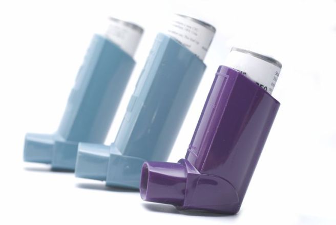
Inhalers. Source: Verywell

The motor features (a resting tremor in one of the limbs, slowness of movement, and rigidity in the limbs) of Parkinson’s disease begin to appear when most of the dopamine producing neurons in the brain have been lost (specifically, >60% of the midbrain dopamine neurons). Thus for the last 50 years the primary means of treating Parkinson’s disease has been via dopamine replacement therapies.

Why don’t we just inject people with dopamine?

The chemical dopamine has a very difficult time crossing the blood-brain barrier, which is a thick membrane surrounding the brain. This barrier protects the brain from unwanted undesirables (think toxic chemicals), but it also blocks the transfer of some chemicals that exert a positive impact (such as dopamine).

When dopamine is blocked from entering the brain, other enzymes can convert it into another chemical called ‘norepinephrine’ (or epinephrine) and this conversion can cause serious side effects in blood pressure and glucose metabolism.

In addition, any dopamine that does find its way into the brain is very quickly broken down by enzymes. Thus, the amount of time that dopamine has to act is reduced, resulting in a very limited outcome. And these reasons are why doctors turned to L-dopa instead of dopamine in the treatment of Parkinson’s disease.

What is L-dopa?

Basically, Levodopa (L-dopa) is a chemical intermediary in the production of dopamine. That is to say, you need L-dopa to make dopamine. L-dopa is very stable inside the body and crosses the blood-brain-barrier very easily.

In the UK, a commonly used version is known as  ‘Sinemet®‘(produced by Merck).

l-dopa_vzorec-svg

The chemical structure of L-dopa. Source: Wikipedia

The best way to understand what L-dopa is probably be to explain the history of this remarkable chemical.

The history of L-dopa

Until the 1950s there were few treatment options for Parkinson’s disease, but a young scientist in Sweden was about to change that.

This is Arvid Carlsson.

Win 1113

Prof Arvid Carlsson. Source: Alchetron

He’s a dude.

In 1957, he discovered that when he injected the brains of rabbits with a neurotoxin (reserpine) it killed the dopamine neurons (and the animals exhibited reduced movement). He also discovered that by injecting the dopamine precursor –L-dopa – into those same animals, he was able to rescue their motor ability. Importantly, he found that the serotonin precursor (called 5-hydroxytryptophan) was not capable of reversing the reduction in motor ability, indicating that the effect was specific to L-dopa.

Here is the 1957 report:

avid

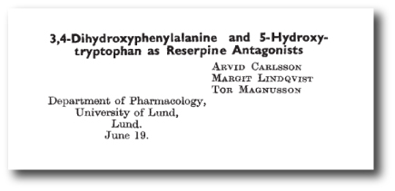
Title: 3,4-Dihydroxyphenylalanine and 5-hydroxytryptophan as reserpine antagonists.
Authors: Carlsson A, Lindqvist M, Magnusson T.
Journal: Nature. 1957 Nov 30;180(4596):1200. No abstract available.
PMID: 13483658       (the article on the Nature website – access required)

This was a fantastic discovery. A Nobel prize winning discovery in fact.

But what to do with it?

At the time, we did not know that dopamine was depleted in Parkinson’s disease. And people with Parkinson’s continued to suffer.

It was not until 1960 that the critical discovery of Parkinson’s disease was made by another young European scientist. Carlsson’s research (and that of others) inspired the Austrian researcher, Oleh Hornykiewicz to look at dopamine levels in people with Parkinson’s disease.

And what he found changed everything.

wi_17_olehhornykiewicz_01

Prof Oleh Hornykiewicz. Source: Kurienwissenschaftundkunst

In his study, Hornykiewicz found very high levels of dopamine in the basal ganglia of normal postmortem adult brains, but a marked and consistent reduction (approx. 10-fold) in six postmortem cases of Parkinsonisms. The basal ganglia is one of the main regions of the brain that dopamine neurons communicate with (releasing dopamine there).


oleh

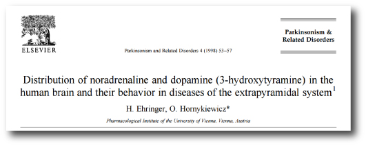
Title: Distribution of noradrenaline and dopamine (3-hydroxytyramine) in the human brain and their behavior in diseases of the extrapyramidal system
Authors: Ehringer H, Hornykiewicz O.
Journal: Parkinsonism Relat Disord. 1998 Aug;4(2):53-7. No abstract available.
PMID: 18591088

Importantly, Hornykiewicz did not stop there.

In November 1960, Hornykiewicz approached Walther Birkmayer, a doctor at a home for the aged in Vienna, and together they began some clinical trials of L-dopa in July 1961. Birkmayer injected 50 to 150 mg intravenously in saline into 20 volunteers with Parkinsonism. In their report, Birkmayer and Hornykiewicz wrote this regarding the results:

“The effect of a single intravenous injection of l-dopa was, in short, a complete abolition or substantial relief of akinesia. Bedridden patients who were unable to sit up, patients who could not stand up when seated, and patients who when standing could not start walking performed after l-dopa all of these activities with ease. They walked around with normal associated movements, and they could even run and jump. The voiceless, aphonic speech, blurred by palilalia and unclear articulation, became forceful and clear as in a normal person. For short periods of time the people were able to perform motor activities, which could not be prompted to any comparable degree by any other known drug”

Despite their initial excitement, Birkmayer and Hornykiewicz found that the response to L-dopa was very limited in its duration. In addition, subsequent trials by others were not able to achieve similar results, with many failing to see any benefit at all.

And that was when George stepped into the picture.

50396550-1200x800

Dr George Cotzias…and yes, he is holding a brain. Source: New Scientist

Dr George Cotzias was a physician working in New York who became very interested in the use of L-dopa for Parkinson’s disease. And he discovered that by starting with very small doses of L-dopa, given orally every two hours and gradually increasing the dose gradually he was able to stabilize patients on large enough doses to cause a dramatic changes in their symptoms. His studies led ultimately to the Food and Drug Administration (FDA) approving the use of L-dopa for use in PD in 1970. Cotzias and his colleagues were also the first to describe L-dopa–induced dyskinesias.

How does L-dopa work?

When you take an L-dopa tablet, the chemical will enter your blood. Via your bloodstream, it arrives in the brain where it will be absorbed by cells. Inside the cells, another chemical (called DOPA decarboxylase) then changes it into dopamine. And that dopamine is released, and that helps to alleviate the motor features of Parkinson’s disease.

dopa3pic-668d0

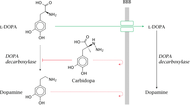
The production of dopamine, using L-dopa. Source: Watcut

Outside the brain, there is a lot of DOPA decarboxylase in other organs of the body, and if this is not blocked then the effect of L-dopa is reduced in the brain, as less L-dopa reaches the brain. To this end, people with Parkinson’s disease are also given Carbidopa (Lodosyn) which inhibits DOPA decarboxylase outside of the brain (Carbidopa does not cross the blood-brain-barrier).

How does the L-dopa inhaler work?

The company behind this new product, Acorda Therapeutics, spent many years developing a powdered version of levodopa that could be delivered to the lungs. Early on in this developmental process the scientists realised a problem: while normal asthma inhalers only need to release micrograms of their medicine to the lungs, a L-dopa inhaler would need to deliver 1,000 times more than that to have any effect. The huge amounts were needed to ensure that enough L-dopa would get from the lungs into the brain to be effective. Thus, the ARCUS inhaler delivers 25 to 50 milligrams in two breaths.

The inhaler contains capsules of L-dopa, which are designed to break open so that the powder can escape. By sucking on the inhaler (see image below), the open capsule starts spinning, releasing the levodopa into the air and subsequently into the lungs.

levodopa-inhaler-lead-658x394

The ARCUS inhalation technology. Source: ParkinsonsLife

Pretty straightforward, right? Nice idea, cool design, easy to use.

But does it work?

What were the results of the clinical trials?

inhaler

Title: Preclinical and clinical assessment of inhaled levodopa for OFF episodes in Parkinson’s disease.
Authors: Lipp MM, Batycky R, Moore J, Leinonen M, Freed MI.
Journal: Sci Transl Med. 2016 Oct 12;8(360):360ra136.
PMID: 27733560     (This article is OPEN ACCESS if you would like to read it)

In their research report, the scientists provided data from three studies: preclinical, phase one clinical, and phase two clinical. In the preclinical work, they measured the levels of L-dopa in dogs who had inhaled levodopa powder. When they looked at blood samples, they found that levodopa levels peaked in all of the animals 2.5 min after administration. This represented a very quick route to the blood system, as dogs that were given levodopa plus carbidopa orally did not exhibit peak blood levodopa levels until 30 min after administration.

In the phase one (safety) clinical trial, 18 healthy persons were enrolled, and again comparisons were made between inhaled CVT-301 and orally administered carbidopa/levodopa. This study demonstrated that CVT-301 was safe and had a similar rapidity of action as in the preclinical dog study.

Next, the researchers conducted a phase two (efficacy) clinical study. This involve 24 people with Parkinson’s disease inhaling CVT-301 as a single 50mg dose during an OFF episode (periods of no prescribed medication). 77% of the CVT-301 treated subjects showed an increase in plasma levodopa within 10 min. By comparison, only 27% of a group of subjects taking oral doses of carbidopa/levodopa at a 25-mg/100-mg dose achieved the same levels within that time. Improvements in timed finger tapping and overall motor function (as measured by the Unified Parkinson’s Disease Rating Scale) were observed between 5 and 15 minutes after administration.

The most common adverse event was cough, but all of the coughing events were considered mild to moderate, generally occurring at the time of inhalation. In most cases, they were resolved rapidly and became less frequent after initial dosing.

So what does it all mean?

Inhalation of L-dopa may represent a novel means of treating people with Parkinson’s disease, especially those who struggle with swallowing pills. The most obvious benefit is the speed with which the subjects see results.

The amount of L-dopa being used is very high, however, and we will be interested to see the results of more long term studies before passing judgement on the inhaler approach. We’ll keep you informed as more information comes to hand.


The banner for today’s post is sourced from the BBC

PAMs for Parkinson’s?

clinicaltrials

In today’s post we are going to review the results of a phase 1 trial for new kind of drug being oriented at Parkinson’s disease. The results were announced in late September, and they indicate that the drug was well tolerated by subjects taking part in the study.


4912prexton

Source: Prexton

Here at the Science of Parkinson’s disease we are always on the look out for novel drug therapies. Many of the treatments currently being tested in the clinic are simply different versions of L-dopa or a dopamine agonist.

So when Prexton Therapeutics recently  announced the results of their phase 1 clinical trial for their lead drug, PXT002331, we sat up and took notes. PXT002331 (formerly called DT1687) is the first drug of its kind to be tested in Parkinson’s disease.

It is a mGluR4 positive allosteric modulator.

What on earth is mGluR4 positive allosteric modulator?
The metabotropic glutamate receptors (mGluR) are an abundant family of receptors in the brain. Proteins bind to these receptors and activate (or block) an associated function. There are many different types of these receptors and mGluR4 is simply a small subset. The mGluR4s, however, are present in the areas affected by Parkinson’s disease, and this is why this particular family of receptors has been the focus of much research attention.

But what about the positive allosteric modulator part of ‘mGluR4 positive allosteric modulator’

Yes, good question.

This is the key part of this new approach. Allosteric modulators are a new class of orally available small molecule therapeutic agents. Traditionally, most marketed drugs bind directly to the same part of receptors that the body’s own natural occurring proteins attach to. This means that those drugs are competing with those endogenous proteins, thus limiting the potential effect of the drug.

Allosteric modulators get around this problem by binding different parts of the receptor. And instead of simply turning on or off the receptor, allosteric modulators can either turn up the volume of the signal being sent by the receptor or decrease the signals. This means that when the body’s naturally occurring protein binds in the receptor, allosteric modulators can either amplify the effect or reduce it depending on which type of allosteric modulators is being administered.

allosteric_modulation_mechanism

How Allosteric modulators work. Source: Addrex Thereapeutics

There are two different types of allosteric modulators: positive and negative. And as the label suggests, positive allosteric modulators (or PAMs) increase the signal from the receptor while negative allosteric modulators (or NAMs) reduce the signal. Thus, mGluR4 PAMS are amplifying the signal of the mGluR4 receptors.

Why do we want an amplification of a particular signal?

That is a hard question to answer.

Here’s the short explanation:

When you are planning to make a movement with your body, the process of actually initiating that movement begins in the cortex, specifically the primary motor cortex:

decisionmaking

A cross section of the human brain illustrating the primary motor cortex. Source: Droso4schools

The primary motor cortex receives information from other regions of the brain (such as the prefrontal cortex where you make a lot of your decisions), and it will then send a signal down into the brain and down the spinal cord telling the limbs to move. On the way down through the brain, the signal will pass through a series of check points that will filter the signal and determine the final strength of it.

basal-ganglia-dbs-figure-1

A schematic of the feedback loop of check points. Source: Parkinson’s Biology

EDITOR’S NOTE: We have borrowed this image from the Parkinson’s biology blog, which we are huge fans of. We highly recommend people visit that site as well as our lovely site. They also provide easy to understand explanations of the biology of Parkinson’s disease.

These checkpoints represent a large feedback loop. The critical step in this process is the processing being conducted in the basal ganglia, which can be broken down into different subregions:

basal-ganglia-dbs-figure-5

A schematic of the components of the basal ganglia. Source: Parkinson’s Biology

The globus pallidus (GPi) is the last area of the basal ganglia that the signal will pass through on it’s way to the thalamus (the ultimate decider of whether you will move or not), so if there is anything going wrong between these two structures the initiation of movement will be disrupted.

In a normal brain, the chemical dopamine is being produced in an area called the substantia nigra pars compacta (say that three times really fast). That dopamine is released in the striatum and other areas of the basal ganglia, and it has a mediating effect on the signal passing through these structures.

basal-ganglia-dbs-figure-3

A schematic of the source of dopamine. Source: Parkinson’s Biology

In Parkinson’s disease, however, the dopamine producing cells of the pars compacta are loss – 60% by the time a person starts to have the clinical motor features appearing. The loss of this dopamine leaves the whole system ‘unmediated’. The feedback loop becomes extremely inhibited, resulting in problems initiating movement.

Deep brain stimulation can un-inhibit the globus pallidus, by mediating the signal passing through that structure. But this requires surgery and the implanting of probes deep inside the brain.

basal-ganglia-dbs-figure-7

A schematic of deep brain stimulation of the globus pallidus. Source: Parkinson’s Biology (great website!)

A better way of reducing the inhibition in this feedback loop is the replacement of dopamine (which we do via the taking of treatments like L-dopa). This has been the standard approach for more than 50 years.

A new method of reducing the inhibition in the feedback loop would be to chemically targeting the globus pallidus, and this is what scientists are trying to do with the mGluR4 PAMS. By amplifying the signal of mGluR4s in the globus pallidus, the scientists believe that they can reduce the level of inhibition in the feedback loop.

The hope is that this approach is a less Parkinson’s disease-affected treatment. That is to say, the globus pallidus is structurally less affected by Parkinson’s disease than the substantia nigra pars compacta, and thus any treatment of the globus pallidus should be more stable over time (as the disease progresses).

That said, it is acknowledged that mGluR4 PAMS are NOT a potential cure for Parkinson’s disease, but rather a better way of treating the condition.

What research has been done on mGluR4 PAMS and Parkinson’s disease?

In August of 2003, some researchers at the pharmaceutical company Merck published a study which indicated that activation of mGluR4 could decrease the excessive levels of inhibition in the  globus pallidus.

jns

Title: Group III metabotropic glutamate receptor-mediated modulation of the striatopallidal synapse.
Authors: Valenti O, Marino MJ, Wittmann M, Lis E, DiLella AG, Kinney GG, Conn PJ.
Journal: Journal of Neuroscience. 2003 Aug 6;23(18):7218-26.
PMID: 12904482      (This article is OPEN ACCESS if you would like to read it)

The researchers found that an mGluR4 agonist (a protein that binds to the receptor directly, encouraging the associated action) reduced inhibitory signal being produced in the globus pallidus (through a presynaptic mechanism of action). They next demonstrated that the effect did not happen in mice which do not have mGluR4s, illustrating the specificity of the effect. They finished the study by injecting the mGluR4 agonist into a rodent model of Parkinson’s disease and found beneficial effects – that were equivalent to L-dopa.

Based on this research, the scientists at Merck next turned their attention to modulating the mGluR4s in the globus pallidus using allosteric modulators:

pnastitle

Title: Allosteric modulation of group III metabotropic glutamate receptor 4: a potential approach to Parkinson’s disease treatment.
Authors: Marino MJ, Williams DL Jr, O’Brien JA, Valenti O, McDonald TP, Clements MK, Wang R, DiLella AG, Hess JF, Kinney GG, Conn PJ.
Journal: Proc Natl Acad Sci U S A. 2003 Nov 11;100(23):13668-73.
PMID: 14593202           (This article is OPEN ACCESS if you would like to read it)

In this article, the same researchers introduce a positive allosteric modulator called ‘PHCCC’ which has a preference for binding to mGluR4. They found that when they put PHCCC – in combination with the mGluR4 agonist used in the previous study – onto cells in petri dishes, they got an amplification of the reduction in inhibition in the cells. Administered alone, PHCCC also produced a marked reversal of the motor deficit observed in a rodent model of Parkinson’s disease.

With these results, the scientists could begin building the justification for taking mGluR4 PAMs to the clinic. They were interested, however, in what impact mGluR4 PAMs could have on the involuntary motor problems associated with long-term L-dopa use, called dyskinesias (we have previously written about these – click here to read that post). So they decided to investigate whether mGluR4 PAMs may have an impact on dyskinesias:

dyskotitle

Title: Pharmacological stimulation of metabotropic glutamate receptor type 4 in a rat model of Parkinson’s disease and L-DOPA-induced dyskinesia: Comparison between a positive allosteric modulator and an orthosteric agonist.
Authors: Iderberg H, Maslava N, Thompson AD, Bubser M, Niswender CM, Hopkins CR, Lindsley CW, Conn PJ, Jones CK, Cenci MA.
Journal: Neuropharmacology. 2015 Aug;95:121-9.
PMID: 25749357          (This article is OPEN ACCESS if you would like to read it)

In this study, the investigators compared a mGluR4 PAM with a mGluR4 agonist (similar to that used in the previous studies) in rodent models of L-dopa induced dyskinesias. They found that the neither of the two drugs modified the development of dyskinetic behaviours, nor could they modify the behaviours when given together with L-dopa. In fact, when a low dose of L-dopa was given to the animals (resulting in only mild dyskinesias), the researchers found that by adding mGluR4 PAM the dyskinetic behaviours became more exaggerated. The investigators concluded that stimulation of mGluR4 does not have anti-dyskinetic activity. This is an important characteristic to determine before taking a drug to the clinic for Parkinson’s disease.

So what were the results of the phase 1 clinical trial?

In July of 2012, Merck spun off the research into a new company called Prexton Therapeutics. The company almost immediately started setting up a phase 1 safety clinical trial for its lead compound, the mGluR4 PAM: PXT002331. A total of 64 healthy volunteers were enrolled to evaluate the safety and tolerability of several different doses of orally taken PXT002331. The study was completed on time and demonstrated that PXT002331 is safe and well tolerated (at doses well above those that produce robust effects in Parkinson’s disease animal models).

Very positive news.

The planning of a phase 2 clinical trial in people with Parkinson’s disease is now underway. It will take place in the first half of 2017, and this study will provide the first indications as to whether this new treatment approach will be effective in human at treating the features of Parkinson’s disease. We will keep you posted on the success of that study when the results become available.

Are other biotech companies using this approach?

Yes, PAM-based therapies for Parkinson’s disease are very much in vogue at the moment.

Just this month, the biotech company Asceneuron received a grant from The Michael J. Fox Foundation for Parkinson’s Research for the development of positive allosteric modulators of the M1 muscarinic acetylcholine receptor (M1 PAMs). So we can hopefully expect more from this approach to therapies.

Interesting times. And hopefully positive results to come.

EDITOR’S NOTE: It is important to remember that any clinical trial research discussed on this blog is of an educational nature. Nothing written here can or should be mistaken as medical advice. All of these drugs are still experimental and require extensive testing before being offered to the general population. Please speak with a certified clinician before attempting any change to your current medical treatment regime.


The image used in the banner of today’s post was sourced from MedTechBoston

The curious case of Bulgarian Gypsies and the incidence of Parkinson’s disease

balkan-music-night-background-images-i

In 2000, a research paper investigating the incidence of Parkinson’s disease in Bulgaria was published in the journal Neuroepidemiology.

The results were rather startling.

In their study, the researchers included a subpopulation of over 6,000 gypsies. In a population of that size they had expected to find 10-30 cases of Parkinson’s disease (based on the incidence in other populations of people).

What they actually found didn’t make any sense.

In this post we will look at the incidence of Parkinson’s disease around the world and why the Bulgarian gypsies are unique in the data.


maxresdefault

Bulgarian gypsies. Source: Youtube

Trying to determine how frequently a particular phenomenon occurs within a given population sounds like a pretty straightforward task, right?

In practise, however, it proves to be very difficult. In some cases, almost impossible. In the western/developed world – where the medical records databases exist – the task of determining certain medical characteristics within a population of interest is slightly easier, but most experts will agree that most measures of incidence still include a pinch of error and a smidgen of guesstimating.

Beyond the developed world, determining incidence in a population is a ‘door-knocking’ job. Researchers literally have to go from house to house and asking for a survey to be filled in, or conduct doorstep evaluations of the inhabitants. A much harder task and cultural characteristics begin to play a role in the outcomes (such as lower incidence of a particular disease in communities that don’t  like to ‘lose face’).

Additional problems with measuring incidence 

Other problems with measuring incidence within a population include:

  • Unimpeded access to the population (eg. some people live in isolated locations/communities)
  • Accurate measures/criteria of the disease (eg. remember we don’t have an accurate diagnostic test for Parkinson’s disease)
  • No response bias (posted surveys receive a limited response, and many affected individuals within a community will live with a condition without alerting their doctor)
  • The size of the effect (if only one or two people are affected by a characteristic, the task of determining incidence becomes much harder – consider the very low incidence of juvenile onset Parkinson’s disease – Click here for more on this)

With all of that said, many efforts have been made in trying to determine the incidence of Parkinson’s disease. Some consensus has become apparent, but there are some interesting differences.

The incidence of Parkinson’s disease

The incidence of Parkinson’s disease varies around the world and there are some interesting differences.

Most studies agree, however, that the incidence of Parkinson’s disease is approximately 0.3% of the general population in industrialized countries. That is, 1 person in every 2-300. As we are all aware, Parkinson’s disease is more common in the elderly, and as such the incidence rises to about 1% (or 1 in 100) in those over 60 years of age. The incidence rate continues to rise with age to 4% of the population over 80 years of age (almost 1 in every 20 people over 80 year of age).

In 2009, Parkinson’s UK published their report on the incidence of Parkinson’s disease within the UK and their numbers are very similar to those summarised above (Click here for a PDF file of that report).

Disease burden – another way of measuring a disease

Many epidemiologists (the people who measure all of this incidence stuff) now incorporate a different kind of population-disease measurement into their analysis: ‘Disease burden’.

Below is a map of ‘hotspot’ countries (in red) around the world that have the disease burden due to Parkinson’s disease according to the World Health Organisation (WHO) (click here for their raw data – Microsoft Excel file).

2000px-parkinson_disease_world_map_-_daly_-_who2004-svg

A world map of Parkinson’s disease burden (red = high incidence). Source: Wikipedia

 The map illustrates the disability-adjusted life year (DALY) rates from Parkinson disease by country (per 100,000 inhabitants).

Yeah I know. It sound complicated, but it isn’t really.

The DALY is simply a measure of the overall disease burden that a population experiences, and it is expressed as the number of years lost due to ill-health, disability or early death. Put another way, the DALY for any given country is calculated by taking the total number of the years of life lost due to dying early and adding it to the number of years lost due to disability. So for the map above, the Maldives (dark red dot in the Indian Ocean) exhibits the highest burden with the country loses 557 years per 100,000 inhabitants.

And importantly these measures are ‘age adjusted’, so that countries with a higher proportion of elderly people (such as Japan) do not appear to have a higher burden due to Parkinson’s disease than a country with a younger population. The WHO numbers are provided by the government health services in each country.

The highest incidence of Parkinson’s disease

Ok, so if we leave the global/macro world of Parkinson’s disease incidence and focus on particular nations/communities of people, what does the research literature tell us about the incidence of Parkinson’s disease?

Well, one of the highest incidence occurs in the Amish community of the US midwest.

disc_parkinsons_01

The Amish communities of the American midwest. Source: DartMed

The Amish community started in Switzerland in the 17th century. In the 18th and 19th centuries, many adherents
immigrated to the USA in an attempt to flee religious persecution. They now live in communities rather culturally isolated from society – maintaining a traditional way of life, ignoring the modern conveniences, and
marrying strictly within their religion (maintaining strict endogamy). They are not completely isolated, however, as they are work/conduct business with mainstream society. From a scientific standpoint, the Amish are a wonderful cases study. They have diligently kept meticulous family records dating far back in history. In addition, they forbid consumption of alcohol or use of tobacco.

Many years ago, researchers began to notice a high incidence of Parkinson’s features within the community. Several population studies have been conducted on the Amish, including this one:

amishtitle

Title: A population-based study of parkinsonism in an Amish community.
Authors: Racette BA, Good LM, Kissel AM, Criswell SR, Perlmutter JS.
Journal: Neuroepidemiology. 2009;33(3):225-30.
PMID: 19641327       (This article is OPEN ACCESS if you would like to read it)

The researcher in this study tried to recruit all of the individuals over the age of 60 (total 262 people) in an Old-Order Amish community of 4,369 people. Of the 213 subjects who agreed to participate, 15 had Parkinson’s disease while a further 73 individuals had a UPDRS (Unified Parkinson’s Disease Rating Scale) motor score of >9 (indicating early stages of Parkinson’s). The researchers calculated the prevalence of Parkinson’s disease in this population of people at 5,703/100,000 or 5% of the population over 60 years of age. This was far higher than the 1% of the 60+ years population in the rest of the world.

There are over 200,000 Amish in North America, and they have played a prominent historical role in Parkinson’s disease research – the first Parkinson’s-related genetic mutations were identified in genetically isolated Amish populations (Click here for more on this). The genetics of Parkinson’s disease in the Amish is not clear, however, as a recent large population analysis demonstrated:

amish2

Title: Parkinson disease loci in the mid-western Amish.
Authors: Davis MF, Cummings AC, D’Aoust LN, Jiang L, Velez Edwards DR, Laux R, Reinhart-Mercer L, Fuzzell D, Scott WK, Pericak-Vance MA, Lee SL, Haines JL.
Journal: Hum Genet. 2013 Nov;132(11):1213-21.
PMID: 23793441     (This article is OPEN ACCESS if you would like to read it)

The scientists behind this study collected DNA samples from 798 individuals (31 with diagnosed Parkinson’s disease) who are part of a 4,998 individuals living in the Amish communities of Indiana and Ohio. Although there were a couple of areas of DNA that may confer susceptibility towards Parkinson’s disease, the researchers did not find any major/significant regions (or loci) suggesting that even within the Amish the genetics of Parkinson’s disease may be more extensive than previously appreciated.

Is there a gender bias in the incidence of Parkinson’s disease?

Yes there is.

On average women have a later onset of Parkinson’s disease than men. In addition, around the world, men are more likely to be affected by Parkinson’s disease than women by a ratio of approximately 2:1.

Curiously, there is one country that bucks this trend: Japan

c2e9ac90-75a3-459d-987d-dfa037582d6d

Source: Emaze

There are now several studies that find the incidence of Parkinson’s disease in Japan is higher in females than males (Click here for more on this), and we have previously looked at this curious difference in a previous post (Click here to read that post)

Is there any evidence that the incidence of Parkinson’s disease is increasing?

Interesting question, and yes there is:

jama1

Title: Time Trends in the Incidence of Parkinson Disease
Authors: Savica R, Grossardt BR, Bower JH, Ahlskog JE, Rocca WA.
Journal: JAMA Neurol. 2016 Aug 1;73(8):981-9.
PMID: 27323276

This very recent study analysed the incidence of Parkinson’s disease by using medical records from the Rochester Epidemiology Project to identify incidence cases of Parkinson’s disease and other types of parkinsonism in Olmsted County (Minnesota) between 1976 to 2005. And the researchers made an interesting discovery: between 1976 and 2005, the incidence of Parkinson’s disease has increased, particularly in men 70 years and older. The researchers speculate as to whether this increase is associated with a dramatic decrease in the rates of smoking or other environmental/life styles changes.

We should add that there is some research that refutes this finding and we are waiting to see what follow up analysis shows us – we will report that when it is available.

So what about the Bulgarian gypsies?

Oh yeah, almost forgot.

gyspy

Title: Prevalence of Parkinson’s disease in Bulgarian Gypsies.
Authors: Milanov I, Kmetski TS, Lyons KE, Koller WC.
Journal: Neuroepidemiology. 2000 Jul-Aug;19(4):206-9.
PMID: 10859500

So between January and November of 1997, the Bulgarian scientists sent out their questionnaire, and they conducted door-to-door visits, eventually collecting a pool of over 6,000 people of gypsy descent. They were trying to determine the incidence of Parkinson’s disease within this community, but what they discovered was not what they expected:

Just one case of Parkinson’s disease.

A 61 year old man.

Given the incidence in most other communities, in a population of 6,000 people one might expect to see maybe 20 cases. Not just one!

The researchers concluded that the prevalence of Parkinson’s disease in the Gypsies was found to be 16/100,000 (based on that 1 case out of 6163 people), compared to 137/100,000 for Caucasians (based on 119 cases from 87,025 people). This means that Bulgarian gypsies have the lowest incidence of Parkinson’s disease in the world.

What? How?

Our answer: ????

We really do not know. No one does.

The authors of the research paper suggest that gypsies are believed to originate from North India, and given that the inhabitants of Asia have a lower rate of Parkinson’s disease than their western counterparts, this may partly explain the low frequency in the Bulgarian gypsies. This is only applicable, however, if similar low rates of Parkinson’s disease are found in other gypsy populations. To our knowledge, these studies have not been done (please feel free to correct us on this matter).


The banner for today’s post was sourced from BalkanMusicNight

Cannabis and Parkinson’s disease

medimarijuana

This is the kind of post that can really get someone in quite a bit of trouble.

Both the legal kind of trouble and the social media type of trouble.

Given the online excitement surrounding a particular video that appeared on the internet last week, however, we thought that it would be useful to have a look at the research that has been done on the medicinal use of Cannabis and Parkinson’s disease.

In addition, we will assess the legal status regarding the medicinal use of Cannabis (in the UK at least).


medicinal

Cannabis being grown for medicinal use. Source: BusinessWire

This week a video appeared online that caused a bit of interest (and hopefully not too many arrests) in the Parkinson’s community.

Here is the video in question:

The video was posted by Ian Frizell, a 55 year old man with early onset Parkinson’s disease. He has recently had deep brain stimulation (DBS) surgery to help control his tremors and he has also posted a video regarding that DBS surgery which people might find useful (Click here to see this).

In the video, Ian turns off his DBS stimulator and his tremors quickly become apparent. He then ‘self medicates’ with cannabis off camera and begins filming again some 20-30 minutes later to show the difference. The change with regards to his tremor are very clear and quite striking.

Here at the SoPD, we find the video very interesting, but we have two immediate questions:

  1. How is this reduction in tremors working?
  2. Would everyone experience the same effect?

We have previously seen many miraculous treatments online (such as coloured glasses controlling dyskinesias video from a few years ago) which have failed when tested under controlled conditions (the coloured glasses did not elicit any effect in the clinical setting – click here to read more). Some of these amazing results can simply be put down to the notorious placebo effect (we have previously discussed this in relation to Parkinson’s disease – click here to read the post), while others may vary on a person to person basis.

Thus, while we applaud Mr Frizell for sharing his finding with the Parkinson’s community, we are weary that the effect may not be applicable to everyone. For this reason, we have made a review of the scientific literature surrounding Cannabis and Parkinson’s disease.

But first:

What exactly is Cannabis?

cannabis

Drawings of the Hemp plant, from  Franz Eugen Köhler’s ‘Medizinal-Pflantzen’. Source: Wikipedia

Cannabis (also known as marijuana) is a family of flowering plants that can be found in three types: sativa, indica, and ruderalis. Cannabis is widely used as a recreational drug, behind only alcohol, caffeine and tobacco in its usage. It typically consumed as dried flower buds (marijuana), as a resin (hashish), or as various extracts which are collectively known as hashish oil.

While the three varieties of cannabis (sativa, indica, and ruderalis) may look very similar, pharmacologically they have very different properties. Cannabis sativa is often reported to cause a “spacey” or heady feeling, while Cannabis indica causes more of a “body high”.  Cannabis ruderalis, by contrast, is less well used due to its low Tetrahydrocannabinol levels.

What is Tetrahydrocannabinol?

thc-structure

Tetrahydrocannabinol (or THC) is one of the principle psychoactive components in Cannabis. It a chemical that is believed to be a plant defensive mechanism against herbivores. THC is a cannabinoid, a type of chemical that attaches to the cannabinoid receptors in the body, and it is this pathway that many scientists are exploring for future neuroprotective therapies for Parkinson’s disease (For a good review on the potential cannabinoid-based therapies for Parkinson’s disease, click here).

A second type of cannabinoid is Cannabidiol (or CBD). CBD is considered to have a wider scope for potential medical applications. This is largely due to clinical reports suggesting reduced side effects compared to THC, in particular a lack of psychoactivity.

So what research has been done regarding Cannabis and Parkinson’s disease?

In 2004, a group of scientists in Prague (Czech Republic) were curious to determine cannabis use in people with Parkinson’s disease, so they conducted a study and published their results:

survey

Title: Survey on cannabis use in Parkinson’s disease: subjective improvement of motor symptoms.
Authors: Venderová K, Růzicka E, Vorísek V, Visnovský P.
Journal: Mov Disord. 2004 Sep;19(9):1102-6.
PMID: 15372606

The researchers posted out 630 questionnaires to people with Parkinson’s disease in Prague.  In total, 339 (53.8%) completed questionnaires were returned to them. Of these, 85 people reported Cannabis use (25.1% of returned questionnaires). They usually consumed it with meals (43.5%), and most of them were taking it once a day (52.9%).

After consuming cannabis, 39 responders (45.9%) described mild or substantial alleviation of their Parkinson’s symptoms in general, 26 (30.6%) improvement of rest tremor, 38 (44.7%) alleviation of rigidity (bradykinesia), 32 (37.7%) alleviation of muscle rigidity, and 12 (14.1%) improvement of L-dopa-induced dyskinesias.

Importantly, half of the people who consumed cannabis experience no effect on their Parkinson’s disease features, and four responders (4.7%) reported that cannabis actually worsened their symptoms. So while this survey suggested some positive effects of cannabis in the treatment of Parkinson’s disease, it is apparent that the effect is different between people.

Additional surveys have been conducted around the world, with similar results (Click here to read more on this).

Have there been any clinical trials?

Yes, there have.

In the 1990s, there was a very small clinical study of cannabis use as a treatment option for Parkinson’s disease, and this study failed to demonstrate any positive outcome. In the study, none of the 5 people with Parkinson’s disease experienced any effect on their Parkinson’s motor features after a week of smoking cannabis (click here for more on this).

This study was followed up by a larger study:

cannabistitle

Title: Cannabis for dyskinesia in Parkinson disease: a randomized double-blind crossover study.
Authors: Carroll CB, Bain PG, Teare L, Liu X, Joint C, Wroath C, Parkin SG, Fox P, Wright D, Hobart J, Zajicek JP.
Journal: Neurology. 2004 Oct 12;63(7):1245-50.
PMID: 15477546

In this randomized, double-blind, placebo-controlled study, 19 people with Parkinson’s disease randomly received either oral cannabis extract or a placebo (twice daily) for 4 weeks. They then took no treatment for an intervening 2-week ‘washout’ period, before they were given the opposite treatment for 4 weeks (so if they received the cannabis extract during the first 4 weeks, they would be given the placebo during the second 4 weeks). In all cases, the participants and the researchers were ‘blind’ to (unaware of) which treatment was being given.

The results indicated that cannabis was well tolerated by all of the participants in the study, but that it had no pro- or anti-Parkinsonian actions. The researchers found no evidence for a treatment effect on levodopa-induced dyskinesia.

In addition to this study, there has been a recent double-blind clinical study of cannabidiol (CBD, mentioned above) in the treatment of Parkinson’s disease:

cbd

Title: Effects of cannabidiol in the treatment of patients with Parkinson’s disease: an exploratory double-blind trial.
Authors: Chagas MH, Zuardi AW, Tumas V, Pena-Pereira MA, Sobreira ET, Bergamaschi MM, dos Santos AC, Teixeira AL, Hallak JE, Crippa JA.
Journal: J Psychopharmacol. 2014 Nov;28(11):1088-98.
PMID: 25237116

The Brazilian researchers who conducted the study took 21 people with Parkinson’s disease and assigned them to one of three groups which were treated with placebo, small dose of CBD (75 mg/day) or high dose of CBD (300 mg/day). They found that there was no positive effects by administering CBD to people with Parkinson’s disease, except in their self-reported measures on ‘quality of life’.

So what does all of this mean?

Firstly, let us be clear that we are not trying to discredit Mr Frizell or suggest that what he is experiencing is not a real effect. The video he has uploaded suggests that he is experiencing very positive benefits by consuming cannabis to help treat his tremors.

Having said that, based on the studies we have reviewed above we (here at the SoPD) have to conclude that the clinical evidence supporting the idea of cannabis as a treatment for Parkinson’s disease is inconclusive. There does appear to be some individuals (like Mr Frizell) who may experience some positive outcomes by consuming the drug, but there are also individuals for whom cannabis has no effect.

One of the reasons that cannabis may not be having an effect on everyone with Parkinson’s disease is that many people with Parkinson’s disease actually have a reduction in the cannabis receptors in the brain (click here for more on this). This reduction is believed to be due to the course of the disease. If there are less receptors for cannabis to bind to, there will be less effect of the drug.

Ok, but how might cannabis be having a positive effect on the guy in the video?

Cannabis is known to cause the release of dopamine in the brain – the chemical classically associated with Parkinson’s disease (Click here and here for more on this). Thus the positive effects that Mr Frizell is experiencing may simply be the result of more dopamine in his brain, similar to taking an L-dopa tablet. Whether enough dopamine is being released to explain the full effect is questionable, but this is still one possible explanation.

There could be questions regarding the long term benefits of Mr Frizell’s cannabis use, as long term users of cannabis generally have reduced levels of dopamine being released in the brain (Click here for more on this). Although the drug initially causes higher levels of dopamine to be released, over time (with long term use) the levels of dopamine in the brain gradually reduce.

I live in the UK. Is it legal for me to try using Cannabis for my Parkinson’s disease?

legality_of_cannabis_for_medical_purposes_new

National status on Cannabis possession for medical purposes. Source: Wikipedia

The map above is incorrect, with regards to the UK at least (and may be incorrect for other regions as well).

According to the Home Office, it is illegal for UK residents to possess cannabis in any form (including medicinal).

Cannabis is illegal to possess, grow, distribute or sell in the UK without the appropriate licences. It is a Class B drug, which carries penalties for unlicensed dealing, unlicensed production and unlicensed trafficking of up to 14 years in prison (Source: Wikipedia; and if you don’t trust Wikipedia, here is the official UK Government website).

In 1999, a major House of Lords inquiry made the recommendation that cannabis should be made available with a doctor’s prescription. The government of the U.K., however, has not accepted the recommendations. Cannabis is not recognised as having any therapeutic value under the law in England and Wales.

Having said all of this, there has recently been an all-party group calls for the legalisation regarding cannabis for medicinal uses to be changed (click here for more on this). Whether this will happen is yet to be seen.

So the answer is “No, you are not allowed to use cannabis to treat your Parkinson’s disease”.

Except…

(And here is where things get a really grey)

There is a cannabis-based product – Sativex – which can be legally prescribed and supplied under special circumstances. Sativex is a mouth spray developed and manufactured by GW Pharmaceuticals in the UK. It is derived from two strains of cannabis leaf and flower, cultivated for their controlled proportions of the active compounds
THC and CBD.

In 2006, the Home Office licensed Sativex so that:

  • Doctors could privately prescribe it (at their own risk)
  • Pharmacists could possess and dispense it
  • Named patients with a prescription could possess

In June 2010 the Medicines Healthcare Regulatory products Agency (MHRA) authorised Sativex as an extra treatment for patients with spasticity due to Multiple Sclerosis (MS). Importantly, doctors can also prescribe it for other things outside of the authorisation, but (again) this is at their own risk.


EDITORIAL NOTE: Given that possessing cannabis is illegal and that more research into the medicinal benefits of cannabis for Parkinson’s disease is required, we here are the SoPD can not endorse the use of cannabis for treating Parkinson’s disease. 

While we are deeply sympathetic to the needs of many individuals within the Parkinson’s community and agree with a reconsideration of the laws surrounding the medicinal use of cannabis, we are also aware of the negative consequences of cannabis use (which can differ from person to person).

If a person with Parkinson’s disease is considering a change in their treatment regime for any reason, we must insist that they first discuss the matter with their trained medical physician before undertaking any changes.

The information provided here is strictly for educational purposes only.


The banner for today’s post was sourced from the IBTimes.

The mystery deepens – Melanoma and Parkinson’s disease

o-melanoma-facebook

Interesting new data published today regarding the curious connection between Parkinson’s disease and melanoma.

It was interesting because the data suggests that there is no genetic connection. No obvious connection that is.

In this post we will review the study and discuss what it all means.


Melanoma

Melanoma. Source: Wikipedia

Question 1.: why are people with Parkinson’s disease are 2-8 times more likely to develop melanoma – the skin cancer – than people without Parkinson’s?

Question 2.: why are people with melanoma almost 3 times more likely to develop Parkinson’s disease than someone without melanoma?

This topic is an old favourite here at the SoPD, and we have discussed it several times in previous posts (Click here and here to read those posts). It is a really good mystery. A lot of people have looked at this issue and the connection between the two conditions has not been immediately forthcoming.

When the genetics mutations of both conditions were previously analysed, it was apparent that none of the known Parkinson’s mutations make someone more susceptible to melanoma, and likewise none of the melanoma-associated genetic mutations make a person vulnerable to Parkinson’s disease (Meng et al 2012;Dong et al 2014; Elincx-Benizri et al 2014).

So what was published today?

New genetic data! Rather than simplifying things, however, this new data has simply made the mystery….well, more of a mystery. The publication in question is:

skin

Title: Rare variants analysis of cutaneous malignant melanoma genes in Parkinson’s disease.
Authors: Lubbe SJ, Escott-Price V, Brice A, Gasser T, Pittman AM, Bras J, Hardy J, Heutink P, Wood NM, Singleton AB, Grosset DG, Carroll CB, Law MH, Demenais F, Iles MM; Melanoma Meta-Analysis Consortium, Bishop DT, Newton-Bishop J, Williams NM, Morris HR; International Parkinson’s Disease Genomics Consortium.
Journal: Neurobiol Aging. 2016 Jul 28.
PMID: 27640074             (This article is OPEN ACCESS if you would like to read it)

Given that previous studies have suggested that there are no obvious genetic mutations connecting Parkinson’s disease with melanoma, the researchers in this study looked for very rare genetic variations in 29 melanoma-associated genes. They did this analysis on a very large pool of genetic data, from 6875 people with Parkinson’s disease and 6065 normal healthy control subjects.

What the researchers found was only very weak connections between two conditions, based on only a few of these genetic mutations (none of which were statistically significant, which means that they could be due to chance).

One very rare genetic mutation in a gene called TYR p.V275F is very interesting. It was found to be more common in people with Parkinson’s disease than controls in 3 independent groups of data. The gene TYR produces a protein called Tyrosinase, which an important ‘rate-limiting enzyme’ in biological production in both neuromelanin and dopamine (the chemical critically associated with Parkinson’s disease).

So what does this mean?

This data suggests that the connection between Parkinson’s disease and melanoma is not due to a known shared genetic mutation. This conclusion, however, leaves open many alternative possibilities. One such possibility involves the vast pieces of human DNA that are described as ‘non-coding‘. These are sections of DNA that will produce a piece of RNA, but that RNA will not be used to produce a protein (as is normal in biology 101). That non-coding RNA will, however, have functions in regulating the activity on sections of DNA or other RNAs (yeah, I know. It’s complicated. Even for me!). Importantly, these non-coding RNA can play a role in diseases. For example, it was discovered a few years ago that a non-coding RNA called BACE1-AS is produced in very large amounts in patients with Alzheimer’s disease (Click here for more on this). We are simply speculating here though.

The scientists who published the research today suggest that they will further investigate and better characterise the interesting link between the gene TYR and Parkinson’s disease, and they will now broaden their analysis of genetic regions that could be influencing the curious connection between Parkinson’s disease and melanoma. Rather than simply focusing on known genetic mutations (common or rare), they will start to dig deeper into our DNA to see what else may underlie the connection between these two conditions.

Watch this space.


The banner for today’s post was sourced from the Huffington Post

First film footage of Parkinson’s disease

f4-large

Something different today.

I have recently been made aware of the work of Arthur Van Gehuchten (1861–1914), a Belgian anatomist who provided the world with some of the earliest films of Parkinson’s disease. The collection of footage is very precious.

In this post we share a couple of the films, which should be of interest to scientists, clinicians, historian and layman alike.


dopamine2016_webbanner_v6

Dopamine 2016 conference in Vienna. Source: Dopamine 2016

Two weeks ago, I attended a 4 days conference in Vienna (Austria) where everyone in the world of Dopamine (the chemical in the brain that plays such a critical role in Parkinson’s disease) collected and compared notes. It was a very interesting meeting (set in a spectacular city), with lots of fascinating new discoveries.

During the third afternoon, I was particularly intrigued by something in the introduction of a presentation. The speaker (Dr Eugene Mosharov from Columbia University) displayed some historical film footage and spoke briefly of the contribution of Arthur Van Gehuchten. I was mesmerised and I asked Dr Mosharov after the talk about the source of his film. He kindly shared the information.

The footage is part of an article published in the journal ‘Lancet’ which reviewed the work of Van Gehuchten, but also provides a nice bit of historical background which gives the films some context:

art1

Title: Moving pictures of Parkinson’s disease.
Authors: Jeanjean A, Aubert G.
Journal: Lancet. 2011 Nov 19;378(9805):1773-4.
PMID: 22106466     (This article is OPEN ACCESS if you would like to read it)

The films can be downloaded from the Lancet website, or they can be found on Youtube. Here is the first film:

And the second film:

And the third film:

Six film clips are presented with the article, but Arthur Van Gehuchten actually filmed and collected over 3 hours of short sequences, which are now housed at the Cinematek (Royal Belgian Film Archive) in Brussels.

It is remarkable to consider that there were very few treatment options available when these films were made – L-dopa was still 50 years away! While some of the content is difficult to watch considering this fact, I thought it would be of interest to acknowledge Gehuchten’s contribution and to share the films here today.


The banner of today’s post was sourced from the journal ‘Neurology

Update: Stem cells trial for Parkinson’s disease

Deep-Brain-Stimulation-60pghfsukanm4j4bljb8mbq9hyafm3pj0e6t4iuyndm

Last night surgeons at the Royal Melbourne Hospital, conducted an 8 hour surgery during which stem cells were injected into 14 sites in the brain of a 64 year old person with Parkinson’s disease. This was the first of 12 surgeries being conducted in a phase 1 clinical trial that will assess the safety of this particular type of stem cell in human.

6380900-3x2-940x627

Surgeons at work. Source: Reuters

Some media outlets have reported the surgery as taking us ‘one step closer to a cure for Parkinson’s disease’ (Click here, here, and here to see their reports). We here at the SoP.com are less excited by this new development, having previously expressed serious concerns about this trial (Click here for that post). We believe that the preclinical data presented thus far does not support going forward to the clinic prematurely with this particular type of stem cell.

Our primary concerns arise from some of the most recent preclinical work published by the company – International Stem Cell Corporation (ISCO) – behind the trial, particularly their preclinical non-human primate work:

stem

Title: Neural Stem Cells Derived from Human Parthenogenetic Stem Cells Engraft and Promote Recovery in a Nonhuman Primate Model of Parkinson’s Disease.
Authors: Gonzalez R, Garitaonandia I, Poustovoitov M, Abramihina T, McEntire C, Culp B, Attwood J, Noskov A, Christiansen-Weber T, Khater M, Mora-Castilla S, To C, Crain A, Sherman G, Semechkin A, Laurent LC, Elsworth JD, Sladek J, Snyder EY, Jr DE, Kern RA.
Journal: Cell Transplant. 2016, 25 (11), 1945-1966.
PMID: 27213850     (This article is OPEN ACCESS if you would like to read it)

In this study, 12 African Green monkeys with induced Parkinson’s disease (caused by the neurotoxin MPTP) were injected in the brain with the ISCO’s stem cells (called hpNSCs). The cells are injected into two areas of the brain: the midbrain (where the dopamine cell that are lost in Parkinson’s disease normal reside) and the striatum (where the dopamine cells release their dopamine). Six additional monkeys with induced Parkinson’s disease received saline as a control condition. Behavioural testing was conducted and the brains were inspected at 6 and 12 months.

When the brains were analysed at 12 months post surgery, the researchers found that less than 2% of the transplanted cells actually developed into dopamine neurons. While this is a very low number of dopamine neurons, of greater concern is that we don’t know what became of the remaining transplanted cells.

More disturbing, however, is that the authors noted extensive migration of the cells into other areas of the brain. They have also reported this phenomena in their previous study involving mice. This is represents a major concern regarding the move to the clinic. The goal of the surgery is to inject the cells into a specific region of the brain for a specific reason  – localised production of dopamine. The surgeons want the cells to stay where they are placed and for them to produce dopamine in that location. If cells are migrating away from that location and the dopamine is being produced in different areas of the brain, the therapeutic effect of the cell transplantation procedure may be reduced and there could also be unexpected side-effects (for example, dopamine being produced in the wrong areas of the brain – areas where dopamine should not be produced). Based on these findings, we still believe that proceeding to the clinic with these particular types of stem cells is premature and unwise.

ISCO is yet to make a press release about this overnight surgery (we can hopefully expect it later today given US time zones). The surgeons who conducted the surgery, however, have reiterated that this study is just a phase 1 trial to determine the safety of these cells in human. The transplanted subjects will be monitored for 12 months.

We will follow the proceedings here at the Science of Parkinson’s and keep you updated.


FULL DISCLOSURE – The author of this blog is associated with research groups conducting the current Transeuro transplantation trials and the proposed G-Force embryonic stem cell trials planned for 2018. He shares the concerns of the Parkinson’s scientific community that the research supporting the current Australian trial does not support the trial moving into the clinic. 

EDITORIAL NOTE – It is important for all readers of this post to appreciate that cell transplantation for Parkinson’s disease is still experimental. Anyone declaring otherwise (or selling a procedure based on this approach) should not be trusted. While we appreciate the desperate desire of the Parkinson’s community to treat the disease ‘by any means possible’, bad or poor outcomes at the clinical trial stage for this technology could have serious consequences for the individuals receiving the procedure and negative ramifications for all future research in the stem cell transplantation area. 


The banner for today’s post is of a scan of a brain after surgery. Source: Bionews-tx

Vaccine for Parkinson’s – AFFiRiS update


affiris_logo

Interest press release from the biotech company AFFiRiS last week (Click here for the press release) regarding their clinical trial of a vaccine for Parkinson’s disease. We have previously outlined the idea behind the trial (Click here for that post) and the team at Michael J Fox foundation also provide a great overview (Click here for that – MJF are partly funding the trial). In today’s post we will briefly review what results AFFiRiS has shared.


getty_rf_photo_of_senior_man_getting_pneumonia_vaccination

Vaccination. Source: WebMD

Vaccination represents an efficient way of boosting the immune system in the targeting of foreign or problematic agents in the body. For a long time it has been believed that the protein Alpha Synuclein is the ‘problematic agent’ involved in the spread of Parkinson’s disease inside the brain. Alpha synuclein is required inside brain cells for various normal functions. In Parkinson’s disease, however, this protein aggregates for some reason and forms circular clusters inside cells called Lewy bodies.

Fig2_v1c

A lewy body (brown with a black arrow) inside a cell. Source: Cure Dementia

It has been hypothesized (and there is a lot of experimental evidence available to support the idea) that released alpha synuclein – freely floating between brain cells  – may be one method by which Parkinson’s disease spread through the brain. With this in mind, groups of scientists (like those at AFFiRiS) are attempting to halt the spread of the condition, by training the immune system to target free-floating alpha synuclein. Vaccination is one method by which this is being attempted.

AFFiRiS is a small biotech company in Vienna (Austria) that has an ongoing clinical trial program for a vaccine (called ‘AFFITOPE® PD01A’) against alpha synuclein. The subjects in the study (22 people with Parkinson’s disease) received four vaccinations – each injection given four-weeks apart – and then the subjects were observed for 2-3 years (6 additional subjects were included in the study for comparative sake, but they did not receive the vaccine.

 Last week the company issued a press release regarding a phase 1 trial (AFF008), which indicated that PD01A is safe and well tolerated, and causing an immune response (which is a good thing) in 19 of 22 (86%) of vaccinated subjects. In 12 of those 19 (63%) participants with and immune response, the researchers found alpha-synuclein antibodies in the blood, suggesting that the body was reacting to the injected vaccine and producing antibodies against alpha synuclein (for more on what antibodies are, click here).

The scientists also conducted some exploratory efficacy assessments – to determine if they could see if the vaccine was working clinically and slowing down the disease. Eight of the 19 (42%) subjects with an immune response, had no increase of their dopaminergic medication (eg. L-Dopa) over the course of the observational period (average three years per subject). And five of those eight subjects had stable clinical motor scores at the end of the study.

The company also conducted parallel laboratory-based experiments which indicate that AFFITOPE® PD01A-induced antibodies are binding to alpha-synuclein in various models of Parkinson’s disease.

The company will be presenting the results on a poster at the 4th World Parkinson Congress in Portland, Oregon, USA on September 21.

So this is a good result right?

It is easy to get excited by the results announced in the press release, but they must be taken with a grain of salt. This is a Phase I trial which is only designed to test the safety of a new therapeutic agent in humans. From this point of view: Yes, the study produced a good result – the vaccine was well tolerated by the trial subjects.

Drawing any other conclusions, however, is not really possible – the study was not double-blind and the assignment of subjects to the treatment groups was not randomize. In addition, the small sample size makes it very difficult to make any definitive conclusions. It must be noted that of the 22 people with Parkinson’s disease that started the study, only five exhibited stabilized clinical motor scores at the end of the study. It may be too soon to tell if the vaccine is having an effect in most of the people involved in the study. Thus longer observation periods are required – which the company is currently undertaking with their follow-up study, AFF008AA. The results of that study are expected in middle-late 2017.

We shall keep you posted.


The banner for today’s post was sourced from AFFiRiS

One year in

happy_birthday

On the 8th September 2015, we kicked this blog off with the goals of:

  • Trying to answer any questions you may have about Parkinson’s disease.
  • Report each week on interesting/exciting research in the world of Parkinson’s disease.
  • Interview Parkinson’s disease researchers, providing a face to the people doing the work.
  • Help you to understand this disease better.

On the first goal, we have fielded many questions and hope that we have provided satisfactory answers (thus far, no complaints). On the second, we are pleased to have published 63 posts over the past year – more than once per week (rather miraculous considering the requirements of work and family). As for interviewing researchers, we have held back on this, but will be looking to initiate something in this next 12 months – it is a question of format rather than availability of interviewees. We are thinking about posting some videos and this may be a better format for readers to meet the researchers behind the science.

On the final goal, well… only you the reader can judge how we are doing in that regard. We hope that we are providing useful information.

Looking forward to another year of Parkinson’s Science!

The team@SoPD


The banner for today’s post was sourced from PlusQuotes


Game changer for Alzheimer’s?

TOP-L-Concussion Front Page

Exciting results published this week regarding a small phase 1b clinical trial of a new treatment for Alzheimer’s disease. In this post, we shall review the findings of the study and consider what they may mean for Parkinson’s disease.


4-studyingdown

An Alzheimer’s brain scans on the left, compared to a normal brain (right). Source: MedicalExpress

Alzheimer’s disease is the most common neurodegenerative disease, accounting for 60% to 70% of all cases of dementia. It is a progressive neurodegenerative condition, like Parkinson’s disease, affecting approximately 30 million people around the world.

Inside the brain, in addition to cellular loss, Alzheimer’s is characterised by the increasing presence of two features:

  • Neurofibrillary tangles
  • Amyloid plaques


F1.large

A schematic demonstrating the difference between healthy and Alzheimer’s affected brains. Source: MmcNeuro

The tangles are aggregations of a protein called ‘Tau’ (we’ll comeback to Tau in a future post). These tangles reside within neurons initially, but as the disease progresses the tangles can be found in the space between cells – believed to be the last remains of a dying cell.

Amyloid plaques are clusters of proteins that outside the cells. A key component of the plaque is beta amyloid. Beta-amyloid is a piece of a larger protein that sits in the outer wall of nerve cells where it has certain functions. In certain circumstances, specific enzymes can cut it off and it floats away.


Amyloid-plaque_formation-big

The releasing of Beta-Amyloid. Source: Wikimedia

Beta-amyloid is a very “sticky” protein and it has been believed that free floating beta-amyloid proteins begin sticking together, gradually building up into the large amyloid plaques. And these large plaques were considered to be involved in the neurodegenerative process of Alzheimer’s disease. Thus, for a long time scientists have attempted to reduce the amount of free-floating beta-amyloid in the brain. One of the main ways they do this is with antibodies.

What are antibodies?

An antibody is the foundation of our immune system. It is a Y-shaped structure, that is used to alert the body when a foreign or unhealthy agent is present.

cq5dam.web.1200.YTE_Chapter_31

An artist’s impression of a Y-shaped antibody. Source: Medimmune

Two arms off the Y-shaped antibody have what is called ‘Antigen binding sites‘. An antigen is a molecule that is capable of inducing a response from the immune system (usually a foreign agent, but it can be a sick/dying cell).

2000px-Antibody.svg

A schematic representation of an antibody. Source: Wikipedia

There are currently billions of antibodies in your body -each with specific sets of antigen binding sites – awaiting the presence of their antigen. Antibodies are present in two forms: secreted, free floating antibodies, and membrane-bound antibodies. Secreted antibodies are produced by B-cells, which are part of the immune system. And it’s this secreted form of antibody that modern science has used to produce new medicines.

Really? How does that work?

Scientists can make antibodies in the lab that target specific proteins and then inject those antibodies into a patient’s body and trick the immune system into removing that particular protein. This can be very tricky, and one has to be absolutely sure of the design of the antibody because you do not want any ‘off-target’ effects – the immune system removing a protein that looks very similar to the one you are actually targeting.

These manufactured antibodies are used in many different areas of medicine, particularly cancer (over 40 antibody preparations have been approved by the U.S. Food and Drug Administration for use in humans against cancers). Recently, large pharmaceutical companies (like Biogen) have been attempting to use these manufactured antibodies against other conditions, like Alzheimer’s disease.

Which brings us to the study published this week:

Abeta

Title: The antibody aducanumab reduces Aβ plaques in Alzheimer’s disease.
Authors: Sevigny J, Chiao P, Bussière T, Weinreb PH, Williams L, Maier M, Dunstan R, Salloway S, Chen T, Ling Y, O’Gorman J, Qian F, Arastu M, Li M, Chollate S, Brennan MS, Quintero-Monzon O, Scannevin RH, Arnold HM, Engber T, Rhodes K, Ferrero J, Hang Y, Mikulskis A, Grimm J, Hock C, Nitsch RM, Sandrock A.
Journal: Nature. 2016 Aug 31;537(7618):50-6.
PMID: 27582220

In this study, the researcher conducted a 12-month, double-blind, placebo-controlled trial of the antibody Aducanumab. This antibody specifically binds to potentially harmful beta-amyloid aggregates (both small and large). At the very start of the trial, each participants was given a brain scan which allowed the researchers to determine the baseline level of beta-amyloid in the brains of the subjects. 

All together the study involved 165 people, randomly divided into five different groups: 4 groups received the 4 different concentrations of the drug (1, 3, 6 or 10 mg per kg) and 1 group which received a placebo treatment. Of these, 125 people completed the study which was 12 months long. Each month they received an injection of the respective treatment (remember these are manufactured antibodies, the body can’t make this particular antibody so it has to be repeated injected).

After 12 months of treatment, the subjects in the  3, 6 and 10 mg per kg groups exhibited a significant reduction in the levels of beta-amyloid protein in the brain (according to brain scan images), indicating that Aducanumab – the injected antibody – was doing it’s job. Individuals who received the highest doses of Aducanumab had the biggest reductions in beta-amyloid in the brain. Interestingly, this reduction in beta-amyloid in the brain was accompanied by a slowing of the clinical decline as measured by tests of dementia.  Individuals treated with the placebo saw neither any reduction in their brain levels of beta amyloid nor their clinical decline.

The authors considered this study strong justification for larger phase III trials. Two of them are now in progress, with completion dates expected around 2020.

So this is a good thing right?

Yes, this is a very exciting result for the Alzheimer’s community. But the results must be taken with a grain of salt. We have discussed beta-amyloid in a previous post (Click here for that post). While it has long been considered the bad boy of the Alzheimer’s world, the function of beta-amyloid remains the subject of debate. Some researchers worry about the medical removal of it from the brain, especially if it has positive functions like anti-microbial (or disease fighting) properties.

Given that the treatment is given monthly and can thus be controlled, we can sleep easy knowing that disaster won’t befall the patients receiving the antibody. And if they continue to demonstrate a slowing/halting of the disease, it would represent a MASSIVE step forward in the neurodegenerative field. I guess what I am saying is that it is too soon to say. It will be interesting, however, to see what happens as these patients are followed up over time. And the two phase 3 clinical trials currently ongoing, which involve hundreds of participants, will provide a more definitive idea of how well the treatment is working.

So what does this have to do with Parkinson’s disease?

Yeah, so let’s get back to our area of interest: Parkinson’s disease. Biogen is the pharmaceutical company that makes the Alzheimer’s antibody (Aducanumab) discussed above. Biogen is also currently conducting a phase 1 safety trial (on normal healthy adults) of an antibody that targets the Parkinson’s disease associated protein, alpha synuclein. We are currently waiting to hear the results of that trial.

Several other companies have antibody-based approaches for Parkinson’s disease (all of them targeting the protein alpha synuclein). These companies include:

There are some worries regarding this approach, however. For example, alpha synuclein is highly expressed in red blood cells, and some researchers worry about what affects the antibodies may have on their function. In addition, alpha synuclein has been suspected of having anti-viral properties – reducing viruses ability to infect a cell and replicate (click here to read more on this). Thus, removal of alpha synuclein by injecting antibodies may not necessarily be a good thing for the brain’s defense system.

Unlike beta-amyloid, however, most of alpha synuclein’s activities seem to be conducted within the walls of brain cells, where antibodies can’t touch it. Thus the hope is that the only alpha synuclein being affected by the antibody treatment is the variety that is free floating around the brain.

The results of the Alzheimer’s study are a tremendous boost to the antibody approach to treating neurodegenerative diseases and it will be very interesting to watch how this plays out for Parkinson’s disease in the near future.

Watch this space!


The banner for today’s post was sourced from TheNewsHerald