
|
# # # # Alpha synuclein is a protein that is closely associated with Parkinson’s. It was the first gene to be associated with increased risk of developing Parkinson’s, and the alpha synuclein protein was found to be present in Lewy bodies – a characteristic feature of the Parkinson’s brain. As a result of this association, researcher have used high levels of this protein to model Parkinson’s in cell culture and animal experiments. Recently, scientists have reported that high levels of alpha synuclein can cause shrinkage of motorneurons, resulting in a reduction of gut motility in mice – potentially connecting multiple features of Parkinson’s in one study. In today’s post, we will review the results of this new study and consider what could happen next. # # # # |
 Channelopathy conditions. Source: Frontiers
A reader recently emailed me to ask if Parkinson’s is a channelopathy?
It’s a good question.
What is a channelopathy?
Channelopathies are conditions caused by disruption of the function of proteins involved in ion channels or the proteins that regulate them. These diseases can be either congenital (present from birth, often resulting from a genetic mutation) or acquired (often resulting from an insult such as autoimmune attack or toxin on a particular type of ion channel – click here to read a good review on this topic).
Hang on a second, what are ion channels?
Ion channels are protein structures in membranes that allow certain elements to pass through them into (or out of) the interior of a cell.
 Source: Biologydictionary
These conduits play critical roles in many processes of normal cellular life – from passing signals between cells to general cellular well being (homeostasis). Many of these channels are very selective in what they allow to pass (for example, there are calcium channels and sodium channels which only allow calcium and sodium to pass, respectively).
When components of a channel are disrupted (resulting in dysfunctional activity in that channel), it can have serious implications for cells and the organisms that they inhabit.
Can you give an example of a disease that is a channelopathy?
Spinocerebellar ataxia type 6 (or SCA6) can be used as an example of a channelopathy.
Spinocerebellar ataxia are a collection of rare, genetic condition that is characterized by slowly progressive cerebellar ataxia (a lack of muscle coordination that can make speech and movement difficult) and nystagmus (involuntary, uncontrollable eye movements).
This video explains what spinocerebellar ataxia are:
SCA6 is a late onset form of spinocerebellar ataxia (typically starting after 65 years of age) – many people with SCA6 can be misdiagnosed with ALS or Parkinson’s. SCA6 is caused by mutations in CACNA1A, a gene that provides the instructions for making one part (the alpha-1 subunit) of a calcium channel called CaV2.1.
Very interesting. But how does this relate to Parkinson’s?
Recently researchers from Israel, Germany and Canada have provided evidence questioning whether some cases of Parkinson’s could be channelopathies.
This is the report here:
 Title: alpha-Synuclein-induced Kv4 channelopathy in mouse vagal motoneurons drives nonmotor parkinsonian symptoms.
Authors: Chiu WH, Kovacheva L, Musgrove RE, Arien-Zakay H, Koprich JB, Brotchie JM, Yaka R, Ben-Zvi D, Hanani M, Roeper J, Goldberg JA.
Journal: Sci Adv. 2021 Mar 10;7(11):eabd3994. Print 2021 Mar.
PMID: 33692101 (This report is OPEN ACCESS if you would like to read it)
In this study, the researchers injected a virus that causes cells to produce high levels of the Parkinson’s associated protein alpha synuclein into the vagus nerve of mice.
What is the vagus nerve?
The vagus (Latin, meaning ‘wandering, uncertain’) nerve is the longest of the 12 cranial nerves – these are the nerves which emerge directly from the brain and brainstem, in contrast to the spinal cord. These cranial nerves provide motor and sensory information mainly to and from structures within the head and neck. The olfactory nerve, for example, conveys the sense of smell.
The vagus nerve is slightly different from the other 11 cranial nerves in that it connects with organs outside of the head and neck. It literally ‘wanders’ through the body, and makes connections with many important organs, from the heart to the kidneys:
 The vagus nerve. Source: Edsinfo
As a result of these connections, the vagus is involved with the control many critical functions of the human body.
Why were the researchers injecting the virus into the vagus nerve?
Good question.
This is Prof Heiko Braak:
 Prof Heiko Braak. Source – Memim.com
Many years ago, Prof Braak – a German neuroanatomist – sat down and examined hundreds of postmortem brains from people with Parkinson’s.
He had collected brains from people at different stages of Parkinson’s and was looking for any kind of pattern that might explain where and how the condition starts. His research led to what is referred to as the Braak stages of Parkinson’s – a six step explanation of how the condition spreads up from the brain stem and into the rest of the brain (Click here to read more about this).
 The Braak stages of PD. Source: Nature
Braak’s results also led him to propose that Parkinson’s may actually begin in the gut and then spread to the central nervous system (the brain). He based this on the observation that many brains that exhibited the very early stages of Parkinson’s had disease-related pathology in a population of neurons called the dorsal motor nucleus of the vagus nerve.
This observation led Braak and his colleagues to inspect the vagus nerve and the mesh-like nerve fibres lining the gastrointestinal system (or the gut).
The nerves surrounding the gut are collectively referred to as the enteric nervous system, and they are connected to the brain via vagus nerve (yes again – that vagus nerve).
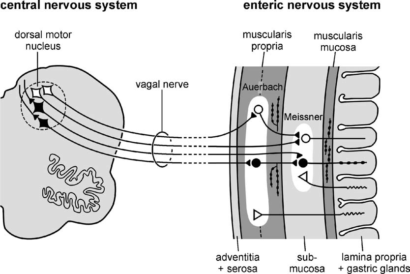 The vagus/vagal nerve connection with the enteric nervous system. Source: Nature
Braak and his colleagues found large deposits of the Parkinson’s-associated protein called alpha synuclein nerves surrounding the gut and the vagus nerve. These deposits were present even at very early stages of the condition, which supported Braak’s theory that maybe the disease was starting (or ‘originating’) in the gut.
So the researchers injected the virus into the vagus nerve in an attempt to model an early stage of Parkinson’s?
Exactly.
Remind me again: What is alpha synuclein? And why is it important to Parkinson’s?
We discuss alpha synuclein a lot on this website – if you are familiar with it, please feel free to jump down to the first recap below.
Alpha synuclein is one of the most common proteins in the brain (making up about 1% of the protein in neurons). The exact function of alpha synuclein is not well understood, but research suggests that it plays a role in multiple cellular functions – including being involved in neurotransmitter release.
But in Parkinson’s, something changes.
 Parkinson’s associated alpha synuclein. Source: Nature
For some reason, in many cases of Parkinson’s alpha synuclein protein starts to cluster and clump together. And this “aggregated” form of alpha synuclein is believed to become toxic, messing with the normal functioning of cells.
For this reason, many efforts to experimentally model Parkinson’s involve generating high levels of alpha synuclein.
|
# RECAP #1: Alpha synuclein is a protein that clusters together in certain neurons in Parkinson’s. Why exactly alpha synuclein does this is unknown, but the protein is used to try and model the condition experimentally. Post-mortem analyses have indicated that in the very early stages of Parkinson’s, clustering of alpha synuclein can be seen in a nerve connection between the brain and the gut called the vagus nerve – suggesting that the condition may start in the gastrointestinal system. As a result, researchers have been experimenting with the vagus nerve to better understand the mechanisms underlying early PD. # |
Ok, so the researchers injected a virus that causes cells to produce high levels of the Parkinson’s associated protein alpha synuclein into the vagus nerve of mice. What did they find?
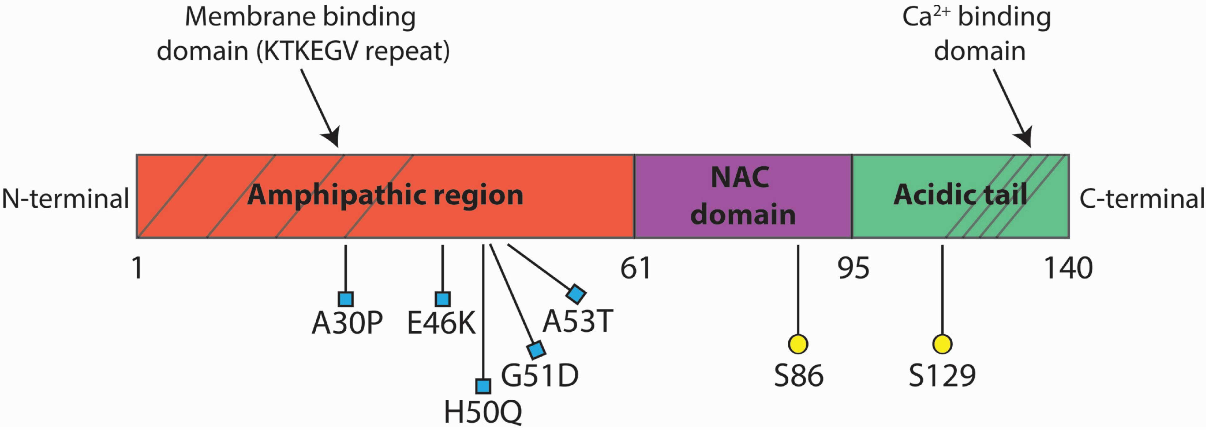
Specifically, the researchers used a virus that produced high levels of a mutant version of alpha synuclein called A53T.
What does that mean? A53T?
The section of DNA that gives rise to alpha synuclein protein is called SNCA.
And there are several genetic variations inside of SNCA that are associated with an increased risk of developing Parkinson’s. A53T is the name of one of those genetic variations.
A53T is a missense point mutation. This means that one amino acid is changed: the 53rd amino acid is changed from alanine (hence the A in “A53T”) to threonine (hence the T in “A53T”). As you can see in the image below, A53T lies in the red (amphipathic) region of SNCA along with several other Parkinson’s-associated variants, such as A30P and E46K:
Mice have been genetically engineered to carry the human SNCA gene with the A53T genetic mutation (Click here to read the original report). These mice initially exhibit hyperactivity and then start to display signs of alpha synuclein protein accumulation and aggregation at about four to six months of age. They also pass away earlier than normal mice (12-14 months of age, compared to 20+ months for normal mice).
Subsequent research involving viruses that produce the A53T version of alpha synuclein have demonstrated that when injected into the brains of mice, this mutant version of alpha synuclein produces more aggregated alpha synuclein that normal alpha synuclein.
The researchers who conducted the study we are reviewing in today’s post found that 6 weeks after they injected a virus that causes cells to produce high levels of the Parkinson’s associated alpha synuclein A53T protein into the vagus nerve of mice, A53T α-synuclein protein could be detected in the dorsal motor nucleus of the vagus nerve.
Not only that, but this increase in levels of alpha synuclein A53T protein in these neurons was associated with a slowing of gastrointestinal tract motility (that is, the movement of ingested food through the gut). Mice injected with an empty (control) version of the virus (AAV-EV) did not exhibit this slowing of gut motility:
 Source: Sciencemag
The researchers next looked at the firing rate of vagal motoneurons that were displaying alpha synuclein A53T protein and found that they were firing 57% slower than the same cells in the control group (injected with an empty virus).
Curiously, this reduction in the firing rate of these vagal motoneurons was specific to them, because in the immediately neighboring region of the brain (called the nucleus of the solitary tract) where alpha synuclein A53T protein was also being produced, the neurons were discharging at their normal rates – there was no reduction.
Motoneurons of the dorsal motor nucleus of the vagus are similar to dopamine neurons (the population of cells in the brain that are particularly vulnerable to Parkinson’s). They are both considered autonomous pacemakers.
What does that mean?
It means that both types of neurons generate repetitive activity even in the absence of synaptic input. That is to say, they are both continuously generating a low frequency of activity, even in the absence of any direct input from other cells.
The researchers found that the high levels of alpha synuclein A53T protein was associated with a reduction in pacemaker activity in the motorneurons.
 Source: Sciencemag
While this spontaneous electrical ‘pacemaker’ activity in dopamine neurons is believed to be dependent on L-type calcium channels (Click here to read a SoPD post about this), the Kv4 channels are believed to play a role in the pacemaker activity in the motorneurons of the dorsal motor nucleus of the vagus.
And so the scientists turned their attention to these channels next.
What are Kv4 channels?
Voltage dependent potassium channels (or Kv – “K” representing potassium and “v” referring to voltage dependent) can be divided into several subtypes. Kv4 is one of those subtypes.
The Kv4 subtype of channels function like the brakes on spontaneous the pacemaker activity, slowing the firing down. Through a series of experiments, the investigators found that the alpha synuclein–induced reduction in firing rate was due to dysfunction in the Kv4 channels.
What caused the dysfunction?
The scientists had a close look at the neurons in the dorsal motor nucleus of the vagus, and they noted that the size of the cells in the alpha synuclein A53T treated group were smaller than those of the control group. That is to say, there was some shrinkage.
 Source: Sciencemag
This shrinkage in the size of the neurons (due to alpha synuclein A53T protein) had the effect of increasing the density of Kv4 channels on the cells. This increase in the density of Kv4 channels enhanced their effect, which resulted in a reduction of the pacemaker activity of the dorsal motor nucleus of the vagus motoneurons:
 Source: Sciencemag
And that ensuing reduction in pacemaker activity led to the slowing of gastrointestinal tract motility in the mice.
The researchers concluded their report by saying “a feasible pathophysiological mechanism for constipation in PD—one of its most common, early-onset prodromal symptoms. Elucidating the physiological underpinning of prodromal symptoms paves the way for a rational design of more selective clinical biomarkers for early diagnosis of the PD“.
|
# # RECAP #2: Researchers have reported that an increase in alpha synuclein protein levels can cause shrinkage of neurons connected to the vagus nerve in mice. This reduction in size results in an increased density of a particular channel on the surface of the cells which results a reduction of the firing rate of the neurons. This reduction in firing rate is associated with a slowing gut motility in the mice. # # |
That’s really interesting. But what did they mean by “prodromal symptoms”?
The prodromal phase of a medical condition is the period of time before diagnosis. The symptoms observed during that phase can be indicators/biomarkers of what may be coming next.
For Parkinson’s, the prodromal symptoms include loss of a sense of smell (hyposmia), the acting out of dreams in a condition called REM sleep disorder (or RBD), and constipation (slow gut motility). In fact, constipation can be a problem decades in advances of any motor symptoms of Parkinson’s:
 Source: Guidelinesinpractice
The researchers believe that they have “provided compelling evidence, through a combination of complementary in vivo and in vitro experiments, for a chain of events leading from α-synucleinopathy, via the selective Kv4 channelopathy to the impaired gastrointestinal motility (e.g., constipation) and dysautonomia that are prevalent in prodromal PD”.
So could this research lead to new treatment options?
The investigators do discuss the clinical implications of their research in the discussion of their report. They note that “there is currently no selective Kv4 channel modulator in the clinic“, but then suggest that “the A-type Kv channel blocker 4-AP – used to treat multiple sclerosis and Lambert-Eaton myasthenic syndrome – could potentially be useful“.
They cite a clinical trial in a Parkinson’s cohort that tested the voltage-dependent potassium channel blocker dalfampridine on gait dysfunction (Click here to read more about the details of this study). Also known as fampridine, this medication is approved for use in improving the walking ability of people with multiple sclerosis.
 Source: Dailymed
The results of the clinical trial in Parkinson’s indicated that 4 weeks of dalfampridine (10mg twice daily) was well tolerated in Parkinson’s patients, but it did not show evidence of any significant benefits for improving gait impairment (compared to a placebo treated group – click here to read more about this). I am not sure if they asked the participants about constipation or gut motility).
But if the current alpha synuclein-induced shrinkage effect can be independently replicated (including the context of a human cell model) perhaps it would be interesting to assess a drug like dalfampridine in a prodromal cohort (that is before Parkinson’s has been diagnosed). Could inhibition of specific sets of potassium channels able to slow/stop the conversion of prodromal PD to fully manifest Parkinson’s? Obviously more data is required, however, before we make this leap.
So is Parkinson’s a channelopathy?
This new data is extremely intriguing, but whether Parkinson’s is a channelopathy is still to be determined.
We really need to see some in depth analyses of the postmortem Parkinson’s brain – getting up close and personal with some of the motorneurons of the dorsal motor nucleus of the vagus nerve to determine if there is shrinkage or an increase in the density of Kv4 channels (or other channels). Observing similar results in the human brain to those observed in this new report would certainly strengthen the hypothesis. Likewise, human cell culture experiments could provide further supportive data and represent a useful assay for drug screening.
We have seen data indicating that the use of L-type calcium channel blockers (dihydropyridines) is associated with a reduced risk of developing Parkinson’s (Click here to read a previous SoPD post on this topic). It would certainly be interesting to know if there is any epidemiological data indicating that clinically used potassium channel blockers are associated with a reduced risk of PD – I am not aware of any (and I would be happy to be corrected on this).
It will also be interesting to see if some of the alpha synuclein targeting approaches that are currently in the clinic (such as MODAG’s Anle138b – click here to read a previous SoPD post about this) have any impact on gut motility or motorneuron/vagus nerve function.
All of these ideas still needs to be examined.
So what does it all mean?
Researchers have reported that the introduction of high levels of mutant alpha synuclein A53T protein to the mouse motorneurons of the dorsal motor nucleus of the vagus nerve results in shrinkage of the cells. This shrinkage has the effect of increasing the density of Kv4 channels, which causes a reduction of cell firing that is associated with a slowing of gut motility in the mice.
The research report discussed in today’s post provides an intriguing set of data that provides a compelling explanation of an early phase of Parkinson’s, in which alpha synuclein is found to aggregate in the motorneurons of the dorsal motor nucleus of the vagus nerve and a slowing of gut motility (seen in the form of constipation) occurs at the same time. The work provides a potential mechanism connecting various components of the overall picture. A pleasing idea, but it will be necessary to explore this further.
And long time readers will appreciate that I liked this study also from the standpoint of seeing some Parkinson’s research focused on a cell type other than the usual dopamine neurons. The condition we call PD is multi-faceted and so our research must be as well. Many different regions of the brain are affected and they all need to be explored in order to get a better understanding of the condition.
And with greater understanding, we will hopefully be in a better place for treatment, both of Parkinson’s and the prodromal phase – stopping the condition before it is diagnosed.
 All of the material on this website is licensed under a
Creative Commons Attribution 4.0 International License
You can do whatever you like with it!
EDITOR’S NOTE: The information provided by the SoPD website is for information and educational purposes only. Under no circumstances should it ever be considered medical or actionable advice. It is provided by research scientists, not medical practitioners. Any actions taken – based on what has been read on the website – are the sole responsibility of the reader. Any actions being contemplated by readers should firstly be discussed with a qualified healthcare professional who is aware of your medical history. While some of the information discussed in this post may cause concern, please speak with your medical physician before attempting any change in an existing treatment regime.
The banner for today’s post was sourced from phys.org

One thought on “Synucleinopathy begets channelopathy?”