Game changer for Alzheimer’s?

TOP-L-Concussion Front Page

Exciting results published this week regarding a small phase 1b clinical trial of a new treatment for Alzheimer’s disease. In this post, we shall review the findings of the study and consider what they may mean for Parkinson’s disease.


4-studyingdown

An Alzheimer’s brain scans on the left, compared to a normal brain (right). Source: MedicalExpress

Alzheimer’s disease is the most common neurodegenerative disease, accounting for 60% to 70% of all cases of dementia. It is a progressive neurodegenerative condition, like Parkinson’s disease, affecting approximately 30 million people around the world.

Inside the brain, in addition to cellular loss, Alzheimer’s is characterised by the increasing presence of two features:

  • Neurofibrillary tangles
  • Amyloid plaques


F1.large

A schematic demonstrating the difference between healthy and Alzheimer’s affected brains. Source: MmcNeuro

The tangles are aggregations of a protein called ‘Tau’ (we’ll comeback to Tau in a future post). These tangles reside within neurons initially, but as the disease progresses the tangles can be found in the space between cells – believed to be the last remains of a dying cell.

Amyloid plaques are clusters of proteins that outside the cells. A key component of the plaque is beta amyloid. Beta-amyloid is a piece of a larger protein that sits in the outer wall of nerve cells where it has certain functions. In certain circumstances, specific enzymes can cut it off and it floats away.


Amyloid-plaque_formation-big

The releasing of Beta-Amyloid. Source: Wikimedia

Beta-amyloid is a very “sticky” protein and it has been believed that free floating beta-amyloid proteins begin sticking together, gradually building up into the large amyloid plaques. And these large plaques were considered to be involved in the neurodegenerative process of Alzheimer’s disease. Thus, for a long time scientists have attempted to reduce the amount of free-floating beta-amyloid in the brain. One of the main ways they do this is with antibodies.

What are antibodies?

An antibody is the foundation of our immune system. It is a Y-shaped structure, that is used to alert the body when a foreign or unhealthy agent is present.

cq5dam.web.1200.YTE_Chapter_31

An artist’s impression of a Y-shaped antibody. Source: Medimmune

Two arms off the Y-shaped antibody have what is called ‘Antigen binding sites‘. An antigen is a molecule that is capable of inducing a response from the immune system (usually a foreign agent, but it can be a sick/dying cell).

2000px-Antibody.svg

A schematic representation of an antibody. Source: Wikipedia

There are currently billions of antibodies in your body -each with specific sets of antigen binding sites – awaiting the presence of their antigen. Antibodies are present in two forms: secreted, free floating antibodies, and membrane-bound antibodies. Secreted antibodies are produced by B-cells, which are part of the immune system. And it’s this secreted form of antibody that modern science has used to produce new medicines.

Really? How does that work?

Scientists can make antibodies in the lab that target specific proteins and then inject those antibodies into a patient’s body and trick the immune system into removing that particular protein. This can be very tricky, and one has to be absolutely sure of the design of the antibody because you do not want any ‘off-target’ effects – the immune system removing a protein that looks very similar to the one you are actually targeting.

These manufactured antibodies are used in many different areas of medicine, particularly cancer (over 40 antibody preparations have been approved by the U.S. Food and Drug Administration for use in humans against cancers). Recently, large pharmaceutical companies (like Biogen) have been attempting to use these manufactured antibodies against other conditions, like Alzheimer’s disease.

Which brings us to the study published this week:

Abeta

Title: The antibody aducanumab reduces Aβ plaques in Alzheimer’s disease.
Authors: Sevigny J, Chiao P, Bussière T, Weinreb PH, Williams L, Maier M, Dunstan R, Salloway S, Chen T, Ling Y, O’Gorman J, Qian F, Arastu M, Li M, Chollate S, Brennan MS, Quintero-Monzon O, Scannevin RH, Arnold HM, Engber T, Rhodes K, Ferrero J, Hang Y, Mikulskis A, Grimm J, Hock C, Nitsch RM, Sandrock A.
Journal: Nature. 2016 Aug 31;537(7618):50-6.
PMID: 27582220

In this study, the researcher conducted a 12-month, double-blind, placebo-controlled trial of the antibody Aducanumab. This antibody specifically binds to potentially harmful beta-amyloid aggregates (both small and large). At the very start of the trial, each participants was given a brain scan which allowed the researchers to determine the baseline level of beta-amyloid in the brains of the subjects. 

All together the study involved 165 people, randomly divided into five different groups: 4 groups received the 4 different concentrations of the drug (1, 3, 6 or 10 mg per kg) and 1 group which received a placebo treatment. Of these, 125 people completed the study which was 12 months long. Each month they received an injection of the respective treatment (remember these are manufactured antibodies, the body can’t make this particular antibody so it has to be repeated injected).

After 12 months of treatment, the subjects in the  3, 6 and 10 mg per kg groups exhibited a significant reduction in the levels of beta-amyloid protein in the brain (according to brain scan images), indicating that Aducanumab – the injected antibody – was doing it’s job. Individuals who received the highest doses of Aducanumab had the biggest reductions in beta-amyloid in the brain. Interestingly, this reduction in beta-amyloid in the brain was accompanied by a slowing of the clinical decline as measured by tests of dementia.  Individuals treated with the placebo saw neither any reduction in their brain levels of beta amyloid nor their clinical decline.

The authors considered this study strong justification for larger phase III trials. Two of them are now in progress, with completion dates expected around 2020.

So this is a good thing right?

Yes, this is a very exciting result for the Alzheimer’s community. But the results must be taken with a grain of salt. We have discussed beta-amyloid in a previous post (Click here for that post). While it has long been considered the bad boy of the Alzheimer’s world, the function of beta-amyloid remains the subject of debate. Some researchers worry about the medical removal of it from the brain, especially if it has positive functions like anti-microbial (or disease fighting) properties.

Given that the treatment is given monthly and can thus be controlled, we can sleep easy knowing that disaster won’t befall the patients receiving the antibody. And if they continue to demonstrate a slowing/halting of the disease, it would represent a MASSIVE step forward in the neurodegenerative field. I guess what I am saying is that it is too soon to say. It will be interesting, however, to see what happens as these patients are followed up over time. And the two phase 3 clinical trials currently ongoing, which involve hundreds of participants, will provide a more definitive idea of how well the treatment is working.

So what does this have to do with Parkinson’s disease?

Yeah, so let’s get back to our area of interest: Parkinson’s disease. Biogen is the pharmaceutical company that makes the Alzheimer’s antibody (Aducanumab) discussed above. Biogen is also currently conducting a phase 1 safety trial (on normal healthy adults) of an antibody that targets the Parkinson’s disease associated protein, alpha synuclein. We are currently waiting to hear the results of that trial.

Several other companies have antibody-based approaches for Parkinson’s disease (all of them targeting the protein alpha synuclein). These companies include:

There are some worries regarding this approach, however. For example, alpha synuclein is highly expressed in red blood cells, and some researchers worry about what affects the antibodies may have on their function. In addition, alpha synuclein has been suspected of having anti-viral properties – reducing viruses ability to infect a cell and replicate (click here to read more on this). Thus, removal of alpha synuclein by injecting antibodies may not necessarily be a good thing for the brain’s defense system.

Unlike beta-amyloid, however, most of alpha synuclein’s activities seem to be conducted within the walls of brain cells, where antibodies can’t touch it. Thus the hope is that the only alpha synuclein being affected by the antibody treatment is the variety that is free floating around the brain.

The results of the Alzheimer’s study are a tremendous boost to the antibody approach to treating neurodegenerative diseases and it will be very interesting to watch how this plays out for Parkinson’s disease in the near future.

Watch this space!


The banner for today’s post was sourced from TheNewsHerald

Is there something in my eye?

SashaEyeball

Some people say that the eyes are the gateway to the soul.

Maybe. I don’t know. Poetic stuff though.

Research published recently, however, suggests that the eyes may also provide a useful aid in the diagnosis of Parkinson’s disease. In today’s post we will review what results have been published and try to understand what they mean for our understanding of this condition.


N00156_H

A schematic of the human eyeball. Source: NIDDK image library

The fact that you can see and read this page is a miraculous thing.

Amazing not just because light is entering your eye, being focused on a particular point in the back of the eyeball and then being turned into a signal that is transmitted to your brain for further analysis, but also because of all the other activities involved with sight. The muscle movements, for example, which are required for turning the eyeball the small fractions necessary for reading this sentence from left to right.

And then there is also the blood supply, keeping the whole system working. This feature is of particular interest to today’s post, as research published last week suggests that there are differences in the blood flow of the eyeball between people with and without Parkinson’s disease.

The anatomy of an eyeball

The human eyeball is – on the macro level – a fairly simple structure.

You have the Iris, which regulates the amount of light entering the eye. At the centre of the iris, you have a central opening called the pupil, which can dilate and constrict as required. Covering these is the cornea, a transparent circular skin. These structures all sit over the lens which helps to refract incoming light and focus it onto the retina. And the retina, of course, is the light sensitive layer that lines the interior of the eye – allowing us to see.

Anatomy

The anatomy of the eye. Source: GemClinic

Within the retina are specialised cells of two sorts:

  • Rod cells (about 125 million of them per eye) which are necessary for seeing in dim light.
  • Cone cells (6-7 million of these) which can be further divided into three types, each sensitive to different primary colours – red, green or blue.

These specialised ‘photoreceptive’ cells send signals down through the layers of the retina to what are called retinal ganglion cells which are the key conduits in the sending of information to the brain.

All of these cells require a constant blood supply, from arteries and veins spreading across the retina, and this a key part of our discussion today (see below).

So what have eyeballs got to do with Parkinson’s disease?

Good question. People with Parkinson’s disease often complain of from visual issues, such as reduced visual acuity, low contrast sensitivity and disturbed colour vision.

And there has been some research into the eyes with regards to Parkinson’s disease. A few weeks ago, this particular study was published:

Eyes1

Title: The retina as an early biomarker of neurodegeneration in a rotenone-induced model of Parkinson’s disease: evidence for a neuroprotective effect of rosiglitazone in the eye and brain.
Authors: Normando EM, Davis BM, De Groef L, Nizari S, Turner LA, Ravindran N, Pahlitzsch M, Brenton J, Malaguarnera G, Guo L, Somavarapu S, Cordeiro MF.
Journal: Acta Neuropathol Commun. 2016 Aug 18;4(1):86. doi: 10.1186/s40478-016-0346-z.
PMID: 27535749       (This article is OPEN ACCESS if you would like to read it)

The researchers in this study used a rodent model of Parkinson’s disease (rotenone-induced). In this model, the animals started losing dopamine cell loss in the brain at 60 days after the model of Parkinson’s disease was chemically induced.

The scientists examined the eyes of the rats at 10, 20, 40 and 60 days of the study. At the 20 day time point, the researchers began to see increased retinal ganglion cell death and swelling of the retinal layers in the eyes. These changes were obviously occurring well before the cell loss is observed in the brain, which leads the authors to ask whether the eyes could potentially used as an early indicator of Parkinson’s disease.

Of particular interest in this study was the use of Rosiglitazone to protect the retinal cells (AND the dopamine neurons in this rodent model of Parkinson’s disease). Rosiglitazone is an anti-diabetic drug. It works as an insulin sensitizer, by binding to fat cells and making them more responsive to insulin (we have previously discussed the curious relationship between Parkinson’s disease and diabetes (click here for more on this), and this result reinforces that connection). The scientists found that giving the drug once every 3 days had very beneficial effects of the survival of the retinal cells. They also observed significant neuroprotection after delaying the treatment for 10 days and then just giving one round of treatment, suggesting that a lot of the drug is not required for positive results.


EDITORIAL NOTE HERE: Before readers start to get any crazy ideas about sourcing and self medicating with Rosiglitazone, it is important to note that there are serious side effects associated with this class of drug. It has been associated with heart disease and stroke (click here to read more), and it should only be taken by people with diabetes and under the strict supervision of a qualified physician. It it mentioned here purely for educational purposes.


So obviously what is required is an examination of the eyes of people with Parkinson’s disease

Yep. And conveniently, in the same week as the previous study came out, this second study was also published:

Eyes2

Title: Evaluation of Retinal Vessel Morphology in Patients with Parkinson’s Disease Using Optical Coherence Tomography.
Authors: Kromer R, Buhmann C, Hidding U, Keserü M, Keserü D, Hassenstein A, Stemplewitz B.
Journal: PLoS One. 2016 Aug 15;11(8):e0161136.
PMID: 27525728          (This article is OPEN ACCESS if you would like to read it)

The researchers examined 49 people with Parkinson’s disease and 49 age- and sex-matched healthy controls. Blood vessels within the retina were identified and then divided into arteries and veins, based on their shape (using computer software). The results of the study indicate significant differences in the morphology of retinal veins in people with Parkinson’s disease when compared to controls.

Interestingly, the retinal effect was more significant on the side of the body firstly affected by Parkinson’s disease (a very common feature of Parkinson’s is that initially the condition will affect one side of the body more than the other).

What does it all mean?

For generations, we have focused on the clinical motor features of Parkinson’s disease (slowness, rigidity, and a resting tremor) when trying to determine if someone has the condition. Now we are learning that there may be other parts of the body that we should be investigating, which could not only provide us with novel diagnostic tools for earlier detection of the disease, but those areas may also provide us with new insights into disease onset and spread as well.

I may be getting a bit ahead of myself here but the possibilities are exciting and we’ll keep you abreast of these new findings as they come to us.


The banner for today’s post was sourced from the Photoforum.

Nilotinib update – new trial delayed

DSK_4634s

It is with great frustration that we read today of the delayed start to the phase 2 clinical trial of the re-purposed cancer drug Nilotinib for Parkinson’s disease (click here for a story outlining the background, and click here for the Michael J Fox Foundation statement).

We have previously  discussed both the preclinical and clinical research regarding Nilotinib and its use in Parkinson’s disease (click here and here for those posts). And the Parkinson’s community certainly got very excited about the findings of the small phase 1 unblinded clinical trial conducted by researchers at Georgetown University in 2015.

With the recent failure of the GDNF trial in Bristol, what the Parkinson’s community (both suffers and researchers alike) needs to do is refocus on moving ahead with exciting new projects, like Nilotinib. To hear that the follow-up trials for Nilotinib, however, will be delayed until 2017 (TWO YEARS after the initial results were announced) due to disagreements regarding the design of the study and who is seemingly in charge of the project, is both baffling and deeply disappointing.

Currently it appears that parties involved in the follow-up clinical trial have decided to go their separate ways, with the researchers at Georgetown University looking to conduct a single site phase 2 study of 75 subjects (if they can access the drug from supplier Novartis), while the Michael J Fox backed consortium will set up a multi-site phase 2 study.

We will continue to follow this situation as it develops and will report events as they happen.

Coffee and Parkinson’s disease – it’s not just caffeine

coffee-beans-1400

Numerous epidemiologic studies have indicated that coffee consumption reduces the risk of Parkinson’s disease. For a long time, efforts have been made to determine what the magic ingredient in this popular beverage is. Many people have speculated that the stimulant caffeine is the critical active ingredient in this neuroprotective effect.

New research, however, suggests that this may not be the case.

Today’s post will review recently published results suggesting that Quercetin (and not caffeine) is the neuroprotective component in coffee.


kaldi-adapted-from-uker

Kaldi the goat herder. Source: CoffeeCrossroads

Legend has it that in 800AD, an Ethiopian goat herder called Kaldi noticed that his animals were “dancing” after eating some berries from a tree that he did not recognise. Fascinated by the happy behaviour of his goats, Kaldi naturally decided to eat the berries for himself and he subsequently became “the happiest herder in happy Arabia”.

This amusing encounter was apparently how humans discovered coffee. It is most likely a fiction as the earliest credible accounts of coffee-consumption emerge from the 15th century in the Sufi shrines of Yemen, but since then coffee has gone on to become one of the most popular drinks in the world.

Stupid question: what exactly is coffee?

For a person who doesn’t drink coffee (like myself), this is actually a really interesting question. Coffee is a beverage made from ground up roasted beans, which are the seeds of berries from the Coffea plant. These are the berries:

coffeeberry_1338562952_62

Coffea berries. Source: About.me

And these are the beans (unroasted):

unroasted-coffee-beans-shelf-life

Unroasted Coffee beans. Source: Kopiholic

Coffee production also makes for fascinating reading (Click here for more) and why we roast the beans is equally interesting (Click here for that), but they are taking us off the topic here.

There are basically two types of coffee beans: Arabica and Robusta.

Approximately 70 percent of the coffee beans we use are Arabica. Surprisingly, the less popular Robusta actually has twice as much caffeine as Arabica. And caffeine is the stimulant that rewards people for drinking this beverage.

Caffeine is also the chemical that has long been thought to have positive effects on Parkinson’s disease, possibly even reducing the risk of the condition (more on that below).

coffee-cup-images-5

Fancy a cuppa? Source: Science-All

What does coffee have to do with Parkinson’s disease?

We have previously discussed the enormous contribution that the Honolulu Heart Study has made to our understanding of Parkinson’s disease (click here to read that post). Many of the earliest associations with the condition were found in that large epidemiologic study. One of those findings was that the consumption of coffee reduced one’s risk of developing Parkinson’s disease.

Coffee-title2

Title: Association of coffee and caffeine intake with the risk of Parkinson disease.
Authors: Ross GW, Abbott RD, Petrovitch H, Morens DM, Grandinetti A, Tung KH, Tanner CM, Masaki KH, Blanchette PL, Curb JD, Popper JS, White LR.
Journal: JAMA. 2000 May 24-31;283(20):2674-9.
PMID: 10819950    (This article is OPEN ACCESS if you would like to read it)

The researchers behind this article analysed the data from the Honolulu Heart Study – an epidemiological study of 8,006 “non-institutionalized men of Japanese ancestry, born 1900-1919, resident on the island of Oahu” – and found that the age-adjusted incidence of Parkinson’s disease declined consistently with increased amounts of coffee intake (from 10.4 per 10,000 person-years in men who drank no coffee to 1.9 per 10,000 person-years in men who drank at least 28 oz/d). This and other findings in their analysis indicated that higher coffee (and caffeine) intake is associated with a significantly lower incidence of Parkinson’s disease.

Subsequent studies have replicated this association, and several have demonstrated the neuroprotective effects of caffeine (click here for a review on this topic).

So what new data has been published?

This is Prof Patrick and Prof Edith McGeer:

ss-alz-research McGeer

Prof Patrick and Prof Edith McGeer. Source: Mcgeerandassociates

This husband and wife team of scientists are well recognised figures within the Parkinson’s disease research work, having produced many seminal scientific reports. Patrick is a particularly interesting character having played basketball for Canada in the 1948 Olympics and then a politician in the British Columbia legislature (1962-1986).

They are also authors on the article we are going to review today:

Coffee-title

Title: Quercetin, not caffeine, is a major neuroprotective component in coffee.
Authors: Lee M, McGeer EG, McGeer PL.
Journal: Neurobiol Aging. 2016 Jul 5;46:113-123.
PMID: 27479153

As we said above, for the longest time people have believed that caffeine was the active ingredient in the miraculous ability of coffee to reduce the risk of Parkinson’s disease. The researchers who published this report were particularly interested in the neuroprotective role for coffee in Parkinson’s disease and they decided to break coffee down into some of its basic components. Specifically:

  • Caffeine
  • quercetin
  • flavone
  • Chlorogenic acids (CGAs)

They tested each of these coffee components on cells (grown in petri dishes) that had been exposed to a toxin, and then assessed cell survival. Curiously, although caffeine did exhibit neuroprotective effects on the cells, it was beaten by the far superior protective effects of quercetin.

What is quercetin?

Quercetin is a flavonoid (a type of plant pigment) that is found in many fruits, vegetables, leaves and grains. Flavonoids are potent antioxidants. Antioxidants scavenge particles (called free radicals) in the body which can damage cell membranes, affect DNA, and even cause cell death. Antioxidants neutralize these free radicals. (For more on flavonoids – click here).

What does this mean?

The results are very interesting, especially if they provide us with a new potential target for therapeutic drug development. It also raises the age-old idea of antioxidants being potentially useful in the treatment of Parkinson’s disease (the previous history of this therapeutic approach has been disappointing – click here to read more on this).

But before you rush out and load up on quercetin, there are a few things to consider:

Quercetin is generally considered pretty safe. Fruits and vegetables are the primary dietary sources of quercetin, particularly citrus fruits, apples, onions, parsley, sage, tea, and red wine.

That said: excessive use of quercetin can have side effects, which may include headache and upset stomach. Very high doses of quercetin can cause damage to the kidneys (doses greater than 1 g per day), and regular periodic breaks from taking quercetin is advised. Importantly, pregnant women, breastfeeding women, and people with kidney disease should avoid quercetin.

EDITOR’S NOTE: If you are considering supplementing your diet with quercetin (or any other potential therapeutic agents) please firstly discuss this change of lifestyle with your medical physician. Information provided here can under no circumstances be considered medical advice.

Having said that we shall keep an eye out for any new research of quercetin and Parkinson’s disease, and report it here.


The banner for today’s post was sourced from Phoxpopmagazine

Identical twins and Parkinson’s disease

Twins

The influence of genetics in  Parkinson’s disease is difficult to determine. If it was simply a genetic disease, identical twins – who share identical DNA – should show no difference in their susceptibility to Parkinson’s disease. They should either both develop the condition, or not. Right?

But this is not the case.

In today’s post we will review a particularly interesting pair of identical twins.


500x500_144169027

Jeff & Jack Gernsheimer in 1982. Source: ReadingEagle 

When people ask the obvious question about the cause of Parkinson’s disease – ‘is it genetics or is it environment?’ – I have a standard answer: ‘it’s complicated’. I then tell them the curious story of identical twins Jeff and Jack Gernsheimer from eastern Pennsylvania. No other case better demonstrates the strange question of what causes Parkinson’s disease.

For almost their entire lives (69 years), Jeff and Jack have lived no more than half a mile apart. Breathing the same air, drinking the same water. They are literally neighbours – just a five-minute walk between their homes. In addition, since 1971 they have worked in the same office at a graphic design firm that they started together. The brothers were the focus of a story in the online magazine Nautilus last year. It’s a fantastic article and I fully recommend you read it.

So here’s the thing: In 2009 Jack was diagnosed with Parkinson’s disease.

To date, Jeff is yet to exhibit any signs of the condition.

Strange huh?

Two genetically identical people, living in the exact same environment and one of them develops Parkinson’s disease.

Ok, how do we explain this?

Hang on a second, slow down. I haven’t even got to the really interesting part yet:

After being diagnosed, Jack had his genome sequenced to see if there were any particular genetic mutations that might make him vulnerable to Parkinson’s disease. That analysis determined that Jack has a mutation in the most common Parkinson’s disease-associated gene: Glucocerebrosidase or GBA (which we have discussed in a previous blog post).

Interesting. So that explains the Parkinson’s disease?

No. Jack’s identical twin brother, Jeff, also has that exact same mutation.

So now we have a pair of identical twins who share the identical genetic code, live in the same environment, and have a genetic mutation associated with Parkinson’s disease, but only Jack has developed the condition while Jeff has not.

I think you will agree, it’s a really interesting tale… and with the help of modern science, it gets even more interesting.

How so?

In 2014, a research paper was published that utilized cells from both Jack & Jeff to determine what differences existed between them:

twinstitle2

Title: iPSC-derived dopamine neurons reveal differences between monozygotic twins discordant for Parkinson’s disease.
Authors: Woodard CM, Campos BA, Kuo SH, Nirenberg MJ, Nestor MW, Zimmer M, Mosharov EV, Sulzer D, Zhou H, Paull D, Clark L, Schadt EE, Sardi SP, Rubin L, Eggan K, Brock M, Lipnick S, Rao M, Chang S, Li A, Noggle SA.
Journal: Cell Reports. 2014 Nov 20;9(4):1173-82.
PMID: 25456120        (this article is OPEN ACCESS if you would like to read it)

EDITOR’S NOTE HERE: Monozygotic means twins from the same egg, (as opposed to dizygotic meaning twins from two eggs). And discordant means ‘at variance, or at odds’ – in medicine it is used when one identical twin has a condition and the other does not.

The researchers conducting this study took skin cells from the brothers and they turned them into brain cells via a miraculous Nobel-prize winning approach. The technique firstly involves turning the skin cells into induced pluripotent stem cells (or iPS cells).

IPS-cells

Source: Csiro

iPS cells can be used to make any cell you wish, and the researchers encouraged Jack and Jeff’s iPS cells to develop into dopamine neurons (one of the types of cells that are vulnerable in Parkinson’s disease).

When the researchers analysed the dopamine neurons from both twins, they found that both had half the normal levels GBA protein activity (an enzymatic reaction) due to the mutation in the GBA gene. The brother’s dopamine neurons also had approximately three times the normal levels of alpha-synuclein protein, and a reduced capacity to synthesize and release dopamine.

fce5078fbe8ec74e95c8a435e607ad11

Dopamine neurons. Source: MindsofMalady

Then the researchers noticed something interesting: the dopamine cells from Jack (the affected twin) had lower dopamine levels than Jeff’s cells. This was rather strange: identical twins should actually have similar levels – all things being equal. The researchers attributed this decrease in dopamine to an increase in the levels of monoamine oxidase B (MAO-B) in Jack’s cells.

What is MAO-B?

Good question. MAO-B is an enzyme in dopamine neurons that helps to break down excess dopamine. After a cell releases dopamine, the cell will re-collect and recycle leftover/unused dopamine. MAO-B is the enzyme that breaks dopamine down. MAO-B inhibitors (such as Rasagiline or Azilect) have been used for some time as a therapy in Parkinson’s disease. By blocking MAO-B with MAO-B inhibitors, people with Parkinson’s disease can have increased levels of dopamine as the remaining dopamine does not get broken down so quickly.

The researchers studying Jack and Jeff’s iPS dopamine neurons found that by replacing the reduced GBA and inhibiting the oversupply of MAO-B (with MAO-B inhibitors) they made the dopamine neurons healthier – with an increase in dopamine levels and increased removal of excessive alpha-synuclein (the protein that is associated with Parkinson’s disease).

Are Jeff and Jack in a unique situation?

Nope. Not at all.

Here are some other examples:

ID-twin3

Title: Pathology of PD in monozygotic twins with a 20-year discordance interval.
Author: Dickson D, Farrer M, Lincoln S, Mason RP, Zimmerman TR Jr, Golbe LI, Hardy J.
Journal: Neurology. 2001 Apr 10;56(7):981-2.
PMID: 11294946

This was a case study in which a pair of identical twins both developed Parkinson’s disease, but one of the twins was diagnosed 20 years before the other.

ID-twin1

Title: Identical twins with Leucine rich repeat kinase type 2 mutations discordant for Parkinson’s disease.
Authors: Xiromerisiou G, Houlden H, Sailer A, Silveira-Moriyama L, Hardy J, Lees AJ.
Journal: Movement Disord. 2012 Sep 1;27(10):1323.
PMID: 22488887                (This article is OPEN ACCESS if you would like to read it)

This second case study involved a pair of twins who both carried a mutation in the Parkinson’s associated gene, Lrrk2 (click here to read more about this gene). They both developed Parkinson’s disease, but 10 years separated their diagnoses.

Twins1

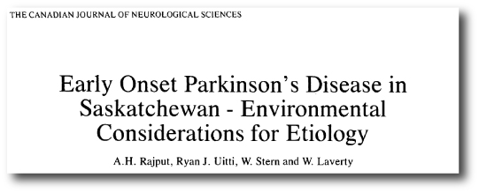
Title: Parkinson disease in twins: an etiologic study.
Authors: Tanner CM, Ottman R, Goldman SM, Ellenberg J, Chan P, Mayeux R, Langston JW.
Journal: JAMA. 1999 Jan 27;281(4):341-6.
PMID: 9929087     (This article is OPEN ACCESS if you would like to read it)

In this study, the scientists screened 19,842 white male twins enrolled in the National Academy of Sciences/National Research Council World War II Veteran Twins Registry. 163 pairs of twin were identified in which at least 1 twin had Parkinson’s disease (and medical records were available).

When diagnosis was made over the age of 50 years of age, approximately 10% of the twin pairs both had Parkinson’s disease (for both monozygotic and dizygotic twins). But when diagnosis was made under the age of 50, the monozygotic concordance was 100% – that is, all of the identical twins diagnosed under the age of 50 had Parkinson’s disease – while the dizygotic concordance remained around 10-20%. The researchers concluded that ‘this pattern strongly supports a primarily inherited cause of early-onset Parkinson’s disease’.

So how do we explain the difference seen in Jack and Jeff?

Some twins may be born with a vulnerability for Parkinson’s disease (like a genetic mutation, in the GBA or Lrrk2 gene for example), but there is some other factor/s that is influential in the initiation of the disease. And this is where scientists start talking about something called epigenetics (Epi, Greek for ‘over’ or ‘above’ and Genetics,…well, you should be able to work that one out).

Epigenetics is the study of changes in an organism that are caused by modifications or variations of gene expression rather than alteration of the genetic code itself. These variations may result from external factors that cause genes to turn on and off.

sep-05-living-well-weizmann-epigenetics-robichek-dna

Source: 2ndActHealth

In the case of the Gernsheimer twins, if you read the story in the online magazine Nautilus you will find that their lives were not entirely the same. There were basic differences, for examples they went to different universities and in the 1970’s Jack enlisted in the army. But there were also some larger, life-altering differences: in the late 1980’s Jack lost a son in tragic circumstances. The brothers speculate that the stress/suffering associated with that particular event may have been a catalyst for the Parkinson’s that followed. Many researchers in the Parkinson’s disease field have speculated on whether a stressful/traumatic event in their lives was the causative agent for their Parkinson’s disease.

So what does it all mean?

It means that the answer is more complicated than first assumed.

And unfortunately, this is where I end up when people ask me about ‘genetics vs environment’ in the cause of Parkinson’s disease: a qualified we really don’t know. But I do always suggest that ‘Genetics vs environment’ may be too simplistic.

To finish, here is a nice, short video of the Gernsheimer twins discussing why they got involved in research:


The source of today’s banner was the AutismBlog.

Pesticides and Parkinson’s disease

1016238_tcm9-156853

Farming and country living (more specifically exposure to pesticides) has often associated with Parkinson’s disease. The findings of numerous epidemiological studies support this connection.

Recently a new study was published that lends considerable support to this idea of factors involved in causing Parkinson’s disease.


In 1986, a group of researchers in the province of Saskatchewan (Canada) made an interesting observation:

Farm

Title: Early onset Parkinson’s disease in Saskatchewan–environmental considerations for etiology.
Author: Rajput AH, Uitti RJ, Stern W, Laverty W.
Journal: Can J Neurol Sci. 1986 Nov;13(4):312-6.
PMID: 3779530

They collected the medical details of 21 people who were born & raised Saskatchewan and that later went on to be diagnosed with early onset Parkinson’s disease (that is diagnosis before the age of 40 years). When evaluating the childhood environments of those 21 people, the researchers immediately noticed that 19 of them spent the first 15 years of their lives exclusively in rural Saskatchewan.

This finding has been replicated in different parts of the world. It has also been expanded upon and there is now solid evidence pesticides, but not fungicides, were associated with Parkinson’s disease (click here to read more on this).

So what research has been done recently?

A few weeks ago, some researchers from North Carolina published interesting new results that further supports the association between exposure to pesticides and Parkinson’s disease:

Farmtitle

Title: Identification of chemicals that mimic transcriptional changes associated with autism, brain aging and neurodegeneration.
Authors: Pearson BL, Simon JM, McCoy ES, Salazar G, Fragola G, Zylka MJ.
Journal: Nat Commun. 2016 Mar 31;7:11173.
PMID: 27029645      (This report is OPEN ACCESS if you would like to read it)

The researchers grew mouse brain cells in petri dishes, and exposed them to hundreds of chemicals commonly found in the environment and on food. Each petri dish was exposed to just one chemical (for 24 hours), and this was done across many individual petri dishes so that many different chemicals could be tested. They then collected and analysed the RNA of the cells exposed to these chemical, allowing the researchers to access what was happening inside those cells – the molecular reactions to the chemicals. Importantly, they also compared the RNA results between samples – that is to say, they compared the effect that the different chemicals had on the cells by comparing the RNA collected from those cells.

What they found was very interesting.

They identified six basic groupings or clusters of chemicals which share similar mechanistic profiles. This means that the chemicals within each group caused similar RNA activity inside the exposed cells. Of particular interest to us here at the Science of Parkinson’s Disease, is that one of these groups of chemicals (cluster 2) exhibited RNA activity similar to that observed in the aged brain and certain neurodegenerative conditions.

Many of the chemicals in cluster 2 (including fenpyroximate, pyridaben and rotenone), are compounds that target the mitochondria – the power generators inside cells. Rotenone in particular has been associated with Parkinson’s disease (Click here for more on this). It would be interesting to investigate if other chemicals in this clustering have similar effects in models of Parkinson’s disease to that caused by Rotenone.

After identifying these chemicals , the researchers next turned their attention to the chemical usage and food commodity residue database collected by the United States Geological Survey, the United States Department of Agriculture (USDA) and the Food and Drug Administration (FDA). The researchers could use this database to see if what the usage trends were for many of the chemicals in cluster 2. While they found that rotenone usage is low and unchanging, many other chemicals have been used with increasing frequency. This lead the scientists to conclude that there is “significant human exposure potential to many of the chemicals in cluster 2”.

So what does it all mean?

There does appear to be a solid connection between country/rural living and Parkinson’s disease. This association has been replicated across continents and over time. And as we discussed above, we have identified chemicals used in the agricultural industry that can increase the risk of developing the condition.

The fact that the majority of the farming community do not go on to develop Parkinson’s disease, however, brings into question the strength of the association. Obviously there are additional aspects (for example, genetics) that are playing an influence.

Caution should be taken when dealing with many of the chemicals used in the agricultural industry, limiting direct exposure to an absolute minimum. It will be interesting to record if there is any decrease in the prevalence of Parkinson’s disease over time with heightened awareness about the dangers of some of the chemicals used down on the farm.


The banner for todays post was sourced from RSPB.

Juvenile-onset Parkinson’s disease

xTgKydA8c

A community in New Brunswick (Canada) was recently shocked to discover that a 2 year old boy in their midst had been diagnosed with Parkinson’s disease (Click here to read more).

Yes, you read that correctly, it’s not a typo: a 2 year old boy.

Juvenile-onset Parkinson’s disease is an extremely rare version of the condition we discuss here at the Science of Parkinson’s. It is loosely defined as being ‘diagnosed with Parkinson’s disease under the age of 20’. The prevalence is unknown, but there is a strong genetic component to form of the condition. In today’s post we will review what is known about Juvenile-onset and look at new research about a gene that has recently been discovered to cause a type of Juvenile-onset Parkinson’s disease.


Henri_Huchard

Dr Henri Huchard. Source: Wikipedia

In 1875, Dr Henri Huchard (1844-1910; a French neurologist and cardiologist) described the first case of a child who, at just 3 years of age, presented all the clinical features of Parkinson’s disease. Since that report, there have been many studies detailing the condition that has become known as ‘juvenile-onset Parkinson’s disease’.

What is juvenile-onset Parkinson’s disease?

Basically, it is a form of Parkinson’s disease that affects children and young people under the age of 20. The defining feature is the age of onset. The average age of onset is approximately 12 years of age (with the majority of cases falling between 7 and 16 years) and males are affected by this condition more than females (at a rate of approximately 5:1).

The actual frequency of juvenile-onset parkinson’s is unknown given how rare it is. When researcher look at people with early onset Parkinson’s disease (that is diagnosis before the age of 40; approximately 5% of the Parkinson’s community), they have found that between 0.5 – 5% of that group of people were diagnosed before 20 years of age. This suggests that within just the Parkinson’s community, the frequency of juvenile-onset parkinson’s is at the most 0.25% (or 2.5 people per 1000 people with Parkinson’s). Thus it is obviously a very rare condition.

It is interesting to note that Lewy bodies (the clusters of aggregated protein that classically characterise the brains of people with Parkinson’s disease) are very rare in cases of juvenile-onset parkinson’s disease. To our knowledge there has been only one case of Lewy bodies in juvenile-onset parkinson’s disease (Click here to read more on this). This suggests that the juvenile-onset form of Parkinson’s disease may differ from other forms of the condition in its underlying biology.

Do we know what causes juvenile-onset parkinson’s disease?

There is a very strong genetic component to juvenile-onset parkinson’s disease. In fact, the incidence of Parkinsonism in relatives of people with juvenile-onset parkinson’s disease is higher than in the general public AND in the relatives of people with other forms of Parkinson’s disease.

Genetic mutations in three genes are recognised as causing juvenile-onset Parkinson’s disease. The three genes are known to the Parkinson’s world as they are all PARK genes (genetic variations that are associated with Parkinson’s). Those three genes are:

  • Parkin (PARK2)
  • PTEN-induced putative kinase 1 (PINK1 or PARK6)
  • DJ1 (PARK7)

In juvenile-onset Parkinson’s disease, all of these mutations are associated with autosomal recessive – meaning that two copies of the genetic variation must be present in order for the disease to develop.

Parkin mutations account for the majority of juvenile-onset Parkinson’s disease cases. Affected individuals have a slowly progressing condition that is L-dopa responsive. Dystonia (abnormal muscle tone resulting in muscular spasm and abnormal posture) is very common at the onset of the condition, particularly in the lower limbs.

Can the condition be treated with L-dopa?

The answer is: ‘Yes, but…’

L-dopa (or dopamine replacement) treatment is the standard therapy for alleviating the motor features of Parkinson’s disease.

The majority of people with juvenile-onset parkinson’s respond well to L-dopa, but in the Parkin mutation version individuals will typically begin to experience L-dopa-induced motor fluctuations (dyskinesias) early in that treatment regime.

What research is currently being done on this condition?

Given that cases are so very rare and so few, it is difficult to conduct research on this population of individuals. Most of the research that is being conducted is focused on the genetics underlying the condition.

And recent that research lead to the discovery of a new genetic variation that causes juvenile-onset Parkinson’s disease:

Juvenile

Title: Discovery of a frameshift mutation in podocalyxin-like (PODXL) gene, coding for a neural adhesion molecule, as causal for autosomal-recessive juvenile Parkinsonism.
Authors: Sudhaman S, Prasad K, Behari M, Muthane UB, Juyal RC, Thelma BK.
Journal: Journal Med Genet. 2016 Jul;53(7):450-6.
PMID: 26864383           (This article is OPEN ACCESS if you would like to read it)

The researchers who wrote this article were presented with a 10 member Indian family from Aligarh, Uttar Pradesh. Of the 8 children in the  family, 3 were affected by Parkinsonian features (tremor, slowness, rigidity and gait problems) that began between 13 and 17 years of age. The researchers conducted DNA sequencing and found that none of the three affected siblings had any of the known Juvenile-onset Parkinson’s disease genetic mutations (specifically, mutations in the genes PARK2, PINK1and DJ1).

They then compared the DNA from the three siblings with the rest of the family and found a genetic variant in a gene called podocalyxin-like (or PODXL). It must be noted that PODXL is a completely novel gene in the world of Parkinson’s disease research, which makes it very interesting. PODXL has never previously been associated with any kind of Parkinson’s disease, though it has been connected with two types of cancer (embryonal carcinoma and periampullary adenocarcinoma).

The researchers then turned to their genetic database of 280 people with Parkinson’s disease have had their genomes sequenced. The researchers wanted to determine if any genetic variants in the PODXL gene were present in other suffers of Parkinson’s disease, but had not been picked up as a major contributing factor. They found three unrelated people with PODXL mutations. All three had classical Parkinson’s features, and were negative for mutations in the Parkin, PINK1 and DJ1 genes.

The researchers concluded that the PODXL gene may be considered as a fourth causal gene for Juvenile-onset Parkinson’s disease, but they indicated that further investigations in other ethnic groups are required.


The banner for today’s post was sourced from ClipArtBest

Editorial note

Cambridge University applications

The scientists behind the Science of Parkinson’s disease website work at Cambridge University and are associated – through their research – with the Wellcome Trust/MRC Cambridge Stem cell institute.

sci-logo

Throughout July, the Stem cell institute is running the #MyView campaign which aims to raise awareness about all of the latest developments in stem cell research.

Of particular interest to the Parkinson’s community will be the work being conducted in Prof Roger Barker’s lab (he of the 2016 Gretschen Amphlet Memorial lecture). The Stem cell institute made a video about the research being conducted in Prof Barker’s lab – viewed through the eyes of someone with Parkinson’s disease. It provides an interesting view of the working being carried out:

We encourage all of our readers to get involved with the #myview discussion and to follow the campaign of social media via Youtube, Facebook, & Twitter.

As scientists we are always very keen to hear the views of people in the Parkinson’s community (both sufferers and carers). It is through campaigns like this that we can gain new insight from different view points.


The banner for today’s post was sourced from the Huffington Post.

Traumatic brain injury and Parkinson’s disease – an association

o-TRAUMATIC-BRAIN-INJURY-facebook

A new study has found traumatic brain injury with loss of consciousness is associated with the risk of Parkinson’s disease, but (interestingly) not Alzheimer’s disease. In this post we will review the study and its findings, before considering the implications of the results.


football-brain-ocotber-2009.jpg

Image sourced from GQ

There has been a lot of talk on the interweb and various media outlets recently about the long term consequences of head injuries associated with physical sports like boxing, rugby, ice hockey and American football (click here for more on this).

Of particular concern is when individuals lose consciousness at the time of the head injury, which has been associated with worse outcomes than simply suffering a bang on the head.

A group of American researchers recently decided to assess whether there was any association between traumatic head injury with loss of consciousness and the increased risk for Alzheimer’s disease.

What they found may have profound implications for Parkinson’s disease.

TBI-title

Title: Association of Traumatic Brain Injury With Late-Life Neurodegenerative Conditions and Neuropathologic Findings.
Authors: Crane PK, Gibbons LE, Dams-O’Connor K, Trittschuh E, Leverenz JB, Keene CD, Sonnen J, Montine TJ, Bennett DA, Leurgans S, Schneider JA, Larson EB.
Journal: JAMA Neurol. 2016 Jul 11. doi: 10.1001/jamaneurol.2016.1948.
PMID: 27400367       (This study is OPEN ACCESS if you would like to read it)

The researchers collected the results of 3 large studies, collectively involving 7130 participants who had head injury data (2879 men and 4251 women; average age of 79.9 years). Of these 845 had suffered traumatic brain injuries with loss of consciousness for at least 1 hour. Interestingly, the researchers found no statistically significant association between traumatic brain injuries with loss of consciousness and risk of Alzheimer’s disease.

Next they looked at Parkinson’s disease and found that people who suffered traumatic brain injuries with loss of consciousness of more than 1 hour had a statistically significant increase in developing Parkinson’s disease (2-3 times more than normal controls).

Of the 7130 participants in the study, postmortem autopsy analysis reports were available for 1589 of the subjects. The researchers looked for the neuropathological hallmarks of Parkinson’s disease, called Lewy bodies, and they found no correlation between people who suffered traumatic brain injuries with loss of consciousness of less than 1 hour and the presence of Lewy bodies. When they looked in the brains of people who suffered traumatic brain injuries with loss of consciousness of more than 1 hour, they did find a correlation. And importantly these neuropathological events were not associated with genetic mutations.

So what does it all mean? 


The results indicate that traumatic brain injuries with loss of consciousness of more than 1 hour could significantly increase a person’s risk of Parkinson’s disease. The crucial  detail in the results is the ‘loss of consciousness of more than 1 hour’. Traumatic head injury can often result in disruption to the blood-brain-barrier (the protective film surrounding the brain), which may result in certain pathogens entering the brain. So the more severe the injury, perhaps the longer the barrier is disrupted. Why this event may relate solely to Parkinson’s disease and not Alzheimer’s disease, however, remains to be determined.

It would be interesting to assess how this finding relates to the greater Parkinson’s community. That is to say, determine how many of the people with Parkinson’s disease have a head injury with loss of consciousness in their past medical records?

Reading this study, one cannot help thinking of the recent passing of Boxing great Muhammad Ali. Ali died this year having spent the last third of his life living with Parkinson’s disease. Many boxing careers have probably involved one or two severe head injuries with loss of consciousness, so why are there not more cases of Parkinson’s disease in the boxing community? Many retired boxers suffer from what is called Dementia pugilistica – a neurodegenerative condition with Alzheimer’s-like dementia. Some estimates suggest that 15-20% of boxers may be affected, with symptoms usually starting 12-16 years after the start of a career in boxing. Some very famous boxers have been diagnosed with this condition, including world champions Floyd Patterson, Joe Louis, Sugar Ray Robinson and boxer/coach Freddie Roach.

The difference between the results of today’s study and dementia pugilistica may lie in the repeated nature of the injuries in boxers and the length of time individuals were unconscious. It will be interesting to see what becomes of this research.


The banner for today’s post was sourced from the Huffington Post

Bees! Bees! Hark to your bees!

Bee-Wallpapers-Desktop-HD-Pictures-3

The title of today’s post comes from a Rudyard Kipling poem, and it seemed appropriate as we review the results of a clinical study for Parkinson’s disease…involving bees!  Much has been written about the medicinal properties of the lovely honey that bees make. The healing properties of the sweet produce of our little friends seems to cure all ailments.

Today’s post, however, is not about honey.

No, today’s post is about the other thing bees are known for: their sting!


ofdbubwgay8bjrwbdvhz

Source: Gizmodo

Earlier today a group of researchers in Paris (France), published the results of a clinical trial in which they gave bee venom to people with Parkinson’s disease for 11 months.

Beetitle

Title: Bee Venom for the Treatment of Parkinson Disease – A Randomized Controlled Clinical Trial.
Authors: Hartmann A, Müllner J, Meier N, Hesekamp H, van Meerbeeck P, Habert MO, Kas A, Tanguy ML, Mazmanian M, Oya H, Abuaf N, Gaouar H, Salhi S, Charbonnier-Beaupel F, Fievet MH, Galanaud D, Arguillere S, Roze E, Degos B, Grabli D, Lacomblez L, Hubsch C, Vidailhet M, Bonnet AM, Corvol JC, Schüpbach M.
Journal: PLoS One. 2016 Jul 12;11(7):e0158235.
PMID: 27403743        (This study is OPEN ACCESS if you would like to read it)

No! What? Bee Venom? Really?

Yeah, I know. Weird, right? But there is actually some logic to the idea.

Bee venom has recently become in vogue for all kinds of health associated products (eg. face masks, etc – click here for more on this), but preclinical experiments have also demonstrated that it can have beneficial effects in models of Parkinson’s disease.

Bee venom (or Apitoxin as it known to the science types) contains some interesting components, one of which is called Apamin.

800px-Apamin.svg

Apamin. Source: Wikipedia

Apamin is the only component of bees venom that can pass through the blood-brain-barrier and enter into the brain. Once inside the brain, Apamin selectively blocks structures on the membrane of cells called ‘calcium channels’.

Calcium channels allow calcium (surprise) to enter a cell, and control many physiological functions including neurotransmitter release, muscle contraction and cell survival. It has already been demonstrated in models of Parkinson’s disease that Apamin has neuroprotective properties on the dopamine neurons in the brain:

bees

Title: Bee venom and its component apamin as neuroprotective agents in a Parkinson disease mouse model.
Authors: Alvarez-Fischer D, Noelker C, Vulinović F, Grünewald A, Chevarin C, Klein C, Oertel WH, Hirsch EC, Michel PP, Hartmann A.
Journal: PLoS One. 2013 Apr 18;8(4):e61700.
PMID: 23637888               (this article is OPEN ACCESS if you would like to read it)

The researchers in this preclinical study demonstrated that bee venom was protective in models of Parkinson’s disease. When they tested Apamin alone, however, they found that it reproduced the protective effects only partially. They concluded that other components of bee venom must be enhancing the protective action of Apamin.

So what happened in the clinical trial?

The researchers in France conducted a randomized double-blind study (meaning that nobody – researchers included – knew who was getting the bee venom or the saline control solution). They took 40 people with early stage Parkinson disease (Hoehn & Yahr stages 1.5 to 3) and assigned them to either monthly bee venom injections or equivalent injections of saline.

After 11 months of monthly injections, the researchers found that bee venom did not significantly decrease the motor features of Parkinson’s disease (as judged by UPDRS III scores during ‘off’ condition). In addition, brain imaging (DAT-scan) did not differ significantly between treatment groups over the 11 months.

The researchers did, however, see improvements in some of the cognitive measures in subjects receiving the bee venom (albeit non-significant).

In their concluding remarks, the researchers questioned whether lack of significant effect was due to the low frequency of injections (once per month). Maybe the subjects in the trial were simply receiving too little of the treatment for it to have an effect. With the support of more preclinical experimental results, they propose that a larger study is warranted with a higher administration frequency and possibly higher individual doses of bee venom.

Will this happen?

It is unclear at present.

In the study, 4 subjects had immune system responses to the bee venom, so it may be wise to firstly establish what components of bee venom are having any beneficial effect before proceeding with further clinical trials.


The banner for today’s post was sourced from modern.scot