Milk (Yes, milk) and Parkinson’s disease

gotmilk

We have previously written about the enormous contribution that the ‘Honolulu Heart Study’ has made to our understanding of Parkinson’s disease. This longitudinal study of 8006 “non-institutionalized men of Japanese ancestry, born 1900-1919, resident on the island of Oahu” has provided some with amazing insight to this condition by  allowing us to go back and look at what variables were apparent before people were diagnosed with Parkinson’s disease (Click here to read that post).

Earlier this year, some researchers associated with the study reported an interesting observation.

It involved milk.

In today’s post, we’ll discuss what milk might taught us about Parkinson’s disease.


1016238_tcm9-156853

United Providers of Milk. Source: RSPB

In essence, milk is a pale liquid extracted from the mammary glands of mammals.

Riveting stuff, huh?

Ever since glandular skin secretions began with the evolutionary precursors to mammals – the synapsids – milk has remained the primary source of nutrition for infants. In addition to providing sustenance during early life, however, milk also contains colostrum which transfers elements of the mother’s own immune system (specifically antibodies) to the offspring. This exchange gives junior some extra help in strengthening their own developing immune system.

wip-synapsids_poster

The synapsids family – proto mammals. Source: Feenixx

As infants grow, there is the process of weaning which gradually introduces the infant to a proper diet and reduces the need for the mother’s milk.

o-baby-food-mess-facebook

A proper diet. Source: Huffington Post

Now this basic idea of milk sustaining and aiding infants worked just fine until about 10,000 years ago, when we humans began doing something rather different:

We began drinking the milk from other mammals.

Sounds disgusting when you write it like that, I know, but between 7000-9000 years ago in south west Asia humans began drinking a lot more milk. Initially sheep’s milk, as it wasn’t until the 14th century that cow’s milk actually became more popular. But today there are more than 250 million cow producing milk for a dairy consuming population of over 6 billion people (despite the fact that milk can be be made in a laboratory – read more here: Cow-less milk).

Drinking milk certainly has it’s benefits:

  • one of the best sources of calcium for the body.
  • filled with Vitamin D that helps the body absorb calcium.
  • contributes to stronger and healthier bones/teeth
  • rehydration

But have you ever considered whether there is any downside to drinking milk?

Because there are.

For example, drinking too much milk can impair a child’s ability to absorb iron and given that milk has virtually no iron in it, this can result in increased risk of iron deficiency.

And then, of course, there is that thing that the Honolulu Heart Study told us about milk and Parkinson’s disease.

What did the Honolulu Heart Study tell us about milk and Parkinson’s disease?

The Honolulu Heart Study – a longitudinal study of “non-institutionalized men of Japanese ancestry, born 1900-1919, resident on the island of Oahu” –  began in October 1965. In all, 8,006 participants were studied and followed for the rest of their lives (Click here for more on this). 128 of the 8006 individuals enrolled in the study went on to develop Parkinson’s disease, and when the researchers went back and looked at the detail of their lives, they noticed something interesting about milk.

milk-title-2

Title: Consumption of milk and calcium in midlife and the future risk of Parkinson disease
Authors: Park M, Ross GW, Petrovitch H, White LR, Masaki KH, Nelson JS, Tanner CM, Curb JD, Blanchette PL, Abbott RD.
Journal: Neurology. 2005 Mar 22;64(6):1047-51.
PMID: 15781824

The researcher found that the incidence of Parkinson’s disease increased with milk intake. In fact, it jumped from 6.9/10,000 person-years in men who consumed no milk to 14.9/10,000 person-years in men who consumed >16 oz/day (approx. 1/2 a litre; p = 0.017). This result suggested that drinking a large cup of milk per day doubled your chances of developing Parkinson’s disease. The researchers noted that this effect was independent of calcium intake. Calcium (from both dairy and nondairy sources) inferred no increase/decrease in the risk of developing Parkinson’s disease. The effect was specific to milk.

Has anyone replicated this finding?

Unfortunately, yes. Two independent groups have found similar results:

milk3-title

Title: Consumption of dairy products and risk of Parkinson’s disease.
Authors: Chen H, O’Reilly E, McCullough ML, Rodriguez C, Schwarzschild MA, Calle EE, Thun MJ, Ascherio A.
Journal: Am J Epidemiol. 2007 May 1;165(9):998-1006.
PMID: 17272289               (This article is OPEN ACCESS if you would like to read it)

These researchers looked at the subjects (57,689 men and 73,175 women) enrolled in the American Cancer Society’s Cancer Prevention Study II Nutrition Cohort, and found a total of 250 men and 138 women with Parkinson’s disease. Dairy product consumption was positively associated with risk of Parkinson’s disease, 1.8 times that of normal in men and 1.3 times in women. When the dairy products were divided into milk, cheese, yogurt and ice cream, only milk remained significantly associated with an increased risk of developing Parkinson’s disease. 

milk4-title

Title: Dietary and lifestyle variables in relation to incidence of Parkinson’s disease in Greece.
Authors: Kyrozis A, Ghika A, Stathopoulos P, Vassilopoulos D, Trichopoulos D, Trichopoulou A.
Journal: Eur J Epidemiol. 2013 Jan;28(1):67-77.
PMID: 23377703

In this third study, the researchers conducted a population-based prospective cohort study involving 26,173 participants in the EPIC-Greece cohort. After analysing their data the investigators also found a strong positive association between the consumption of milk and Parkinson’s disease. And like the previous study, there was no association with cheese or yoghurt. The effect was again specific to milk.

So what is there something in particular in milk causing this effect?

That is the assumption, but we are not clear on what it is exactly. There is some new evidence, however, hinting that certain contaminants.

And this brings us to the research report from earlier this year:

milk-title-1

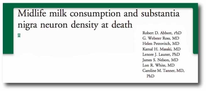
Title: Midlife milk consumption and substantia nigra neuron density at death
Authors: Abbott RD, Ross GW, Petrovitch H, Masaki KH, Launer LJ, Nelson JS, White LR, Tanner CM.
Journal: Neurology. 2016 Feb 9;86(6):512-9.
PMID: 26658906

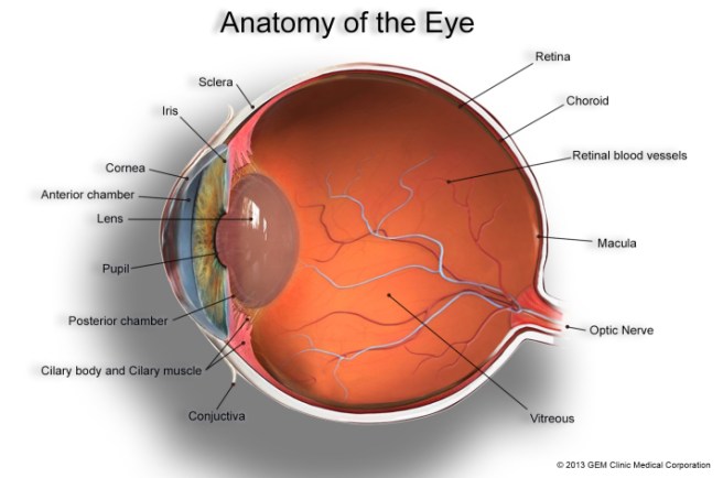
In this study, the researchers looked at the milk intake data for 449 men in the Honolulu Heart Study (which were collected from 1965 to 1968), and then conducted postmortem examinations of their brains (between 1992 to 2004). The researchers found that subjects who drank more than 2 cups of milk per day during their midlife years had approximately 40% fewer dopamine neurons (in certain areas of a region called the substantia nigra where the dopamine neurons live).

But here is the interesting twist in the story:

None of these 449 subjects were diagnosed with Parkinson’s disease

These were all neurologically normal/healthy individuals.

Plus this particular effect was only observed among the milk drinking, non-smokers. The milk drinking smokers did not have this cell loss (smoking is associated with a reduced risk of developing Parkinson’s disease – click here for more on this).

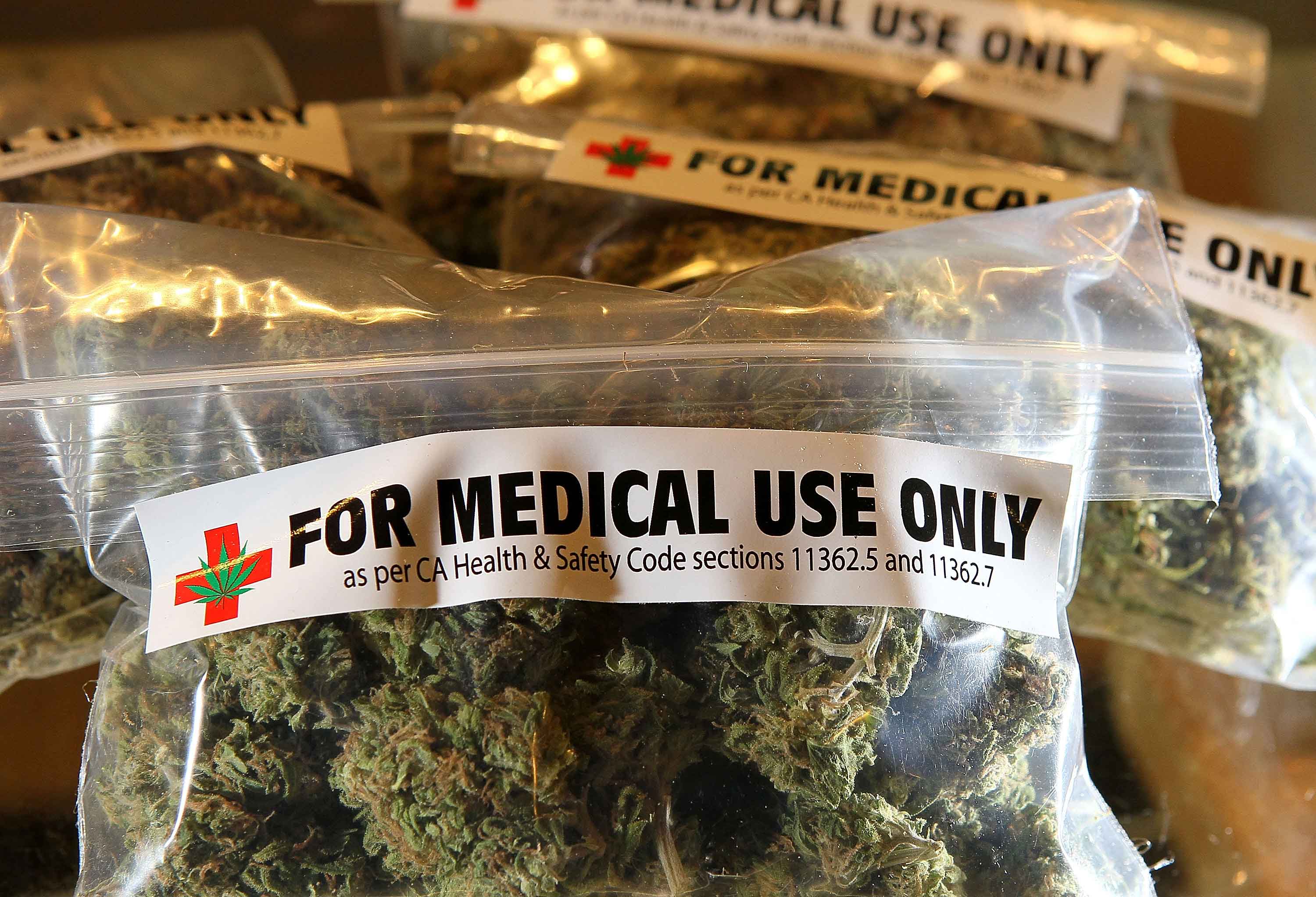
The researchers then took the study a step further. They  noticed that the cell loss effect was also associated with the presence of heptachlor epoxide in the brain.

What is heptac..whatever?

Heptachlor is an organochlorine compound that was used as an insecticide. Pesticides and insecticides have long been associated with increased risk of Parkinson’s disease (click here to read that post).

In this study, of the 116 brains analysed, heptachlor epoxide was found in 90% of the non-smokers who drank the most milk, but only 63% of those subjects who drank no milk. This lead the researchers to speculate as to whether contamination of milk by heptachlor epoxide could have caused the cell loss. We should point out here that this particular part of the analysis is a wee bit flimsy. The sample size for the non-smoking, high milk consumption group was very small: only 12 individuals.

So what does it all mean?

It means I am now dairy free.

EDITORIAL NOTE HERE: While we do not expect this post to crash the world wide milk market, we did not want to frighten any readers out of their habit of drinking milk. It should be noted here that the daily intake of milk associated with increased risk of Parkinson’s disease is very high (>16 oz/day or 1/2 a litre/day). Having said that lowering ones dairy intake does have many benefits for ones health.

In addition, in our last post, we discussed the microbiome of the gut and how the bacteria there could be influencing Parkinson’s disease. It would be interesting to see whether follow-up studies of that particular study highlight any insecticide/pesticide interactions with the bacteria of the gut.

One last thing: The purpose of today’s post was not to scare people out of drinking milk, but merely to throw a curious observation out there for people to think about. It will be interesting to hear what people think about this, especially any observations based on their own experience.


The banner for today’s post was sourced from AndFarAway

PAMs for Parkinson’s?

clinicaltrials

In today’s post we are going to review the results of a phase 1 trial for new kind of drug being oriented at Parkinson’s disease. The results were announced in late September, and they indicate that the drug was well tolerated by subjects taking part in the study.


4912prexton

Source: Prexton

Here at the Science of Parkinson’s disease we are always on the look out for novel drug therapies. Many of the treatments currently being tested in the clinic are simply different versions of L-dopa or a dopamine agonist.

So when Prexton Therapeutics recently  announced the results of their phase 1 clinical trial for their lead drug, PXT002331, we sat up and took notes. PXT002331 (formerly called DT1687) is the first drug of its kind to be tested in Parkinson’s disease.

It is a mGluR4 positive allosteric modulator.

What on earth is mGluR4 positive allosteric modulator?
The metabotropic glutamate receptors (mGluR) are an abundant family of receptors in the brain. Proteins bind to these receptors and activate (or block) an associated function. There are many different types of these receptors and mGluR4 is simply a small subset. The mGluR4s, however, are present in the areas affected by Parkinson’s disease, and this is why this particular family of receptors has been the focus of much research attention.

But what about the positive allosteric modulator part of ‘mGluR4 positive allosteric modulator’

Yes, good question.

This is the key part of this new approach. Allosteric modulators are a new class of orally available small molecule therapeutic agents. Traditionally, most marketed drugs bind directly to the same part of receptors that the body’s own natural occurring proteins attach to. This means that those drugs are competing with those endogenous proteins, thus limiting the potential effect of the drug.

Allosteric modulators get around this problem by binding different parts of the receptor. And instead of simply turning on or off the receptor, allosteric modulators can either turn up the volume of the signal being sent by the receptor or decrease the signals. This means that when the body’s naturally occurring protein binds in the receptor, allosteric modulators can either amplify the effect or reduce it depending on which type of allosteric modulators is being administered.

allosteric_modulation_mechanism

How Allosteric modulators work. Source: Addrex Thereapeutics

There are two different types of allosteric modulators: positive and negative. And as the label suggests, positive allosteric modulators (or PAMs) increase the signal from the receptor while negative allosteric modulators (or NAMs) reduce the signal. Thus, mGluR4 PAMS are amplifying the signal of the mGluR4 receptors.

Why do we want an amplification of a particular signal?

That is a hard question to answer.

Here’s the short explanation:

When you are planning to make a movement with your body, the process of actually initiating that movement begins in the cortex, specifically the primary motor cortex:

decisionmaking

A cross section of the human brain illustrating the primary motor cortex. Source: Droso4schools

The primary motor cortex receives information from other regions of the brain (such as the prefrontal cortex where you make a lot of your decisions), and it will then send a signal down into the brain and down the spinal cord telling the limbs to move. On the way down through the brain, the signal will pass through a series of check points that will filter the signal and determine the final strength of it.

basal-ganglia-dbs-figure-1

A schematic of the feedback loop of check points. Source: Parkinson’s Biology

EDITOR’S NOTE: We have borrowed this image from the Parkinson’s biology blog, which we are huge fans of. We highly recommend people visit that site as well as our lovely site. They also provide easy to understand explanations of the biology of Parkinson’s disease.

These checkpoints represent a large feedback loop. The critical step in this process is the processing being conducted in the basal ganglia, which can be broken down into different subregions:

basal-ganglia-dbs-figure-5

A schematic of the components of the basal ganglia. Source: Parkinson’s Biology

The globus pallidus (GPi) is the last area of the basal ganglia that the signal will pass through on it’s way to the thalamus (the ultimate decider of whether you will move or not), so if there is anything going wrong between these two structures the initiation of movement will be disrupted.

In a normal brain, the chemical dopamine is being produced in an area called the substantia nigra pars compacta (say that three times really fast). That dopamine is released in the striatum and other areas of the basal ganglia, and it has a mediating effect on the signal passing through these structures.

basal-ganglia-dbs-figure-3

A schematic of the source of dopamine. Source: Parkinson’s Biology

In Parkinson’s disease, however, the dopamine producing cells of the pars compacta are loss – 60% by the time a person starts to have the clinical motor features appearing. The loss of this dopamine leaves the whole system ‘unmediated’. The feedback loop becomes extremely inhibited, resulting in problems initiating movement.

Deep brain stimulation can un-inhibit the globus pallidus, by mediating the signal passing through that structure. But this requires surgery and the implanting of probes deep inside the brain.

basal-ganglia-dbs-figure-7

A schematic of deep brain stimulation of the globus pallidus. Source: Parkinson’s Biology (great website!)

A better way of reducing the inhibition in this feedback loop is the replacement of dopamine (which we do via the taking of treatments like L-dopa). This has been the standard approach for more than 50 years.

A new method of reducing the inhibition in the feedback loop would be to chemically targeting the globus pallidus, and this is what scientists are trying to do with the mGluR4 PAMS. By amplifying the signal of mGluR4s in the globus pallidus, the scientists believe that they can reduce the level of inhibition in the feedback loop.

The hope is that this approach is a less Parkinson’s disease-affected treatment. That is to say, the globus pallidus is structurally less affected by Parkinson’s disease than the substantia nigra pars compacta, and thus any treatment of the globus pallidus should be more stable over time (as the disease progresses).

That said, it is acknowledged that mGluR4 PAMS are NOT a potential cure for Parkinson’s disease, but rather a better way of treating the condition.

What research has been done on mGluR4 PAMS and Parkinson’s disease?

In August of 2003, some researchers at the pharmaceutical company Merck published a study which indicated that activation of mGluR4 could decrease the excessive levels of inhibition in the  globus pallidus.

jns

Title: Group III metabotropic glutamate receptor-mediated modulation of the striatopallidal synapse.
Authors: Valenti O, Marino MJ, Wittmann M, Lis E, DiLella AG, Kinney GG, Conn PJ.
Journal: Journal of Neuroscience. 2003 Aug 6;23(18):7218-26.
PMID: 12904482      (This article is OPEN ACCESS if you would like to read it)

The researchers found that an mGluR4 agonist (a protein that binds to the receptor directly, encouraging the associated action) reduced inhibitory signal being produced in the globus pallidus (through a presynaptic mechanism of action). They next demonstrated that the effect did not happen in mice which do not have mGluR4s, illustrating the specificity of the effect. They finished the study by injecting the mGluR4 agonist into a rodent model of Parkinson’s disease and found beneficial effects – that were equivalent to L-dopa.

Based on this research, the scientists at Merck next turned their attention to modulating the mGluR4s in the globus pallidus using allosteric modulators:

pnastitle

Title: Allosteric modulation of group III metabotropic glutamate receptor 4: a potential approach to Parkinson’s disease treatment.
Authors: Marino MJ, Williams DL Jr, O’Brien JA, Valenti O, McDonald TP, Clements MK, Wang R, DiLella AG, Hess JF, Kinney GG, Conn PJ.
Journal: Proc Natl Acad Sci U S A. 2003 Nov 11;100(23):13668-73.
PMID: 14593202           (This article is OPEN ACCESS if you would like to read it)

In this article, the same researchers introduce a positive allosteric modulator called ‘PHCCC’ which has a preference for binding to mGluR4. They found that when they put PHCCC – in combination with the mGluR4 agonist used in the previous study – onto cells in petri dishes, they got an amplification of the reduction in inhibition in the cells. Administered alone, PHCCC also produced a marked reversal of the motor deficit observed in a rodent model of Parkinson’s disease.

With these results, the scientists could begin building the justification for taking mGluR4 PAMs to the clinic. They were interested, however, in what impact mGluR4 PAMs could have on the involuntary motor problems associated with long-term L-dopa use, called dyskinesias (we have previously written about these – click here to read that post). So they decided to investigate whether mGluR4 PAMs may have an impact on dyskinesias:

dyskotitle

Title: Pharmacological stimulation of metabotropic glutamate receptor type 4 in a rat model of Parkinson’s disease and L-DOPA-induced dyskinesia: Comparison between a positive allosteric modulator and an orthosteric agonist.
Authors: Iderberg H, Maslava N, Thompson AD, Bubser M, Niswender CM, Hopkins CR, Lindsley CW, Conn PJ, Jones CK, Cenci MA.
Journal: Neuropharmacology. 2015 Aug;95:121-9.
PMID: 25749357          (This article is OPEN ACCESS if you would like to read it)

In this study, the investigators compared a mGluR4 PAM with a mGluR4 agonist (similar to that used in the previous studies) in rodent models of L-dopa induced dyskinesias. They found that the neither of the two drugs modified the development of dyskinetic behaviours, nor could they modify the behaviours when given together with L-dopa. In fact, when a low dose of L-dopa was given to the animals (resulting in only mild dyskinesias), the researchers found that by adding mGluR4 PAM the dyskinetic behaviours became more exaggerated. The investigators concluded that stimulation of mGluR4 does not have anti-dyskinetic activity. This is an important characteristic to determine before taking a drug to the clinic for Parkinson’s disease.

So what were the results of the phase 1 clinical trial?

In July of 2012, Merck spun off the research into a new company called Prexton Therapeutics. The company almost immediately started setting up a phase 1 safety clinical trial for its lead compound, the mGluR4 PAM: PXT002331. A total of 64 healthy volunteers were enrolled to evaluate the safety and tolerability of several different doses of orally taken PXT002331. The study was completed on time and demonstrated that PXT002331 is safe and well tolerated (at doses well above those that produce robust effects in Parkinson’s disease animal models).

Very positive news.

The planning of a phase 2 clinical trial in people with Parkinson’s disease is now underway. It will take place in the first half of 2017, and this study will provide the first indications as to whether this new treatment approach will be effective in human at treating the features of Parkinson’s disease. We will keep you posted on the success of that study when the results become available.

Are other biotech companies using this approach?

Yes, PAM-based therapies for Parkinson’s disease are very much in vogue at the moment.

Just this month, the biotech company Asceneuron received a grant from The Michael J. Fox Foundation for Parkinson’s Research for the development of positive allosteric modulators of the M1 muscarinic acetylcholine receptor (M1 PAMs). So we can hopefully expect more from this approach to therapies.

Interesting times. And hopefully positive results to come.

EDITOR’S NOTE: It is important to remember that any clinical trial research discussed on this blog is of an educational nature. Nothing written here can or should be mistaken as medical advice. All of these drugs are still experimental and require extensive testing before being offered to the general population. Please speak with a certified clinician before attempting any change to your current medical treatment regime.


The image used in the banner of today’s post was sourced from MedTechBoston

Cannabis and Parkinson’s disease

medimarijuana

This is the kind of post that can really get someone in quite a bit of trouble.

Both the legal kind of trouble and the social media type of trouble.

Given the online excitement surrounding a particular video that appeared on the internet last week, however, we thought that it would be useful to have a look at the research that has been done on the medicinal use of Cannabis and Parkinson’s disease.

In addition, we will assess the legal status regarding the medicinal use of Cannabis (in the UK at least).


medicinal

Cannabis being grown for medicinal use. Source: BusinessWire

This week a video appeared online that caused a bit of interest (and hopefully not too many arrests) in the Parkinson’s community.

Here is the video in question:

The video was posted by Ian Frizell, a 55 year old man with early onset Parkinson’s disease. He has recently had deep brain stimulation (DBS) surgery to help control his tremors and he has also posted a video regarding that DBS surgery which people might find useful (Click here to see this).

In the video, Ian turns off his DBS stimulator and his tremors quickly become apparent. He then ‘self medicates’ with cannabis off camera and begins filming again some 20-30 minutes later to show the difference. The change with regards to his tremor are very clear and quite striking.

Here at the SoPD, we find the video very interesting, but we have two immediate questions:

  1. How is this reduction in tremors working?
  2. Would everyone experience the same effect?

We have previously seen many miraculous treatments online (such as coloured glasses controlling dyskinesias video from a few years ago) which have failed when tested under controlled conditions (the coloured glasses did not elicit any effect in the clinical setting – click here to read more). Some of these amazing results can simply be put down to the notorious placebo effect (we have previously discussed this in relation to Parkinson’s disease – click here to read the post), while others may vary on a person to person basis.

Thus, while we applaud Mr Frizell for sharing his finding with the Parkinson’s community, we are weary that the effect may not be applicable to everyone. For this reason, we have made a review of the scientific literature surrounding Cannabis and Parkinson’s disease.

But first:

What exactly is Cannabis?

cannabis

Drawings of the Hemp plant, from  Franz Eugen Köhler’s ‘Medizinal-Pflantzen’. Source: Wikipedia

Cannabis (also known as marijuana) is a family of flowering plants that can be found in three types: sativa, indica, and ruderalis. Cannabis is widely used as a recreational drug, behind only alcohol, caffeine and tobacco in its usage. It typically consumed as dried flower buds (marijuana), as a resin (hashish), or as various extracts which are collectively known as hashish oil.

While the three varieties of cannabis (sativa, indica, and ruderalis) may look very similar, pharmacologically they have very different properties. Cannabis sativa is often reported to cause a “spacey” or heady feeling, while Cannabis indica causes more of a “body high”.  Cannabis ruderalis, by contrast, is less well used due to its low Tetrahydrocannabinol levels.

What is Tetrahydrocannabinol?

thc-structure

Tetrahydrocannabinol (or THC) is one of the principle psychoactive components in Cannabis. It a chemical that is believed to be a plant defensive mechanism against herbivores. THC is a cannabinoid, a type of chemical that attaches to the cannabinoid receptors in the body, and it is this pathway that many scientists are exploring for future neuroprotective therapies for Parkinson’s disease (For a good review on the potential cannabinoid-based therapies for Parkinson’s disease, click here).

A second type of cannabinoid is Cannabidiol (or CBD). CBD is considered to have a wider scope for potential medical applications. This is largely due to clinical reports suggesting reduced side effects compared to THC, in particular a lack of psychoactivity.

So what research has been done regarding Cannabis and Parkinson’s disease?

In 2004, a group of scientists in Prague (Czech Republic) were curious to determine cannabis use in people with Parkinson’s disease, so they conducted a study and published their results:

survey

Title: Survey on cannabis use in Parkinson’s disease: subjective improvement of motor symptoms.
Authors: Venderová K, Růzicka E, Vorísek V, Visnovský P.
Journal: Mov Disord. 2004 Sep;19(9):1102-6.
PMID: 15372606

The researchers posted out 630 questionnaires to people with Parkinson’s disease in Prague.  In total, 339 (53.8%) completed questionnaires were returned to them. Of these, 85 people reported Cannabis use (25.1% of returned questionnaires). They usually consumed it with meals (43.5%), and most of them were taking it once a day (52.9%).

After consuming cannabis, 39 responders (45.9%) described mild or substantial alleviation of their Parkinson’s symptoms in general, 26 (30.6%) improvement of rest tremor, 38 (44.7%) alleviation of rigidity (bradykinesia), 32 (37.7%) alleviation of muscle rigidity, and 12 (14.1%) improvement of L-dopa-induced dyskinesias.

Importantly, half of the people who consumed cannabis experience no effect on their Parkinson’s disease features, and four responders (4.7%) reported that cannabis actually worsened their symptoms. So while this survey suggested some positive effects of cannabis in the treatment of Parkinson’s disease, it is apparent that the effect is different between people.

Additional surveys have been conducted around the world, with similar results (Click here to read more on this).

Have there been any clinical trials?

Yes, there have.

In the 1990s, there was a very small clinical study of cannabis use as a treatment option for Parkinson’s disease, and this study failed to demonstrate any positive outcome. In the study, none of the 5 people with Parkinson’s disease experienced any effect on their Parkinson’s motor features after a week of smoking cannabis (click here for more on this).

This study was followed up by a larger study:

cannabistitle

Title: Cannabis for dyskinesia in Parkinson disease: a randomized double-blind crossover study.
Authors: Carroll CB, Bain PG, Teare L, Liu X, Joint C, Wroath C, Parkin SG, Fox P, Wright D, Hobart J, Zajicek JP.
Journal: Neurology. 2004 Oct 12;63(7):1245-50.
PMID: 15477546

In this randomized, double-blind, placebo-controlled study, 19 people with Parkinson’s disease randomly received either oral cannabis extract or a placebo (twice daily) for 4 weeks. They then took no treatment for an intervening 2-week ‘washout’ period, before they were given the opposite treatment for 4 weeks (so if they received the cannabis extract during the first 4 weeks, they would be given the placebo during the second 4 weeks). In all cases, the participants and the researchers were ‘blind’ to (unaware of) which treatment was being given.

The results indicated that cannabis was well tolerated by all of the participants in the study, but that it had no pro- or anti-Parkinsonian actions. The researchers found no evidence for a treatment effect on levodopa-induced dyskinesia.

In addition to this study, there has been a recent double-blind clinical study of cannabidiol (CBD, mentioned above) in the treatment of Parkinson’s disease:

cbd

Title: Effects of cannabidiol in the treatment of patients with Parkinson’s disease: an exploratory double-blind trial.
Authors: Chagas MH, Zuardi AW, Tumas V, Pena-Pereira MA, Sobreira ET, Bergamaschi MM, dos Santos AC, Teixeira AL, Hallak JE, Crippa JA.
Journal: J Psychopharmacol. 2014 Nov;28(11):1088-98.
PMID: 25237116

The Brazilian researchers who conducted the study took 21 people with Parkinson’s disease and assigned them to one of three groups which were treated with placebo, small dose of CBD (75 mg/day) or high dose of CBD (300 mg/day). They found that there was no positive effects by administering CBD to people with Parkinson’s disease, except in their self-reported measures on ‘quality of life’.

So what does all of this mean?

Firstly, let us be clear that we are not trying to discredit Mr Frizell or suggest that what he is experiencing is not a real effect. The video he has uploaded suggests that he is experiencing very positive benefits by consuming cannabis to help treat his tremors.

Having said that, based on the studies we have reviewed above we (here at the SoPD) have to conclude that the clinical evidence supporting the idea of cannabis as a treatment for Parkinson’s disease is inconclusive. There does appear to be some individuals (like Mr Frizell) who may experience some positive outcomes by consuming the drug, but there are also individuals for whom cannabis has no effect.

One of the reasons that cannabis may not be having an effect on everyone with Parkinson’s disease is that many people with Parkinson’s disease actually have a reduction in the cannabis receptors in the brain (click here for more on this). This reduction is believed to be due to the course of the disease. If there are less receptors for cannabis to bind to, there will be less effect of the drug.

Ok, but how might cannabis be having a positive effect on the guy in the video?

Cannabis is known to cause the release of dopamine in the brain – the chemical classically associated with Parkinson’s disease (Click here and here for more on this). Thus the positive effects that Mr Frizell is experiencing may simply be the result of more dopamine in his brain, similar to taking an L-dopa tablet. Whether enough dopamine is being released to explain the full effect is questionable, but this is still one possible explanation.

There could be questions regarding the long term benefits of Mr Frizell’s cannabis use, as long term users of cannabis generally have reduced levels of dopamine being released in the brain (Click here for more on this). Although the drug initially causes higher levels of dopamine to be released, over time (with long term use) the levels of dopamine in the brain gradually reduce.

I live in the UK. Is it legal for me to try using Cannabis for my Parkinson’s disease?

legality_of_cannabis_for_medical_purposes_new

National status on Cannabis possession for medical purposes. Source: Wikipedia

The map above is incorrect, with regards to the UK at least (and may be incorrect for other regions as well).

According to the Home Office, it is illegal for UK residents to possess cannabis in any form (including medicinal).

Cannabis is illegal to possess, grow, distribute or sell in the UK without the appropriate licences. It is a Class B drug, which carries penalties for unlicensed dealing, unlicensed production and unlicensed trafficking of up to 14 years in prison (Source: Wikipedia; and if you don’t trust Wikipedia, here is the official UK Government website).

In 1999, a major House of Lords inquiry made the recommendation that cannabis should be made available with a doctor’s prescription. The government of the U.K., however, has not accepted the recommendations. Cannabis is not recognised as having any therapeutic value under the law in England and Wales.

Having said all of this, there has recently been an all-party group calls for the legalisation regarding cannabis for medicinal uses to be changed (click here for more on this). Whether this will happen is yet to be seen.

So the answer is “No, you are not allowed to use cannabis to treat your Parkinson’s disease”.

Except…

(And here is where things get a really grey)

There is a cannabis-based product – Sativex – which can be legally prescribed and supplied under special circumstances. Sativex is a mouth spray developed and manufactured by GW Pharmaceuticals in the UK. It is derived from two strains of cannabis leaf and flower, cultivated for their controlled proportions of the active compounds
THC and CBD.

In 2006, the Home Office licensed Sativex so that:

  • Doctors could privately prescribe it (at their own risk)
  • Pharmacists could possess and dispense it
  • Named patients with a prescription could possess

In June 2010 the Medicines Healthcare Regulatory products Agency (MHRA) authorised Sativex as an extra treatment for patients with spasticity due to Multiple Sclerosis (MS). Importantly, doctors can also prescribe it for other things outside of the authorisation, but (again) this is at their own risk.


EDITORIAL NOTE: Given that possessing cannabis is illegal and that more research into the medicinal benefits of cannabis for Parkinson’s disease is required, we here are the SoPD can not endorse the use of cannabis for treating Parkinson’s disease. 

While we are deeply sympathetic to the needs of many individuals within the Parkinson’s community and agree with a reconsideration of the laws surrounding the medicinal use of cannabis, we are also aware of the negative consequences of cannabis use (which can differ from person to person).

If a person with Parkinson’s disease is considering a change in their treatment regime for any reason, we must insist that they first discuss the matter with their trained medical physician before undertaking any changes.

The information provided here is strictly for educational purposes only.


The banner for today’s post was sourced from the IBTimes.

Is there something in my eye?

SashaEyeball

Some people say that the eyes are the gateway to the soul.

Maybe. I don’t know. Poetic stuff though.

Research published recently, however, suggests that the eyes may also provide a useful aid in the diagnosis of Parkinson’s disease. In today’s post we will review what results have been published and try to understand what they mean for our understanding of this condition.


N00156_H

A schematic of the human eyeball. Source: NIDDK image library

The fact that you can see and read this page is a miraculous thing.

Amazing not just because light is entering your eye, being focused on a particular point in the back of the eyeball and then being turned into a signal that is transmitted to your brain for further analysis, but also because of all the other activities involved with sight. The muscle movements, for example, which are required for turning the eyeball the small fractions necessary for reading this sentence from left to right.

And then there is also the blood supply, keeping the whole system working. This feature is of particular interest to today’s post, as research published last week suggests that there are differences in the blood flow of the eyeball between people with and without Parkinson’s disease.

The anatomy of an eyeball

The human eyeball is – on the macro level – a fairly simple structure.

You have the Iris, which regulates the amount of light entering the eye. At the centre of the iris, you have a central opening called the pupil, which can dilate and constrict as required. Covering these is the cornea, a transparent circular skin. These structures all sit over the lens which helps to refract incoming light and focus it onto the retina. And the retina, of course, is the light sensitive layer that lines the interior of the eye – allowing us to see.

Anatomy

The anatomy of the eye. Source: GemClinic

Within the retina are specialised cells of two sorts:

  • Rod cells (about 125 million of them per eye) which are necessary for seeing in dim light.
  • Cone cells (6-7 million of these) which can be further divided into three types, each sensitive to different primary colours – red, green or blue.

These specialised ‘photoreceptive’ cells send signals down through the layers of the retina to what are called retinal ganglion cells which are the key conduits in the sending of information to the brain.

All of these cells require a constant blood supply, from arteries and veins spreading across the retina, and this a key part of our discussion today (see below).

So what have eyeballs got to do with Parkinson’s disease?

Good question. People with Parkinson’s disease often complain of from visual issues, such as reduced visual acuity, low contrast sensitivity and disturbed colour vision.

And there has been some research into the eyes with regards to Parkinson’s disease. A few weeks ago, this particular study was published:

Eyes1

Title: The retina as an early biomarker of neurodegeneration in a rotenone-induced model of Parkinson’s disease: evidence for a neuroprotective effect of rosiglitazone in the eye and brain.
Authors: Normando EM, Davis BM, De Groef L, Nizari S, Turner LA, Ravindran N, Pahlitzsch M, Brenton J, Malaguarnera G, Guo L, Somavarapu S, Cordeiro MF.
Journal: Acta Neuropathol Commun. 2016 Aug 18;4(1):86. doi: 10.1186/s40478-016-0346-z.
PMID: 27535749       (This article is OPEN ACCESS if you would like to read it)

The researchers in this study used a rodent model of Parkinson’s disease (rotenone-induced). In this model, the animals started losing dopamine cell loss in the brain at 60 days after the model of Parkinson’s disease was chemically induced.

The scientists examined the eyes of the rats at 10, 20, 40 and 60 days of the study. At the 20 day time point, the researchers began to see increased retinal ganglion cell death and swelling of the retinal layers in the eyes. These changes were obviously occurring well before the cell loss is observed in the brain, which leads the authors to ask whether the eyes could potentially used as an early indicator of Parkinson’s disease.

Of particular interest in this study was the use of Rosiglitazone to protect the retinal cells (AND the dopamine neurons in this rodent model of Parkinson’s disease). Rosiglitazone is an anti-diabetic drug. It works as an insulin sensitizer, by binding to fat cells and making them more responsive to insulin (we have previously discussed the curious relationship between Parkinson’s disease and diabetes (click here for more on this), and this result reinforces that connection). The scientists found that giving the drug once every 3 days had very beneficial effects of the survival of the retinal cells. They also observed significant neuroprotection after delaying the treatment for 10 days and then just giving one round of treatment, suggesting that a lot of the drug is not required for positive results.


EDITORIAL NOTE HERE: Before readers start to get any crazy ideas about sourcing and self medicating with Rosiglitazone, it is important to note that there are serious side effects associated with this class of drug. It has been associated with heart disease and stroke (click here to read more), and it should only be taken by people with diabetes and under the strict supervision of a qualified physician. It it mentioned here purely for educational purposes.


So obviously what is required is an examination of the eyes of people with Parkinson’s disease

Yep. And conveniently, in the same week as the previous study came out, this second study was also published:

Eyes2

Title: Evaluation of Retinal Vessel Morphology in Patients with Parkinson’s Disease Using Optical Coherence Tomography.
Authors: Kromer R, Buhmann C, Hidding U, Keserü M, Keserü D, Hassenstein A, Stemplewitz B.
Journal: PLoS One. 2016 Aug 15;11(8):e0161136.
PMID: 27525728          (This article is OPEN ACCESS if you would like to read it)

The researchers examined 49 people with Parkinson’s disease and 49 age- and sex-matched healthy controls. Blood vessels within the retina were identified and then divided into arteries and veins, based on their shape (using computer software). The results of the study indicate significant differences in the morphology of retinal veins in people with Parkinson’s disease when compared to controls.

Interestingly, the retinal effect was more significant on the side of the body firstly affected by Parkinson’s disease (a very common feature of Parkinson’s is that initially the condition will affect one side of the body more than the other).

What does it all mean?

For generations, we have focused on the clinical motor features of Parkinson’s disease (slowness, rigidity, and a resting tremor) when trying to determine if someone has the condition. Now we are learning that there may be other parts of the body that we should be investigating, which could not only provide us with novel diagnostic tools for earlier detection of the disease, but those areas may also provide us with new insights into disease onset and spread as well.

I may be getting a bit ahead of myself here but the possibilities are exciting and we’ll keep you abreast of these new findings as they come to us.


The banner for today’s post was sourced from the Photoforum.

Juvenile-onset Parkinson’s disease

xTgKydA8c

A community in New Brunswick (Canada) was recently shocked to discover that a 2 year old boy in their midst had been diagnosed with Parkinson’s disease (Click here to read more).

Yes, you read that correctly, it’s not a typo: a 2 year old boy.

Juvenile-onset Parkinson’s disease is an extremely rare version of the condition we discuss here at the Science of Parkinson’s. It is loosely defined as being ‘diagnosed with Parkinson’s disease under the age of 20’. The prevalence is unknown, but there is a strong genetic component to form of the condition. In today’s post we will review what is known about Juvenile-onset and look at new research about a gene that has recently been discovered to cause a type of Juvenile-onset Parkinson’s disease.


Henri_Huchard

Dr Henri Huchard. Source: Wikipedia

In 1875, Dr Henri Huchard (1844-1910; a French neurologist and cardiologist) described the first case of a child who, at just 3 years of age, presented all the clinical features of Parkinson’s disease. Since that report, there have been many studies detailing the condition that has become known as ‘juvenile-onset Parkinson’s disease’.

What is juvenile-onset Parkinson’s disease?

Basically, it is a form of Parkinson’s disease that affects children and young people under the age of 20. The defining feature is the age of onset. The average age of onset is approximately 12 years of age (with the majority of cases falling between 7 and 16 years) and males are affected by this condition more than females (at a rate of approximately 5:1).

The actual frequency of juvenile-onset parkinson’s is unknown given how rare it is. When researcher look at people with early onset Parkinson’s disease (that is diagnosis before the age of 40; approximately 5% of the Parkinson’s community), they have found that between 0.5 – 5% of that group of people were diagnosed before 20 years of age. This suggests that within just the Parkinson’s community, the frequency of juvenile-onset parkinson’s is at the most 0.25% (or 2.5 people per 1000 people with Parkinson’s). Thus it is obviously a very rare condition.

It is interesting to note that Lewy bodies (the clusters of aggregated protein that classically characterise the brains of people with Parkinson’s disease) are very rare in cases of juvenile-onset parkinson’s disease. To our knowledge there has been only one case of Lewy bodies in juvenile-onset parkinson’s disease (Click here to read more on this). This suggests that the juvenile-onset form of Parkinson’s disease may differ from other forms of the condition in its underlying biology.

Do we know what causes juvenile-onset parkinson’s disease?

There is a very strong genetic component to juvenile-onset parkinson’s disease. In fact, the incidence of Parkinsonism in relatives of people with juvenile-onset parkinson’s disease is higher than in the general public AND in the relatives of people with other forms of Parkinson’s disease.

Genetic mutations in three genes are recognised as causing juvenile-onset Parkinson’s disease. The three genes are known to the Parkinson’s world as they are all PARK genes (genetic variations that are associated with Parkinson’s). Those three genes are:

  • Parkin (PARK2)
  • PTEN-induced putative kinase 1 (PINK1 or PARK6)
  • DJ1 (PARK7)

In juvenile-onset Parkinson’s disease, all of these mutations are associated with autosomal recessive – meaning that two copies of the genetic variation must be present in order for the disease to develop.

Parkin mutations account for the majority of juvenile-onset Parkinson’s disease cases. Affected individuals have a slowly progressing condition that is L-dopa responsive. Dystonia (abnormal muscle tone resulting in muscular spasm and abnormal posture) is very common at the onset of the condition, particularly in the lower limbs.

Can the condition be treated with L-dopa?

The answer is: ‘Yes, but…’

L-dopa (or dopamine replacement) treatment is the standard therapy for alleviating the motor features of Parkinson’s disease.

The majority of people with juvenile-onset parkinson’s respond well to L-dopa, but in the Parkin mutation version individuals will typically begin to experience L-dopa-induced motor fluctuations (dyskinesias) early in that treatment regime.

What research is currently being done on this condition?

Given that cases are so very rare and so few, it is difficult to conduct research on this population of individuals. Most of the research that is being conducted is focused on the genetics underlying the condition.

And recent that research lead to the discovery of a new genetic variation that causes juvenile-onset Parkinson’s disease:

Juvenile

Title: Discovery of a frameshift mutation in podocalyxin-like (PODXL) gene, coding for a neural adhesion molecule, as causal for autosomal-recessive juvenile Parkinsonism.
Authors: Sudhaman S, Prasad K, Behari M, Muthane UB, Juyal RC, Thelma BK.
Journal: Journal Med Genet. 2016 Jul;53(7):450-6.
PMID: 26864383           (This article is OPEN ACCESS if you would like to read it)

The researchers who wrote this article were presented with a 10 member Indian family from Aligarh, Uttar Pradesh. Of the 8 children in the  family, 3 were affected by Parkinsonian features (tremor, slowness, rigidity and gait problems) that began between 13 and 17 years of age. The researchers conducted DNA sequencing and found that none of the three affected siblings had any of the known Juvenile-onset Parkinson’s disease genetic mutations (specifically, mutations in the genes PARK2, PINK1and DJ1).

They then compared the DNA from the three siblings with the rest of the family and found a genetic variant in a gene called podocalyxin-like (or PODXL). It must be noted that PODXL is a completely novel gene in the world of Parkinson’s disease research, which makes it very interesting. PODXL has never previously been associated with any kind of Parkinson’s disease, though it has been connected with two types of cancer (embryonal carcinoma and periampullary adenocarcinoma).

The researchers then turned to their genetic database of 280 people with Parkinson’s disease have had their genomes sequenced. The researchers wanted to determine if any genetic variants in the PODXL gene were present in other suffers of Parkinson’s disease, but had not been picked up as a major contributing factor. They found three unrelated people with PODXL mutations. All three had classical Parkinson’s features, and were negative for mutations in the Parkin, PINK1 and DJ1 genes.

The researchers concluded that the PODXL gene may be considered as a fourth causal gene for Juvenile-onset Parkinson’s disease, but they indicated that further investigations in other ethnic groups are required.


The banner for today’s post was sourced from ClipArtBest

Editorial note

Cambridge University applications

The scientists behind the Science of Parkinson’s disease website work at Cambridge University and are associated – through their research – with the Wellcome Trust/MRC Cambridge Stem cell institute.

sci-logo

Throughout July, the Stem cell institute is running the #MyView campaign which aims to raise awareness about all of the latest developments in stem cell research.

Of particular interest to the Parkinson’s community will be the work being conducted in Prof Roger Barker’s lab (he of the 2016 Gretschen Amphlet Memorial lecture). The Stem cell institute made a video about the research being conducted in Prof Barker’s lab – viewed through the eyes of someone with Parkinson’s disease. It provides an interesting view of the working being carried out:

We encourage all of our readers to get involved with the #myview discussion and to follow the campaign of social media via Youtube, Facebook, & Twitter.

As scientists we are always very keen to hear the views of people in the Parkinson’s community (both sufferers and carers). It is through campaigns like this that we can gain new insight from different view points.


The banner for today’s post was sourced from the Huffington Post.

Nilotinib and Parkinson’s disease – an update

2000px-Nilotinib.svg

We have previously discussed news briefings regarding a cancer drug that displayed interesting results in a pilot clinical study of Parkinson’s disease (click here to read that post). Today we will delve more deeply into the results of that particular study and consider what they mean.


DSK_4634s

Nilotinib (Tasigna) from Novartis. Source: William-Jon

In October of last year, at the Society for Neuroscience meeting in Chicago, a presentation of data from a clinical trial got the Parkinson’s community really excited. The study was investigating the effects of a cancer drug called ‘Nilotinib’ (also known as Tasigna) on Parkinson’s disease and the initial results were rather interesting.

The results of the pilot clinical study for Nilotinib were published today in the Journal of Parkinson’s disease:

Nilo-title

Title: Nilotinib Effects in Parkinson’s disease and Dementia with Lewy bodies
Authors: Pagan F, Hebron M, Valadez E, Tores-Yaghi Y,Huang X, Mills R, Wilmarth B, Howard H, Dunn C, Carlson A, Lawler A, Rogers S, Falconer R, Ahn J, Li Z, & Moussa C.
Journal: Journal of Parkinson’s Disease, vol. Preprint
PMID: Yet to be allocated              (This article is OPEN ACCESS if you would like to read it).

The study was setup to determine safety of using Nilotinib in Parkinson’s disease dementia or dementia with Lewy bodies.

What is Nilotinib?

Nilotinib is a drug that can be used to treat a type of leukemia when the other cancer drugs have failed. It was approved for this treating cancer by the FDA in 2007.

The researchers behind the current study believe that Nilotinib works by turning on autophagy – the “garbage disposal machinery” inside brain cells. Autophagy is a process that clears waste and toxic proteins from inside cells, preventing them from accumulating and possibly causing the death of the cell.

Print

The process of autophagy – Source: Wormbook

Waste material inside a cell is collected in membranes that form sacs (called vesicles). These vesicles then bind to another sac (called a lysosome) which contains enzymes that will breakdown and degrade the waste material.

The researchers suggest that Nilotinib may be working in Parkinson’s disease by helping affected cells to better clear away the build up of unnecessary proteins, which helps cells to function more efficiently.

What happened in the clinical study?

Twelve people with either Parkinson’s disease dementia or dementia with Lewy bodies were randomized given either 150 mg (n = 5) or 300 mg (n = 7) daily doses of Nilotinib for 24 weeks. After the treatment period the subjects were followed up for 12 weeks. All of the subjects were considered to have mid to late stage Parkinson’s features (Hoehn and Yahr stage 3–5). One subject was withdrawn from the study at week 4 due to a heart attack and another discontinued at 5 months due to unrelated circumstances.

An important question in the study was whether Nilotinib could actually enter the brain. Various tests conducted on the subjects suggesting that the drug had no problem crossing the ‘blood brain barrier‘ and having an effect in the brain. The levels of Nilotinib in the brain peaked at 2 hrs after taking the drug and the levels of the target protein (called p-Abl) were reduced by 30% at 1 hr. This level of activity remained stable for several hours.

The motor features of Parkinson’s disease were assessed using the Unified Parkinson’s Disease Rating Scale (UPDRS) and the investigators observed an average decrease of 3.4 points and 3.6 points at six months (week 24) compared to the baseline measures (scores from the start of the study) with 150 mg and 300 mg Nilotinib, respectively. A decrease in motor scores represent a reduction in Parkinson’s motor features.

The really remarkable result, however, comes from the testing of cognitive performance, which was monitored with Mini Mental Status Examination (MMSE). The researchers report an average increase of 3.85 and 3.5 points in MMSE at six months (24-week) compared to baseline, for 150 mg and 300 mg of Nilotinib, respectively. This means that the mental processing of the subjects improved across the study.

The motor and cognitive results were complemented by measures of proteins in blood and cerebrospinal fluid samples taken from the subjects. The researchers saw increases in dopamine related proteins (suggesting that more dopamine was present in the brain) and stabilization of alpha synuclein levels.

The researchers concluded that these observations warrant a larger randomized, double-blind, placebo-controlled trial to truly evaluate the safety and efficacy of Nilotinib.

Here at the SoPD, we are inclined to agree.

So what does all this mean?

The results of the study are very interesting, and the researchers should be congratulated on the outcome (and presentation of all the data in the report). As they themselves acknowledge, the study was open labelled – meaning that everyone in the study knew that they were getting the treatment – so the placebo effect could be at play here.

One intriguing note in the report was that most of the participants in the study ‘experienced increased psychotic symptoms (hallucination, paranoia, agitation) and some dyskinesia whilst on Nilotinib’ suggesting an increase in dopamine levels in the brain.

Obviously a larger, double-blind study is required to determine whether the effect of the drug in Parkinson’s disease is real. The Michael J. Fox Foundation, the Van Andel Research Institute (Michigan, USA) and the Cure Parkinson’s Trust are collaborating on the development program for a double-blind, placebo-controlled clinical trial of nilotinib, which it is hoped will begin in 2017.


The banner for today’s banner was sourced from Wikimedia 

A change of dogma for Alzheimer’s disease?

plaque-neuron-connection-loss-cropped

This week an interesting new study dealing with the biology of Alzheimer’s was published in the journal Science Translational Medicine. It has drawn a lot of attention as it may be turning our understanding of Alzheimer’s disease on it’s head. If the results are independently replicated and verified, it could potentially have major implications for Parkinson’s disease.


For the last 30 years, a protein called beta-amyloid has been considered one of the bad boys of the most common neurodegenerative condition, Alzheimer’s disease.

What is Alzheimer’s disease?

Alzheimer’s disease is a progressive neurodegenerative condition that can occur in middle or old age. It involves a generalized degeneration of the brain, not localised to specific regions like Parkinson’s disease.

What happens in the Alzheimer’s brain?

In the brain, in addition to cellular loss, Alzheimer’s is characterised by the presence of two features:

  • Neurofibrillary tangles
  • Amyloid plaques

The tangles are aggregations of a protein called ‘Tau’ (we’ll comeback to Tau in a future post). These tangles reside within neurons initially, but as the disease progresses the tangles can be found in the space between cells – believed to be the last remains of a dying cell.

F1.large

A normal brain vs an Alzheimer’s affected brain. Source: MMCNeuro

Amyloid plaques are clusters of proteins that sit between cells. A key component of the plaque is beta amyloid. Beta-amyloid is a piece of a larger protein that sits in the outer wall of nerve cells where it has certain functions. In certain circumstances, specific enzymes can cut it off and it floats away.

Amyloid-plaque_formation-big

Beta-Amyloid. Source: Wikimedia

Beta-amyloid is a very “sticky” protein and for a long time it has been believed that free floating beta-amyloid proteins begin sticking together, gradually building up into the large amyloid plaques. And these large plaques were considered to be involved in the neurodegenerative process of Alzheimer’s disease.

So what was discovered this week?

This week a study was published that suggests a new (and positive) function for beta amyloid:

BetaAm

Title: Amyloid-β peptide protects against microbial infection in mouse and worm models of Alzheimer’s disease.
Authors: Kumar DK, Choi SH, Washicosky KJ, Eimer WA, Tucker S, Ghofrani J, Lefkowitz A, McColl G, Goldstein LE, Tanzi RE, Moir RD.
Journal: Sci Transl Med. 2016 May 25;8(340):340ra72.
PMID: 27225182

The researchers took three types of mice:

  • genetically normal mice
  • mice with no beta amyloid
  • mice producing a lot of beta amyloid

They infected all of the mice with the microbe that causes meningitis, and they found that the mice producing a lot of beta amyloid lived significantly longer than other groups of mice. They then repeated the experiment in a species of microscopic worm – called C.elegans – and found similar results. These findings suggested that beta amyloid was having a positive effect in the brain.

But then they noticed something strange.

The mice producing a lot of beta amyloid usually do not develop a lot of protein aggregation until old age, but when the researchers looked in the brains of the mice they infected with meningitis, they found significant levels of aggregation in the mice producing a lot of beta amyloid but at a young age..

This led the researchers to conduct some cell culture experiments in which they watched what was happening to the bacteria and beta amyloid. They found that the beta amyloid was sticking to the bacteria and this was leading to the formation of protein aggregates.

The results of these experiments suggested to the researchers an intriguing possibility that beta amyloid may be playing a protective in the brain – acting as an immune system for the brain – against infection.

Thus the aggregations we see in the brains of people with Alzheimer’s may not be the cause of the cell death associated with the disease, but rather evidence of the ‘brain’s immune system’ trying to fight back against unknown infectious agents. The researcher’s of the study were quick to point out that this antimicrobial action of beta amyloid is simply a new function of the protein, and it may have nothing to do with the disease itself. But it will be interesting to see where this research goes next.

What has this got to do with Parkinson’s disease?

Parkinson’s disease is only definitive diagnosed at the postmortem stage. This is done by microscopic examination of the brain. In the brains of people with Parkinson’s disease, there are protein aggregates calls Lewy bodies. These are densely packed clusters of a protein called ‘alpha synuclein‘.

Fig2_v1c

The brown spot is a Lewy body inside of a brain cell. Source: Cure Dementia

If the results of the study presented above are correct and beta amyloid is a protective protein in the brain against infection, could it not be that alpha synuclein may be playing a similar role? It is a fascinating idea that it will be interesting to test.

What are the implications of the study?

Currently, there are numerous clinical trials for Alzheimer’s disease, involving treatments that act against beta amyloid. If the study presented above is correct, and beta amyloid has a role in protecting the brain, these new treatments in clinical trial may actually be weakening the brain’s ability to fight infection.

Similarly, if alpha synuclein is found to exhibit ‘protective’ properties like beta amyloid, then the alpha synuclein vaccine clinical trials currently underway (in which the body’s immune system is primed to remove free floating alpha synuclein, in an attempt to stop the disease from spreading) may need to be reconsidered. At a minimum, investigations into whether alpha synuclein has antimicrobial properties need to be conducted.


Today’s banner was sourced from PBS.

The debate surrounding a new Stem cell transplantation trial for Parkinson’s

Deep-Brain-Stimulation-60pghfsukanm4j4bljb8mbq9hyafm3pj0e6t4iuyndm

In December last year, the Australian government gave official clearance for an American company – International Stem Cell Corporation – to conduct a stem cell based clinical trial at the Royal Melbourne Hospital in Melbourne. This news was greeted with both excited hope from the Parkinson’s support community, but also concern from the Parkinson’s research community. In this post we will explore exactly what is going on.

Before reading on it may be wise for those unfamiliar with transplantation therapy in Parkinson’s disease to read our previous post about the topic, where we discuss the concept and the history of the field. Click here to read that post.


logo

On the 14th December, the ‘Therapeutics Goods Administration’ (TGA) of Australia passed a regulatory submission from International Stem Cell Corporation (ISCO) for its wholly owned subsidiary, Cyto Therapeutics, to conduct a Phase I/II clinical trial of human stem cell-derived neural cells in patients with moderate to severe Parkinson’s disease. The hospital where the trial will be conducted -the  Royal Melbourne Hospital in Melbourne – gave ethical approval in March this year for the trial to start and the company is now recruiting subjects.

What are the details of the trial?

logo

Cyto Therapeutics (the subsidiary of ISCO) is planning a Phase I/IIa clinical study. This will evaluate the safety of the technique and provide some preliminary efficacy results. They are going to transplant human parthenogenetic stem cells-derived neural stem cells (ISC-hpNSC, for an explanation of this, please see below) into the brains of 12 patients with moderate to severe Parkinson’s disease. The study will be:

  • an open-label (meaning that everyone knows what they are being treated with),
  • single center (Royal Melbourne Hospital in Melbourne),
  • uncontrolled (there wil be no sham/placebo treated group for comparison)
  • an evaluation of three different doses of neural cells (from 30,000,000 to 70,000,000)

Following the transplantation procedure, the patients will be monitored for 12 months at specified intervals, to evaluate the safety and biologic activity of ISC-hpNSC. The monitoring process will include various neurological assessments and brain scans (PET) performed at baseline (as part of the initial screening assessment), and at 6 and 12 months post surgery.

What are ISC-hpNSCs?

Transplantation of cells is theoretically a good way of replacing the tissue that is lost in neurodegenerative conditions, like Parkinson’s disease. Previous (and the current Transeuro) clinical trials have usually used tissue dissected from aborted fetuses to supply the dopamine neurons required for the transplantations. Obviously there are major ethic and moral issues/problems with this approach. There are also procedural issues with these trials (surgeries being cancelled as not enough tissue is available – tissue from at least three fetuses is required for each transplant).

Growing dopamine cells in petri dishes solves many of these problems. Millions of cells can be grown from a small number of starting cells, and there are no ethical issues regarding the fetal donors. As a result, there has been a major effort in the research community to push stem cells to become dopamine neurons that can be used in transplantation procedures.

Embryonic stem (ES) cells are of particular interest to researchers as a good starting point because the cells have the potential to become any type of cell in the body – they are ‘pluripotent’. ES cells can be encouraged using specific chemicals to become whatever kind of cell you want.

Humanstemcell

Embryonic stem cells in a petridish. Source: Wikipedia

Embryonic stem cells are derived from a fertilized egg cell. The egg cell will divide, to become two cells, then four, eight, sixteen, etc. Gradually, it enters a stage called the ‘blastocyst’. Inside the blastocyst is a group of cell that are called the ‘inner stem cell mass’, and it is these cells that can be collected and used as ES cells.

stem-cell-cultivation-3

The process of attaining ES cells. Source: Howstuffworks

The human parthenogenetic stem cells-derived neural stem cells (hpNSC) that are going to be used in the Melbourne trial are slightly different. The hpNSCs come from an unfertilized egg – that is to say, no sperm cell is involved. The egg cell is chemically encouraged to start dividing and then becoming a blastocyst. This process is called ‘Parthenogenesis’, and it actually occurs naturally in some plants and animals.  Proponents of the parthenogenic approach suggest that this is a more ethical way of generating ES cells as it does not result in the destruction of a viable organism.

What has been the response to the announced trial?

In general, the response from the Parkinson’s community has been very positive. The announcement of the trial was greeted by numerous support groups as a positive step forward (for some examples see Parkinson’s UK and the stem cellar blog).

So why then is the research community concerned about the study?

Basically the research community is concerned that this trial will be a repeat of the infamous Colorado/Columbia Trial and Tampa Bay trial back in the 1990s (two double-blind studies which initially suggested no positive effect from transplantation). Both of these studies have been criticised for methodological flaws, but more importantly longer term follow-ups with patients have suggested that the period of observation was too short (12-24 months post transplant), and longer term the transplants have had more positive outcomes – the cells simply required a longer period of time to fully develop into mature neurons. This last detail is important when considering the new trial in Australia – the trial will only follow the subjects for a period of one year.

There are concerns that the absence of paternal genes in parthenogenic stem cells has not been thoroughly investigated (remember that these cells only have the genes from the female egg cell). Paternal genes are believed to be more dominant that female genes during development (Click here for more on this). They may play an important role in the development of dopamine neurons, but this has never been investigated. As a result, researchers are asking if it is wise to move to the clinic before such issues are addressed.

There is also concerns that the preclinical research supporting the trial from the companies involved (ISCO and Cyto Therapeutic) is lacking. While there has been some research into the use of parthenogenic stem cells in models of Parkinson’s (Click here for an example), the research from the company involved in this trial is limited to just a couple of peer-reviewed publications.

The research community has begun expressing their concerns in editorial comments in various journals – the most recent being in the Journal of Parkinson’s disease (Click here to read that article – it is open access).

What preclinical research is supporting the trial?

As far as we here at the SoPD are aware (and we would be very pleased to be corrected on this), there is one research article on the company website dealing with the production of dopamine neurons, and that study did not deal with transplantation. It simply described the recipe from making dopamine neurons.

SciRep-title

Title: Deriving dopaminergic neurons for clinical use. A practical approach.
Authors: Gonzalez R, Garitaonandia I, Abramihina T, Wambua GK, Ostrowska A, Brock M, Noskov A, Boscolo FS, Craw JS, Laurent LC, Snyder EY, Semechkin RA.
Journal: Sci Rep. 2013;3:1463.
PMID: 23492920                 (This article is OPEN ACCESS if you would like to read it)

(One important caveat here – the research published in this study was conducted using both embryonic stem cells (WA-09 cell line) and hpNSCs, but there is no indication in the text as to which cells were used for each result or whether the different types of pluripotent cells gave the same results. The text is unclear on this)

The company also published a study last year in which they transplanted the hpNSCs into both a rodent and primate model of Parkinson’s disease:

Gonzalez-title

Title: Proof of concept studies exploring the safety and functional activity of human parthenogenetic-derived neural stem cells for the treatment of Parkinson’s disease.
Authors: Gonzalez R, Garitaonandia I, Crain A, Poustovoitov M, Abramihina T, Noskov A, Jiang C, Morey R, Laurent LC, Elsworth JD, Snyder EY, Redmond DE Jr, Semechkin R.
Journal: Cell Transplant. 2015;24(4):681-90.
PMID: 25839189

The researchers in this study grew the hpNSCs in petridishes and pushed the cells towards becoming dopamine neurons, and then transplanted them into ten Parkinsonian rats and two Parkinsonian primates. Several months after transplantation, the researchers found the hpNSCs inside the brain and some of them had become dopamine neurons. There was, unfortunately, no indication as to how many of the hpNSCs survived the transplantation procedure. Nor any indication as to how many of them actually became dopamine neurons.

In addition, no behavioural data is presented in the study so there is no evidence that the cells had any functional effect. The researchers did measure the amount of dopamine in the brain, but those result suggested that there was only marginally more dopamine in the transplanted animals than the control animals (which had lesioned dopamine systems and saline injections rather than hpNSCs). Thus there is very evidence that the cells are functional inside the brain.

The researchers wrote in the report that “Most of the engrafted hpNSCs were dispersed from the graft site and remained undifferentiated”. This is not an ideal situation for a cell being transplanted into a particular region of the brain. Nor is it ideal for an undifferentiated cell to be going to the clinic.

And given that these two papers form the bulk of what has been published by the company with regards to their Parkinson’s disease work, researchers are concerned that the company is moving so aggressively to trial.

To be completely fair, ISCO has stated in a press release from April 2014, that their hpNSCs have been tested in 18 Parkinsonian primates. They suggested that those transplanted animals presented “significant improvement in the main Parkinson’s rating score”. Given that those results have never been made public, however, we are unclear as to what they actually mean (what is the “main Parkinson’s rating score”?).


We will follow the proceedings here at the Science of Parkinson’s with great interest.


FULL DISCLOSURE – The author of this blog is associated with research groups conducting the current Transeuro transplantation trials and the proposed G-Force embryonic stem cell trials planned for 2018. He has endeavoured to present an unbiased review of the current situation, but ultimately he is human and it is difficult to remain unbiased. He shares the concerns of the Parkinson’s scientific community that the research supporting the current Australian trial is lacking in its thoroughness. 

It is important for all readers of this post to appreciate that cell transplantation for Parkinson’s disease is still experimental. Anyone declaring otherwise (or selling a procedure based on this approach) should not be trusted. While we appreciate the desperate desire of the Parkinson’s community to treat the disease ‘by any means possible’, bad or poor outcomes at the clinical trial stage for this technology could have serious consequences for the individuals receiving the procedure and negative ramifications for all future research in the stem cell transplantation area. 


The header is of a scan of a brain after surgery. Source: Bionews-tx


UPDATE: 26/05/2016
ISCO has published further pre-clinical data this week regarding the cells that will be transplanted in their clinical trial. The data presented is from 18 transplanted monkeys:

Title: Neural Stem Cells Derived from Human Parthenogenetic Stem Cells Engraft and Promote Recovery in a Nonhuman Primate Model of Parkinson’s Disease.
Authors: Gonzalez R, Garitaonandia I, Poustovoitov M, Abramihina T, McEntire C, Culp B, Attwood J, Noskov A, Christiansen-Weber T, Khater M, Mora-Castilla S, To C, Crain A, Sherman G, Semechkin A, Laurent LC, Elsworth JD, Sladek J, Snyder EY, Jr DE, Kern RA.
Journal: Cell Transplant. 2016 May 20. [Epub ahead of print]
PMID: 27213850     (This article is OPEN ACCESS if you would like to read it)

In this study, 12 African Green monkeys with induced Parkinson’s disease (caused by the neurotoxin MPTP) were transplanted with hpNSCs in the midbrain and the striatum. 6 additional monkeys with induced Parkinson’s disease received saline as a control condition. Behavioural testing was conducted and the brains were inspected at 6 and 12 months.

Behaviourally, there was very little difference between the animals that were transplanted versus the control animals when they were compared at 12 months of age. This suggests that the transplant procedure is safe, but may not be having an effect at 12 months.

An inspection of the brain suggested that 10% of the transplanted cells survive to 12 months of age, and a few of them become dopamine neurons.

Some concerns regarding this new study:
Again the researchers have chosen to use saline injections as their control condition. It would be useful to see a comparison of hpNSCs with other types of transplanted cells (eg. fetal tissue or embryonic stem cells) – for a fairer comparison of efficiency.

The biochemical readings (the amount of dopamine in the brain) suggest an small increase in dopamine levels following transplantation, but only in one or two areas of the brain. Most of the analysed regions show no difference. And there is no comparison with a normal brain so it is difficult judge how truly restorative this procedure is. The increases that are observed may be minimal compared to what they should be in a normal brain.

Less than 2% of the transplanted cells became dopamine neurons. This is a bit of a worry given that we don’t know what the rest of the transplanted cells are doing. And the authors noted extensive migration of the cells into other areas of the brain. They reported this in their previous study. This is cause for real concern leading up to their clinical trial. The cells are being transplanted into a specific region of the brain for a specific reason (localised production of dopamine). If that dopamine is being produced in different areas of the brain, there may be unexpected side-effects from the procedure.

Another cause for concern leading up to the clinical trial is that the follow up period for the trial is only 12 months. Given that so little improvement has been seen in these monkeys over 12 months, how do the investigators expect to see significant changes in human over 12 months? The cells may well have an effect long term, but from the behavioural results presented in this new study, it is apparent that it will be extremely difficult to judge efficacy within 12 months.

Even when trying to view the study with an unbiased eye, it is difficult to agree with the researchers conclusion that the results “support the approval of the world’s first pluripotent stem cell based Phase I/IIa study for the treatment of Parkinson’s disease”. The lack of effect over 12 months and the migration of the transplanted cells suggest a serious rethink of the planned clinical study is required.

Finding PARK16

o-GENETICS-facebook

The genetics of any disease is very complicated. We are, however, gradually identifying the genetic mutations/variations that are associated with Parkinson’s disease and coming to understand that role of those genes in the condition. This week, researchers have identified a mutation underlying one form of Parkinson’s disease, which is associated with the name PARK16.

In this post we will review what the scientists have found and what it means.


Parkinson's-disease-regulatory-network-The-genes-and-miRNAs-implicated-in-PD-pathology

A map of some of the genetic interactions associated with Parkinson’s disease. Source: Pubmed

As the image above demonstrates the genetic interactions underlying some forms of Parkinson’s disease are extremely complicated. And it is important to note, dear reader, that that schematic provides only a partially completed picture. It maps out only a portion of the interactions that we know of, and we can only guess at the interactions that we don’t know of. Complicated right?

Approximately 10-15% of cases of Parkinson’s disease are associated with a genetic variation in the DNA that renders an individual vulnerable to the condition.

The region of DNA in which a mutations occurs is called the ‘Locus’. There are more than 20 loci (these  regions of mutations) now associated with Parkinson’s disease. The loci are referred to as ‘PARK genes’.

What are the PARK genes?

Below is a table of the first 15 PARK genes to be associated with Parkinson’s disease:

jkma-54-70-i001-l

A list of the PARK genes. Source: JKMA

The PARK genes in the table are numbered 1 to 15 (16-20 are not mentioned here), and their genetic location is indicated under the label ‘Chromosome’ (this tells us which chromosome the locus is located on and where on that chromosome it is). The specific gene and protein that are affected by the mutation are also labelled (for example the gene (and protein) associated with PARK8 is Lrrk2). It is interesting to note that the gene responsible for making the protein alpha synuclein (SNCA) has two PARK gene loci within it (PARK 1 and PARK4), further emphasizing the importance of this gene in the disease.

You may also notice that there are a lot of unknowns under the labels ‘protein function’ and ‘Pathology’ (with regards to Parkinson’s disease), this is because we are still researching these genes. Furthermore, PARK3 and PARK11 both have question marks beside the genes associated with these loci, indicating that we are still not sure if these are the genes responsible for the dysfunction we observed in these forms of Parkinson’s disease.

Obviously the PARK  genes list is a work in progress.

That said, this week researchers from the University of Tehran (Iran) published a report about the gene they believe is responsible for the dysfunction associated with PARK16 mutations:

Adora1-title

Title: Mutation in ADORA1 identified as likely cause of early-onset parkinsonism and cognitive dysfunction.
Author: Jaberi E, Rohani M, Shahidi GA, Nafissi S, Arefian E, Soleimani M, Moghadam A, Arzenani MK, Keramatian F, Klotzle B, Fan JB, Turk C, Steemers F, Elahi E.
Journal: Mov Disord. 2016 May 2.
PMID: 27134041

The researchers had two siblings (brothers) referred to them that had been diagnosed with early onset Parkinson’s disease (2 siblings from a family of 8 children). Both of the siblings were in their early 30s, but had exhibited Parkinson’s-like features since their early 20s. They had responded to L-dopa therapy, but involuntary movements (L-dopa-induced dyskinesias) had started to appear after just 2 years of treatment.

Naturally the researchers were keen to determine if there was a genetic reason for this situation. To this end, they conducted whole genome analysis to determine what genetic variations the two siblings shared.

They took DNA from white blood cells of the 10 family members (two parents and eight children), and sequenced the genomes for analysis. What they found was two regions of DNA that were the same in the two affected siblings, but different in the rest of the family. In one of these regions was in the gene ADORA1, which encodes a receptor for a particular protein that can influence dopamine release. Importantly, the ADORA1 gene is located within the domain of the PARK16 locus.

When the researcher checked the sibling’s genetic variation inside the ADORA1 gene on a database of 60,000 normal individuals, they found only one other individual who was partially affected by it, suggesting that this mutation is very rare. Based on these findings, the researchers concluded that variations in ADORA1 may explain some of the cases of PARK16 -associated Parkinson’s disease.

So what does it all mean?

It means that we have another piece of the puzzle, and each week other pieces are falling into place. ADORA1 may not be the only genetic variant within the PARK16 locus, but it will explain some cases of PARK16 Parkinson’s disease. Next we need to work out what the variation does to the gene function of ADORA1.

And that will hopefully be a future blog post.