Is there something in my eye?

SashaEyeball

Some people say that the eyes are the gateway to the soul.

Maybe. I don’t know. Poetic stuff though.

Research published recently, however, suggests that the eyes may also provide a useful aid in the diagnosis of Parkinson’s disease. In today’s post we will review what results have been published and try to understand what they mean for our understanding of this condition.


N00156_H

A schematic of the human eyeball. Source: NIDDK image library

The fact that you can see and read this page is a miraculous thing.

Amazing not just because light is entering your eye, being focused on a particular point in the back of the eyeball and then being turned into a signal that is transmitted to your brain for further analysis, but also because of all the other activities involved with sight. The muscle movements, for example, which are required for turning the eyeball the small fractions necessary for reading this sentence from left to right.

And then there is also the blood supply, keeping the whole system working. This feature is of particular interest to today’s post, as research published last week suggests that there are differences in the blood flow of the eyeball between people with and without Parkinson’s disease.

The anatomy of an eyeball

The human eyeball is – on the macro level – a fairly simple structure.

You have the Iris, which regulates the amount of light entering the eye. At the centre of the iris, you have a central opening called the pupil, which can dilate and constrict as required. Covering these is the cornea, a transparent circular skin. These structures all sit over the lens which helps to refract incoming light and focus it onto the retina. And the retina, of course, is the light sensitive layer that lines the interior of the eye – allowing us to see.

Anatomy

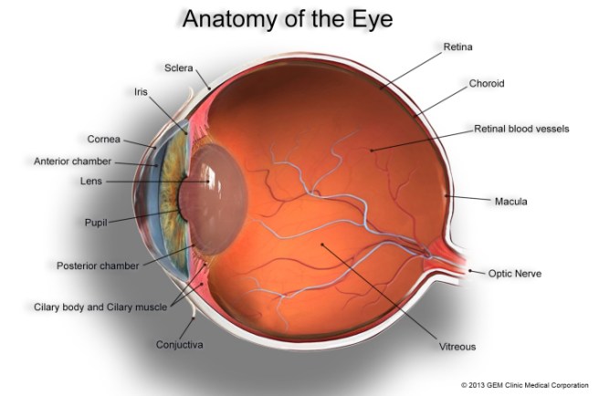
The anatomy of the eye. Source: GemClinic

Within the retina are specialised cells of two sorts:

  • Rod cells (about 125 million of them per eye) which are necessary for seeing in dim light.
  • Cone cells (6-7 million of these) which can be further divided into three types, each sensitive to different primary colours – red, green or blue.

These specialised ‘photoreceptive’ cells send signals down through the layers of the retina to what are called retinal ganglion cells which are the key conduits in the sending of information to the brain.

All of these cells require a constant blood supply, from arteries and veins spreading across the retina, and this a key part of our discussion today (see below).

So what have eyeballs got to do with Parkinson’s disease?

Good question. People with Parkinson’s disease often complain of from visual issues, such as reduced visual acuity, low contrast sensitivity and disturbed colour vision.

And there has been some research into the eyes with regards to Parkinson’s disease. A few weeks ago, this particular study was published:

Eyes1

Title: The retina as an early biomarker of neurodegeneration in a rotenone-induced model of Parkinson’s disease: evidence for a neuroprotective effect of rosiglitazone in the eye and brain.
Authors: Normando EM, Davis BM, De Groef L, Nizari S, Turner LA, Ravindran N, Pahlitzsch M, Brenton J, Malaguarnera G, Guo L, Somavarapu S, Cordeiro MF.
Journal: Acta Neuropathol Commun. 2016 Aug 18;4(1):86. doi: 10.1186/s40478-016-0346-z.
PMID: 27535749       (This article is OPEN ACCESS if you would like to read it)

The researchers in this study used a rodent model of Parkinson’s disease (rotenone-induced). In this model, the animals started losing dopamine cell loss in the brain at 60 days after the model of Parkinson’s disease was chemically induced.

The scientists examined the eyes of the rats at 10, 20, 40 and 60 days of the study. At the 20 day time point, the researchers began to see increased retinal ganglion cell death and swelling of the retinal layers in the eyes. These changes were obviously occurring well before the cell loss is observed in the brain, which leads the authors to ask whether the eyes could potentially used as an early indicator of Parkinson’s disease.

Of particular interest in this study was the use of Rosiglitazone to protect the retinal cells (AND the dopamine neurons in this rodent model of Parkinson’s disease). Rosiglitazone is an anti-diabetic drug. It works as an insulin sensitizer, by binding to fat cells and making them more responsive to insulin (we have previously discussed the curious relationship between Parkinson’s disease and diabetes (click here for more on this), and this result reinforces that connection). The scientists found that giving the drug once every 3 days had very beneficial effects of the survival of the retinal cells. They also observed significant neuroprotection after delaying the treatment for 10 days and then just giving one round of treatment, suggesting that a lot of the drug is not required for positive results.


EDITORIAL NOTE HERE: Before readers start to get any crazy ideas about sourcing and self medicating with Rosiglitazone, it is important to note that there are serious side effects associated with this class of drug. It has been associated with heart disease and stroke (click here to read more), and it should only be taken by people with diabetes and under the strict supervision of a qualified physician. It it mentioned here purely for educational purposes.


So obviously what is required is an examination of the eyes of people with Parkinson’s disease

Yep. And conveniently, in the same week as the previous study came out, this second study was also published:

Eyes2

Title: Evaluation of Retinal Vessel Morphology in Patients with Parkinson’s Disease Using Optical Coherence Tomography.
Authors: Kromer R, Buhmann C, Hidding U, Keserü M, Keserü D, Hassenstein A, Stemplewitz B.
Journal: PLoS One. 2016 Aug 15;11(8):e0161136.
PMID: 27525728          (This article is OPEN ACCESS if you would like to read it)

The researchers examined 49 people with Parkinson’s disease and 49 age- and sex-matched healthy controls. Blood vessels within the retina were identified and then divided into arteries and veins, based on their shape (using computer software). The results of the study indicate significant differences in the morphology of retinal veins in people with Parkinson’s disease when compared to controls.

Interestingly, the retinal effect was more significant on the side of the body firstly affected by Parkinson’s disease (a very common feature of Parkinson’s is that initially the condition will affect one side of the body more than the other).

What does it all mean?

For generations, we have focused on the clinical motor features of Parkinson’s disease (slowness, rigidity, and a resting tremor) when trying to determine if someone has the condition. Now we are learning that there may be other parts of the body that we should be investigating, which could not only provide us with novel diagnostic tools for earlier detection of the disease, but those areas may also provide us with new insights into disease onset and spread as well.

I may be getting a bit ahead of myself here but the possibilities are exciting and we’ll keep you abreast of these new findings as they come to us.


The banner for today’s post was sourced from the Photoforum.

Nilotinib update – new trial delayed

DSK_4634s

It is with great frustration that we read today of the delayed start to the phase 2 clinical trial of the re-purposed cancer drug Nilotinib for Parkinson’s disease (click here for a story outlining the background, and click here for the Michael J Fox Foundation statement).

We have previously  discussed both the preclinical and clinical research regarding Nilotinib and its use in Parkinson’s disease (click here and here for those posts). And the Parkinson’s community certainly got very excited about the findings of the small phase 1 unblinded clinical trial conducted by researchers at Georgetown University in 2015.

With the recent failure of the GDNF trial in Bristol, what the Parkinson’s community (both suffers and researchers alike) needs to do is refocus on moving ahead with exciting new projects, like Nilotinib. To hear that the follow-up trials for Nilotinib, however, will be delayed until 2017 (TWO YEARS after the initial results were announced) due to disagreements regarding the design of the study and who is seemingly in charge of the project, is both baffling and deeply disappointing.

Currently it appears that parties involved in the follow-up clinical trial have decided to go their separate ways, with the researchers at Georgetown University looking to conduct a single site phase 2 study of 75 subjects (if they can access the drug from supplier Novartis), while the Michael J Fox backed consortium will set up a multi-site phase 2 study.

We will continue to follow this situation as it develops and will report events as they happen.

Coffee and Parkinson’s disease – it’s not just caffeine

coffee-beans-1400

Numerous epidemiologic studies have indicated that coffee consumption reduces the risk of Parkinson’s disease. For a long time, efforts have been made to determine what the magic ingredient in this popular beverage is. Many people have speculated that the stimulant caffeine is the critical active ingredient in this neuroprotective effect.

New research, however, suggests that this may not be the case.

Today’s post will review recently published results suggesting that Quercetin (and not caffeine) is the neuroprotective component in coffee.


kaldi-adapted-from-uker

Kaldi the goat herder. Source: CoffeeCrossroads

Legend has it that in 800AD, an Ethiopian goat herder called Kaldi noticed that his animals were “dancing” after eating some berries from a tree that he did not recognise. Fascinated by the happy behaviour of his goats, Kaldi naturally decided to eat the berries for himself and he subsequently became “the happiest herder in happy Arabia”.

This amusing encounter was apparently how humans discovered coffee. It is most likely a fiction as the earliest credible accounts of coffee-consumption emerge from the 15th century in the Sufi shrines of Yemen, but since then coffee has gone on to become one of the most popular drinks in the world.

Stupid question: what exactly is coffee?

For a person who doesn’t drink coffee (like myself), this is actually a really interesting question. Coffee is a beverage made from ground up roasted beans, which are the seeds of berries from the Coffea plant. These are the berries:

coffeeberry_1338562952_62

Coffea berries. Source: About.me

And these are the beans (unroasted):

unroasted-coffee-beans-shelf-life

Unroasted Coffee beans. Source: Kopiholic

Coffee production also makes for fascinating reading (Click here for more) and why we roast the beans is equally interesting (Click here for that), but they are taking us off the topic here.

There are basically two types of coffee beans: Arabica and Robusta.

Approximately 70 percent of the coffee beans we use are Arabica. Surprisingly, the less popular Robusta actually has twice as much caffeine as Arabica. And caffeine is the stimulant that rewards people for drinking this beverage.

Caffeine is also the chemical that has long been thought to have positive effects on Parkinson’s disease, possibly even reducing the risk of the condition (more on that below).

coffee-cup-images-5

Fancy a cuppa? Source: Science-All

What does coffee have to do with Parkinson’s disease?

We have previously discussed the enormous contribution that the Honolulu Heart Study has made to our understanding of Parkinson’s disease (click here to read that post). Many of the earliest associations with the condition were found in that large epidemiologic study. One of those findings was that the consumption of coffee reduced one’s risk of developing Parkinson’s disease.

Coffee-title2

Title: Association of coffee and caffeine intake with the risk of Parkinson disease.
Authors: Ross GW, Abbott RD, Petrovitch H, Morens DM, Grandinetti A, Tung KH, Tanner CM, Masaki KH, Blanchette PL, Curb JD, Popper JS, White LR.
Journal: JAMA. 2000 May 24-31;283(20):2674-9.
PMID: 10819950    (This article is OPEN ACCESS if you would like to read it)

The researchers behind this article analysed the data from the Honolulu Heart Study – an epidemiological study of 8,006 “non-institutionalized men of Japanese ancestry, born 1900-1919, resident on the island of Oahu” – and found that the age-adjusted incidence of Parkinson’s disease declined consistently with increased amounts of coffee intake (from 10.4 per 10,000 person-years in men who drank no coffee to 1.9 per 10,000 person-years in men who drank at least 28 oz/d). This and other findings in their analysis indicated that higher coffee (and caffeine) intake is associated with a significantly lower incidence of Parkinson’s disease.

Subsequent studies have replicated this association, and several have demonstrated the neuroprotective effects of caffeine (click here for a review on this topic).

So what new data has been published?

This is Prof Patrick and Prof Edith McGeer:

ss-alz-research McGeer

Prof Patrick and Prof Edith McGeer. Source: Mcgeerandassociates

This husband and wife team of scientists are well recognised figures within the Parkinson’s disease research work, having produced many seminal scientific reports. Patrick is a particularly interesting character having played basketball for Canada in the 1948 Olympics and then a politician in the British Columbia legislature (1962-1986).

They are also authors on the article we are going to review today:

Coffee-title

Title: Quercetin, not caffeine, is a major neuroprotective component in coffee.
Authors: Lee M, McGeer EG, McGeer PL.
Journal: Neurobiol Aging. 2016 Jul 5;46:113-123.
PMID: 27479153

As we said above, for the longest time people have believed that caffeine was the active ingredient in the miraculous ability of coffee to reduce the risk of Parkinson’s disease. The researchers who published this report were particularly interested in the neuroprotective role for coffee in Parkinson’s disease and they decided to break coffee down into some of its basic components. Specifically:

  • Caffeine
  • quercetin
  • flavone
  • Chlorogenic acids (CGAs)

They tested each of these coffee components on cells (grown in petri dishes) that had been exposed to a toxin, and then assessed cell survival. Curiously, although caffeine did exhibit neuroprotective effects on the cells, it was beaten by the far superior protective effects of quercetin.

What is quercetin?

Quercetin is a flavonoid (a type of plant pigment) that is found in many fruits, vegetables, leaves and grains. Flavonoids are potent antioxidants. Antioxidants scavenge particles (called free radicals) in the body which can damage cell membranes, affect DNA, and even cause cell death. Antioxidants neutralize these free radicals. (For more on flavonoids – click here).

What does this mean?

The results are very interesting, especially if they provide us with a new potential target for therapeutic drug development. It also raises the age-old idea of antioxidants being potentially useful in the treatment of Parkinson’s disease (the previous history of this therapeutic approach has been disappointing – click here to read more on this).

But before you rush out and load up on quercetin, there are a few things to consider:

Quercetin is generally considered pretty safe. Fruits and vegetables are the primary dietary sources of quercetin, particularly citrus fruits, apples, onions, parsley, sage, tea, and red wine.

That said: excessive use of quercetin can have side effects, which may include headache and upset stomach. Very high doses of quercetin can cause damage to the kidneys (doses greater than 1 g per day), and regular periodic breaks from taking quercetin is advised. Importantly, pregnant women, breastfeeding women, and people with kidney disease should avoid quercetin.

EDITOR’S NOTE: If you are considering supplementing your diet with quercetin (or any other potential therapeutic agents) please firstly discuss this change of lifestyle with your medical physician. Information provided here can under no circumstances be considered medical advice.

Having said that we shall keep an eye out for any new research of quercetin and Parkinson’s disease, and report it here.


The banner for today’s post was sourced from Phoxpopmagazine