
|
There is a protein in most of the cells in your body called “PTEN-induced putative kinase 1″ (or simply PINK1). It plays an important role in keeping your cells healthy. Genetic variations in the PINK1 gene have been shown to increase ones risk of developing Parkinson’s. This week researchers have identified a method by which the function of the PINK1 protein can be inhibited and this results in increased vulnerability to Parkinson’s. In this post, we will look at what PINK1 does, how it is inhibited, and what this could mean for the Parkinson’s community. |

Mitochondria (green) in health cells (left) and in unhealthy cells (right).
The nucleus of the cell is in blue. Source: Salk Institute
I have previously spoken a lot about mitochondria and Parkinson’s on this website.
For the uninitiated, mitochondria are the power house of each cell. They help to keep the lights on. Without them, the party is over and the cell dies.

Mitochondria and their location in the cell. Source: NCBI
You may remember from high school biology class that mitochondria are tiny bean-shaped objects within the cell. They convert nutrients from food into Adenosine Triphosphate (or ATP). ATP is the fuel which cells run on. Given their critical role in energy supply, mitochondria are plentiful (some cells have thousands) and highly organised within the cell, being moved around to wherever they are needed.
Like you and I and all other things in life, however, mitochondria have a use-by date.
As mitochondria get old and worn out (or damaged) with time, the cell will recycle them via a process called mitophagy (a blending of the words mitochondria and autophagy which is the waste disposal system of each cell).
What does this have to do with Parkinson’s disease?
Well, about 10% of Parkinson’s cases are associated with particular genetic variations that render people vulnerable to developing the condition. Some of these mutations are in sections of DNA (called genes) that provide the instructions for proteins that are involved in the process of mitophagy. Two genes, in particular, are the focus of a lot of Parkinson’s-related research – they are called PARKIN and PINK1.
What do PARKIN and PINK1 do?
Both proteins appear to have many different functions, but their roles in the process of mitophagy are well understood.
PINK1 acts like a kind of handle on the surface of mitochondria. In normal, healthy cells, the PINK1 protein attaches to the surface of mitochondria and it is slowly absorbed until it completely disappears from the surface and is degraded. In unhealthy cells, however, this process is inhibited and PINK1 starts to accumulate on the outer surface of the mitochondria. Lots of handles poking out of the surface of the mitochondria.
Now, if PINK1 a handle, then PARKIN is a flag that likes to hold onto the PINK1 handle. While exposed on the surface of mitochondria PINK1 starts grabbing the PARKIN protein. This pairing is a signal to the cell that this particular mitochondrion (singular) is not healthy and needs to be removed.

Pink1 and Parkin in normal (right) and unhealthy (left) situations. Source: Hindawi
In the absence of normal PINK1 or PARKIN proteins, there is no handle-flag system and sick/damaged mitochondria start to pile up. They are not disposed of appropriately and as a result the cell getting sick and ultimately dying.

Mitophagy in a nutshell. Source: Frontiersin
People with particular mutations in the PINK1 or PARKIN genes are vulnerable to developing an early onset form of Parkinson’s disease. It is believed that the dysfunctional disposal of (and accumulation of) old mitochondria are part of the reason why these individuals develop the condition at such an early age.
The versions of Parkinson’s associated with these two proteins involve an autosomal recessive mutation – meaning that a copy of the mutation has to be provided by both the parents in order for a condition to develop.

Autosomal recessive genetic transfer. Source: Wikipedia
For a very good review of the genetics of Parkinson’s disease – click here. Alternatively, have a look at our Genetics of Parkinson’s page.
But what does any of this have to do with snow? You mentioned snow in the title – so where does snow come into the picture?
This week this research was published:
 Title: S-Nitrosylation of PINK1 Attenuates PINK1/Parkin-Dependent Mitophagy in hiPSC-Based Parkinson’s Disease Models
Authors: Oh CK, Sultan A, Platzer J, Dolatabadi N, Soldner F, McClatchy DB, Diedrich JK, Yates JR, Ambasudhan R, Nakamura T, Jaenisch R, Lipton SA
Journal: Cell Reports (2017) 21, 8, 2171–2182
PMID: 29166608 (This article is OPEN ACCESS if you would like to read it)
In this study, the researchers were interested in determining whether environmental factors can cause particular modifications to the PINK1 protein that would mimic the effects of the genetic mutations (for example, the accumulation of old mitochondria in cells). Of particular interest to the investigators was something called the S-nitrosylation (or SNO) reaction.
What is the S-nitrosylation reaction?
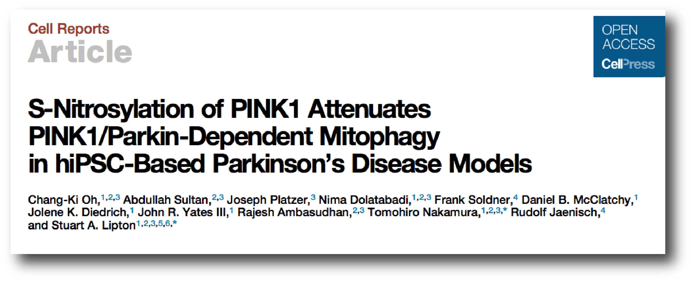
Nitrosylation is a chemical reaction that adds a nitrosyl group (the ‘NO’ part of SNO) to a protein.
S-nitrosylation is a modification of proteins in which specific chemicals associated with the protein are modified by the chemical nitric oxide. This process impacts the proteins function, stability, and localisation within a cell.
Basically, the SNO reaction alters a proteins ability to do its job.
The SNO reaction. Source: Aibolita
The researchers found that the SNO reaction inhibited the ability of PINK1 to do its job in the process of mitophagy. PINK1 was unable to localise to the mitochondria after the SNO reaction, which resulted in an increase of old mitochondria inside cells. And this subsequently causes cells to get sick and die.
Thus the formation of PINK-SNO is definitely harmful to cells in the Parkinson’s brain.

Source: Cell
But this SNO reaction only affect PINK1 right?
Unfortunately, no.
A few years ago these same researchers demonstrated that another Parkinson’s associated protein was affected by SNO. Perhaps you have heard of that protein. It’s called PARKIN:

Title: S-Nitrosylation of parkin as a novel regulator of p53-mediated neuronal cell death in sporadic Parkinson’s disease.
Authors: Sunico CR, Nakamura T, Rockenstein E, Mante M, Adame A, Chan SF, Newmeyer TF, Masliah E, Nakanishi N, Lipton SA.
Journal: Mol Neurodegener. 2013 Aug 28;8:29.
PMID: 23985028 (This article is OPEN ACCESS if you would like to read it)
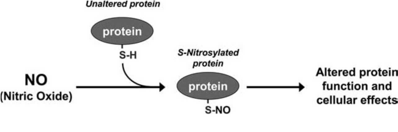
In this research report, the investigators presented data suggesting that S-nitrosylation of PARKIN decreased its activity as a ‘repressor’ of p53 gene activity. This means that while PARKIN usually keeps the activity of the p53 gene very low, in the absence of PARKIN (due to SNO) the levels of p53 protein increase rapidly.
What is p53?
p53 (or TP53) is a gene on chromosome 17 that encodes a protein that has three major functions:
- controlling cell division
- DNA repair
- Apoptosis (or cell death).
p53 (the protein) performs these functions as a transcriptional activator (that is a protein that binds to DNA and helps produce RNA (the process of transcription) – see our previous post explaining this).

p53 protein structure, bound to DNA (in gold). Source: Wikipedia
In regulating the cell division, p53 prevents cells from dividing too much and in this role it is known as a tumour suppression – it suppresses the emergence of cancerous tumours. Genetic mutations in the p53 gene can result in run away cell division, and (surprise!) as many as 50% of all human tumours contain mutations in the p53 gene.

Cancer vs no cancer. Source: Khan Academy
With regards to DNA repair, p53 is sometimes called “the guardian of the genome” as it prevents mutations and helps to conserve stability in the genome. This function also serves to prevent the development of cancer, by helping to repair potentially cancer causing mutations….and in this role it is known as a tumour suppression.
And finally, in cell death, p53 plays a critical role in telling a cell when to die. And (continuing with the cancer theme), if there is a mutation in the p53 gene, fewer cells will be told to die – increasing the risk of cancer occurring. And in this role p53 is known as a tumour suppression. But in the absence of a mutation in the p53 gene or PARKIN to reduce the activity of the gene, p53 levels will gradually rise and instruct the cell to initiate apoptosis.
This is why drugs that act as PARKIN activators would be very beneficial – not only could it help the Parkinson’s community, but they could also be used against many types of cancer.
Ok, so if PARKIN is inhibited by SNO then p53 levels would rise and tell cells to die?
Exactly.
And the researchers found this in both cell cultures and a mouse model of Parkinson’s. In addition, the investigators also looked at levels of SNO-parkin and p53 in postmortem human brains, and they compared Parkinsonian brains to those of unaffected controls. They found levels of SNO-parkin and p53 were simultaneously elevated the Parkinsonian brain.
Thus, a very similar process may be occurring in the PARKIN situation as the PINK1 situation, in that SNO is preventing both of these proteins from doing their jobs.
Source: Ncbi
But I don’t understand how this makes someone vulnerable to Parkinson’s. How does it work?
Each of us inherits two copies of every gene – one from each of our parents.
In this way, we all have at least one copy of a particular gene if the other copy is mutated in some way. It’s a kind of insurance policy for mother nature. Depending on the protein, however, this insurance policy may or may not be sufficient to provide proper compensation.
In the case of PINK1, if only one copy of the PINK1 gene can produce protein and most of that protein is then targeted by SNO, the cells in the body may be left vulnerable to the damaging effect of old mitochondria accumulating. And this may ultimately lead to cells dying. Thus, while people with autosomal recessive PINK1 mutations (a mutated copy of PINK1 from both parents) are vulnerable to Parkinson’s, their siblings who have only 1 copy of the mutated PINK1 gene may also be vulnerable. Such a mechanism could help to explain the frequent family history of Parkinson’s that is seen in cases of idiopathic (or spontaneous) Parkinson’s.
The data supports the idea that pesticide-induced SNO modifications of proteins like PINK1 and PARKIN may contribute to the ‘sporadic’/idiopathic forms of Parkinson’s – mimicking the effects of genetic mutations of these genes.
It is an intriguing idea.
Professor Stuart Lipton of the Scripps Research Institute (and one of the researchers involved in this study suggested that “The take-home message here is that the environment may affect you based on your individual genetics, and thus both are influential in causing diseases like Parkinson’s.” (Source).
So what does it all mean?
We often try to separate nature and nurture.
Genetics and the environment.
But in truth the situation is a lot more complicated than that. This week some interesting new data was published that illustrates how the environment and genetics could be interacting to make cells vulnerable to Parkinson’s.
The next step in this research is to study how one may be able to prevent SNO reactions on particular proteins like PINK1. If a treatment is found, it would probably be beneficial not only for folks in the Parkinson’s community, but also the wider general public. Allowing cells to function more efficiently and not have a build-up of old mitochondria may have health benefits for all of us.
The banner for today’s post was sourced from Moviewriternyu

Simon
Thanks for that very readable explanation.
Keith
LikeLike
You’re very welcome! Glad you liked it.
Simon
LikeLike