|
# # # # Lewy bodies are densely packed aggregates of proteins and lipids that can be found in some neurons in the brain of many people with Parkinson’s. They have long been considered a cardinal feature of the Parkinsonian brain. To date, humans are the only species that have displayed evidence of Lewy bodies. But very recently new data has suggested that Lewy body-like pathology might not be so human-specific. In today’s post, we will discuss what are Lewy bodies are and explore the new research report suggesting that they might not be unique to humans. # # # # |
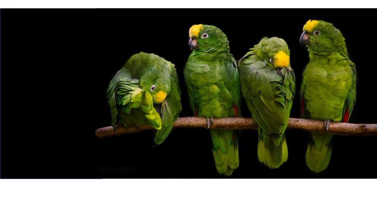 Source: Theanimalfacts
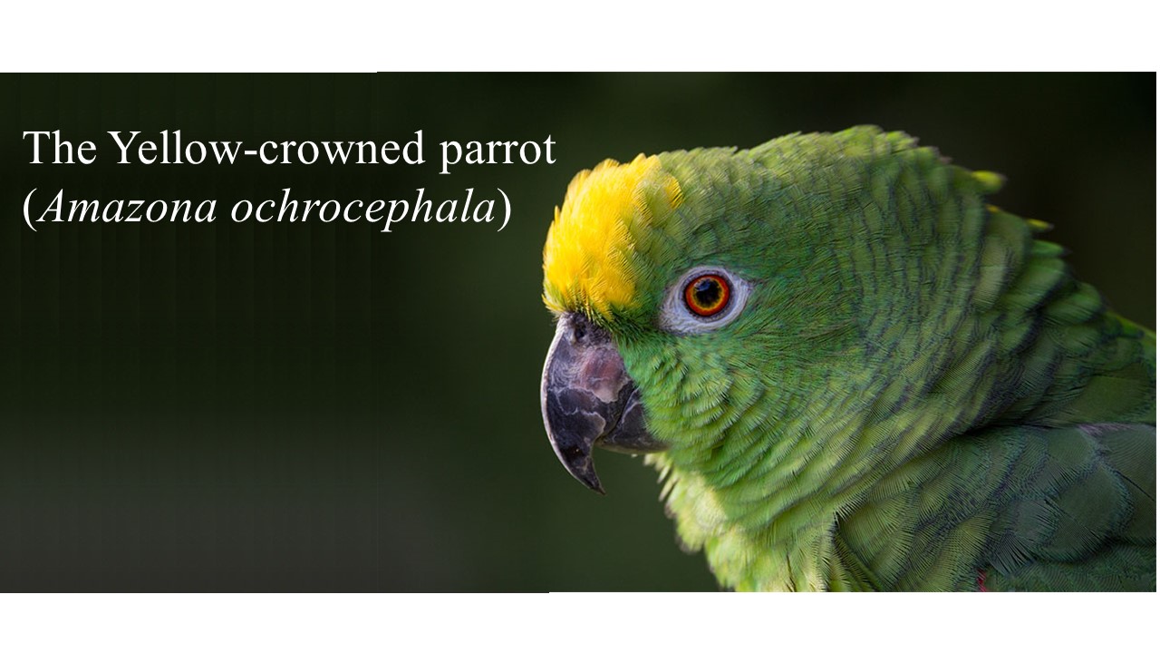
The yellow-crowned parrot are remarkably similar to humans.
They have a lifespan of 60 to 80 years (in captivity; 20 to 30 years in the wild), and they are monogamous in their pairing, with pairs often remaining together for life. And they are rather vocal (but curiously they don’t have vocal cords).
Ok, their courtship is slightly different to humans (it involves lots of bowing, drooping, flicking of wings, raising of feet, and dilating of pupils) and chocolate is extremely poisonous to them.
But by and large, they are remarkably similar to us (at least as far as birds go).
Great, but what does any of this have to do with Parkinson’s?
Very recently, this report was published:
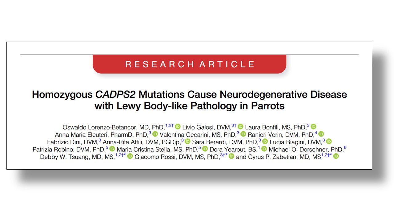 Title: Homozygous CADPS2 Mutations Cause Neurodegenerative Disease with Lewy Body-like Pathology in Parrots.
Authors: Lorenzo-Betancor O, Galosi L, Bonfili L, Eleuteri AM, Cecarini V, Verin R, Dini F, Attili AR, Berardi S, Biagini L, Robino P, Stella MC, Yearout D, Dorschner MO, Tsuang DW, Rossi G, Zabetian CP.
Journal: Mov Disord. 2022 Sep 10. doi: 10.1002/mds.29211. Online ahead of print.
PMID: 36086934 (This report is OPEN ACCESS if you would like to read it)
In this study, the researchers detailed the cases of three 3-month-old hand-reared Yellow-crowned Amazon (Amazona ochrocephala) siblings, which were brought to the School of Biosciences and Veterinary Medicine of the University of Camerino in Italy, displaying severe neurological symptoms.
Specifically, they exhibited uncoordinated movements, with lots of head tilt and ‘stargazing’ (twisted back to staring upwards) from 2 months of age. And there was a tremor.
The breeder said that the parrots had displayed a very early-onset persistent tremor, which was the most noticeable sign that something was wrong. It began periodically in just one wing, but increased considerably when the parrots were tired. It started out asymmetric, but gradually became bilateral and spread to other parts of the body. These young birds were unable to perch and could not eat independently.
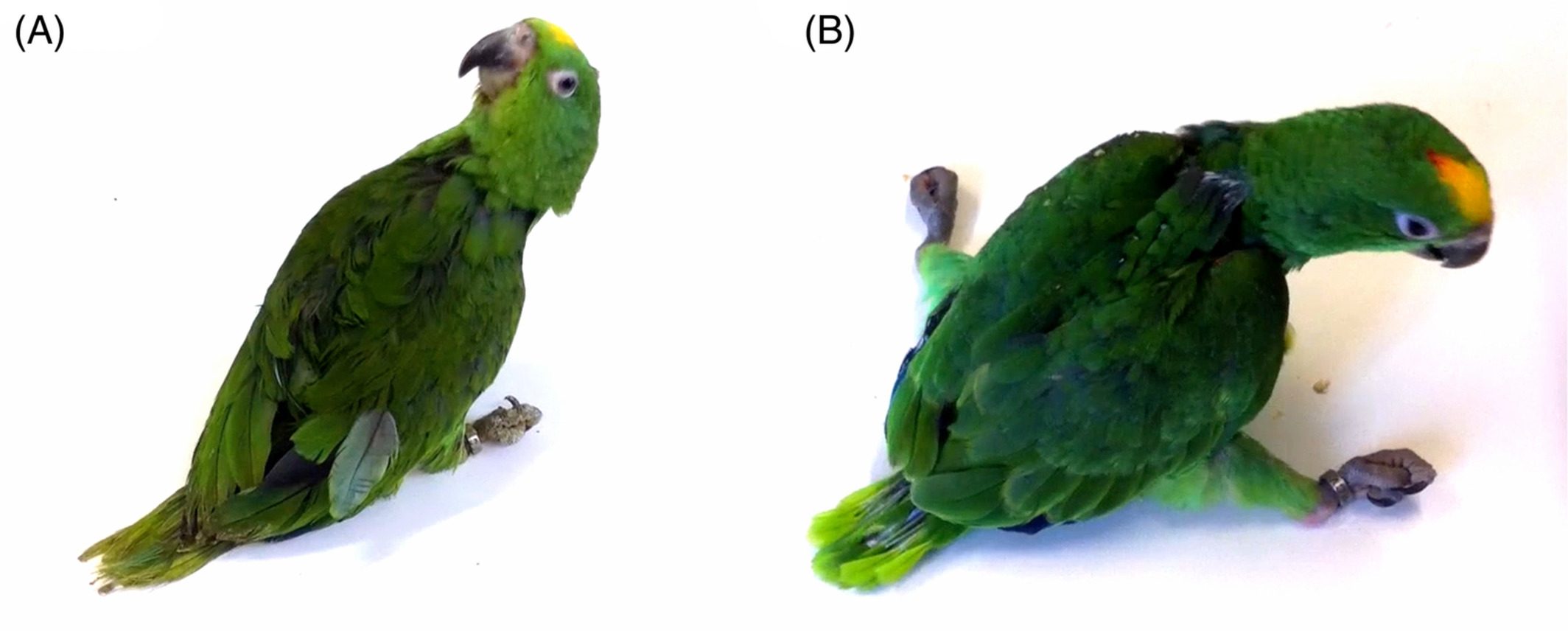 Posturing of the birds. Source: MovementDisoders
The investigators looked at the blood of the animals, but found no abnormalities that could be considered causal (such as viral or bacterial infections). And an MRI of the brain (brain imaging) was negative for any notable pathological structural changes.
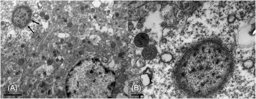
When the researchers conducted a closed examination of the brains, they found “moderate to severe neuronal loss” and evidence of reactive microglia (the resident immune cells of the brain).
But most interesting of all, they also observed “widely distributed variably sized (up to ~100 μm in diameter), round to elongate, well-defined eosinophilic structures“. They did not refer to them as Lewy bodies, but rather Lewy body-like inclusions.
 Source: MovementDisoders
What are Lewy bodies?
A Lewy body is referred to as a cellular inclusion, as they are almost always found inside the cell body. They generally measure between 5–25 microns in diameter (5 microns is 0.005 mm) and thus they are tiny. But when compared to the neuron within which they reside they are rather large (neurons usually measures 40-100 microns in diameter).
 A photo of a Lewy body inside of a neuron. Source: Neuropathology-web
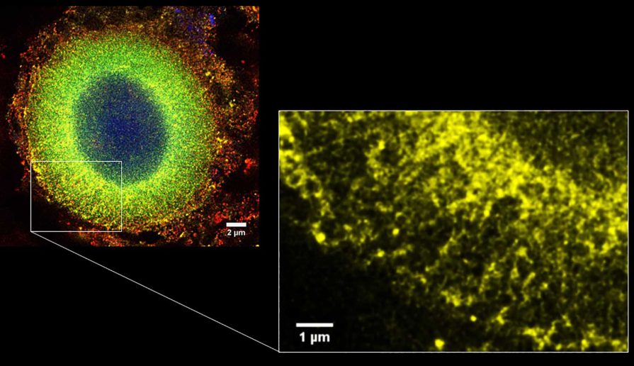
They are densely packed structures made up of all kinds of cellular components (“undigested membrane fragments, damaged organelles, and other cellular garbage” – source). But they do have a key feature which is a halo of the Parkinson’s-associated protein alpha synuclein (Click here to read a previous SoPD post on alpha synuclein). In the image below, you can see a high magnification image of a Lewy body on the left, with alpha synuclein protein stained in yellow. Note the halo-like localization of the synuclein protein around the border of the Lewy body – very little in the center of it. The inset on the right shows an even higher magnification image of the alpha synuclein protein:
 Source: Springer
Now, it should be noted that the researchers looking at the brains of the poor parrots described what they observed in the brains as Lewy body-like inclusions. Firstly, the structures that they observed were 2-3 times larger than your typical Lewy body. In addition, they lacked a distinctive core and did not display the normal halo of Lewy bodies.
But they did contain alpha synuclein protein.
As you can see in the image below, alpha synuclein was stained brown on sections of brain tissue from the poor parrots, and there are lots of brown dots evident across the samples:
 Source: MovementDisoders
Another feature of the brains was changes in the shape of the neurons. The neurons containing one of these Lewy body-like inclusions, the investigators noted that enlargement of the cell body, displacement of the nucleus towards the side of the cell, and a smaller than normal nucleus. This could be considered a sign of a cell under a great deal of stress, and perhaps in the early stages of cell death. To reinforce this view, the investigators also reported that some of these neurons were partially engulfed by glial cells, suggesting that the cells were in serious trouble.
Interesting. So they have no idea about what caused this?
Well, as we mentioned above, there was no sign of viral or bacterial infection, so the researchers turned their attention to genetics.
DNA samples were collected from the parrots parents, two uncles, and their grandparents. And when the researchers carefully compared all of these samples with their three poor case studies, they found a spontaneous genetic mutation in the CADPS2 gene in all of their three parrots that displayed this curious condition.
What is the CADPS2 gene?
The calcium-dependent secretion activator 2 (CADPS2) gene provides the instructions for making a type of calcium binding protein that controls the release of two neurotrophic factors in mice (BDNF and NT3). Neurotrophic factors help to support and nurture neurons, promoting their growth and survival.
Given this function of CADPS2, the researchers proposed that “one mechanism by which mutations in CADPS2 might induce neurodegeneration is by altering the appropriate release of BDNF” and they noted that dopamine neurons (which are highly vulnerable in Parkinson’s) produce high levels of CADPS2 protein.
And before you ask: No, CADPS2 genetic variations have not been reported in neurodegenerative conditions in humans.
But this does not mean that CADPS2 protein could not be playing a role.
What do you mean?
In 2017, this report was published:
 Title: CADPS2 gene expression is oppositely regulated by LRRK2 and alpha-synuclein.
Authors: Obergasteiger J, Überbacher C, Pramstaller PP, Hicks AA, Corti C, Volta M.
Journal: Biochem Biophys Res Commun. 2017 Aug 26;490(3):876-881.
PMID: 28647363
In this study, the researchers grew cells in culture and they engineered the cells to produce high levels of either LRRK2 or alpha synuclein (both are Parkinson’s-associated proteins). They reported that these two situations had very different effects on CADPS2-related activity.
High levels of LRRK2 protein to increased CADPS2 protein levels, while high levels of alpha synuclein reduced CADPS2 protein levels. So it may be the case that CADPS2 protein levels “might be regulated in part by genes that are well-established as playing a role in Parkinson’s pathogenesis” even in the absence of any genetic mutation in the CADPS2 gene.
So genetic mutations in CADPS2 are not associated with Parkinson’s, but other Parkinson’s-associated genes could be influencing CADPS2 activity?
Exactly. And this is how the parrot researchers concluded their report, suggesting that this could be an avenue of future investigation.
Is there any evidence of CADPS2 activity in Parkinson’s cases?
Actually, yes.
Earlier this year, this report was published:
 Title: Single-cell sequencing of human midbrain reveals glial activation and a Parkinson-specific neuronal state.
Authors: Smajić S, Prada-Medina CA, Landoulsi Z, Ghelfi J, Delcambre S, Dietrich C, Jarazo J, Henck J, Balachandran S, Pachchek S, Morris CM, Antony P, Timmermann B, Sauer S, Pereira SL, Schwamborn JC, May P, Grünewald A, Spielmann M.
Journal: Brain. 2022 Apr 29;145(3):964-978. doi: 10.1093/brain/awab446.
PMID: 34919646 (This report is OPEN ACCESS if you would like to read it)
In this study, the researchers analysed single cells collected from the brains of people who had passed away with Parkinson’s. They discovered a dopamine neuron subpopulation that was characterized by high levels of CADPS2 protein and low levels of tyrosine hydroxylase (a enzyme that is critical to the production of dopamine), and this subpopulation was only present in the brains from people with idiopathic Parkinson’s.
So yes, there is evidence CADPS2 potentially playing a role in Parkinson’s.
So what does it all mean?
On the SoPD we have previously explored the idea of Parkinson’s being a uniquely human condition (Click here and here to read previous SoPD posts on this topic), but it often strikes me as a strange comparison for several reasons. If other animals are able to develop a Parkinson’s-like conditions, why should it look like human Parkinson’s at all? These other animals have very different biology to us (for example, as we discussed above chocolate is very poisonous to the yellow-crowned parrot, but it is one of the basic food groups for most humans in western cultures), so surely “Parkinson’s” in another animal might look very different.
For two decades we have genetically engineered mice with Parkinson’s associated genetic variations inserted into their DNA. Despite attempting many different arrangements of these genetic risk factors, the resulting phenotypes (the observable result) have not resembled the condition we know as Parkinson’s (without further manipulation). In fact, many of these genetically engineered mice have appeared outwardly normal with little evidence of neurodegeneration – even in the late stages of aging.
While better animal models of medical condition would be very useful in the development of new therapeutic interventions, it is fair to say that modeling of human conditions should best be viewed as ‘proximate biology‘ (thank you Mark Cookson). Rather than providing us with an artificial model of disease, it points us towards features of the underlying biology that can be better explored.
And this is the case with today’s post. Poor little parrots inherited a genetic variation that caused a catastrophic degenerative condition, which while not directly related to Parkinson’s, certainly suggests some very interesting avenue of investigation related to the biology of the condition. I am really curious where these lines of inquiry will lead.
 All of the material on this website is licensed under a
Creative Commons Attribution 4.0 International License
You can do whatever you like with it!
The banner for today’s post was sourced from Omlet

Thank you for the information about B1 use in Parkinsons.
I am interested in the use of Benfotiamine.
I received this feedback form an avid High Dosage Thiamine follower
I wonder if you can clear this part up for me,
Thanking You
Alan Fraser
”benfothiamine
11/19/2017
Dear Roy,
Sorry for my late, but unfortunately I can not speak English well, so it’s Dr Erika Trevi who is writing these emails to you.
We don’t use benfothiamine because previous trials report it does not enter in the neural cells, that’s why it is not used for the diseases which don’t affect the Central Nervous System (Bettendorff L.).We administer thiamine cloridrate. For your situation 1 intramuscular injection twice a week (or an oral dose of 4 grams each day, two tablets in the morning and two at lunch time) should bring to the complete regression of the symptoms in 1 or 2 months. As an attached link, you’ll find a paper with the possible thiamine side effects.
Best regards,
Antonio Costantini”
LikeLike
I found your article through Google by typing “Can a bird get Parkinson’s Disease?” The reason I wanted to know is because I have a 25 year old Green Cheek Conure (Jade) who has developed some severe neurological symptoms within the past few months. In fact, the image and description you provided regarding “star gazing ” is exactly how I would describe one of his symptoms. He also has a tremor affecting one of his wings (his right wing). He has a wobbly stance and has difficulty perching. He spends most of his day in a “happy hut” type of tent (poor baby!). He typically forages food from the floor and uses a box to climb up to a rope perch to get to his water dish (except I do leave one on the floor for him as well). He can perch on my shoulder as long as he snuggles up to my neck. I’m careful not to bend over or make any quick, sudden movements, or he could lose his balance.
Prior to the onslaught of these symptoms, Jade has been a very healthy bird. In fact, his former cage mate, Jem, had gotten quite ill last year and unfortunately, passed away. Jem was 22 at the time. I had him for 21 of his 22 years. I’ve had Jade for his entire life, since he was weaned. Jem had also been relatively healthy despite having epilepsy his entire life. He’d never gotten sick with anything until the year before last. In 2021, he’d been sick with an upper respiratory infection but had gotten better with medication. Unfortunately, he acquired another infection several months after that in 2022. Again, he got better for a couple of months then his health took a turn for the worse a third, and final, time. Through all this, thankfully Jade didn’t get sick.
So that’s what I mean: Jade has been very healthy his entire life until Jem passed away. Now Jade has these neurological symptoms, which I actually thought might be seizures, initially, because, while Jade has been experiencing tremors daily, they tend to get worse in the evenings before bed. There have been times where the tremors have been so severe, they appeared to be like grand mal seizures, but Jade is fully aware of his surroundings…unlike Jem (and unlike people whom I’ve cared for who also had epilepsy BTW). At those times, they last for about 2 minutes from beginning to end then Jade resumes what he was doing afterwards until another bad one occurs. These happen about 3 times a night now. And, as mentioned, he has a regular tremor and wobbly stance throughout the day.
We live in a rural area without access to an avian veterinarian. I only have a dog/cat vet who has enough knowledge with avian basics like collecting blood work to send to labs to check for infections and to prescribe medications when needed. I realize your article is recent, so, sadly, I have to assume that it’s unlikely that there’s any kind of treatment I can administer to help my little Jade. However, I thought I’d reach out to you regarding my sweet boy in case I could be of assistance in any way and also, if there WAS some way to help my Jade, I hope you would offer some advice.
LikeLike