Multiple System Atrophy: A prion disease?

‘Parkinsonisms’ refer to a group of neurological conditions that cause movement features similar to those observed in Parkinson’s disease. They include multiple system atrophy (MSA) and Progressive supranuclear palsy (PSP) and idiopathic Parkinson’s.

Newly published research now shines a light on a possible mechanism for differentiating between multiple system atrophy and idiopathic Parkinson’s.

In today’s post we will look at what multiple system atrophy is, review the new research report, and discuss what these results could mean for the Parkinson’s community.


Brain immaging of multiple system atrophy–related spatial covariance pattern (MSARP) and Parkinson disease–related spatial covariance pattern (PDRP). Source: Neurology

For a long time I have been looking to write a piece of Multiple system atrophy.

I have been contacted by several readers asking for more information about it, and the only thing really delaying me – other than the tsunami of Parkinson’s related research that I am currently trying to write posts for – was the lack of a really interesting piece of research to base the post around.

Guess what came into my inbox yesterday:

Title: Familial Parkinson’s point mutation abolishes multiple system atrophy prion replication.
Authors: Woerman AL, Kazmi SA, Patel S, Aoyagi A, Oehler A, Widjaja K, Mordes DA, Olson SH, Prusiner SB.
Journal: Proc Natl Acad Sci U S A. 2017 Dec 26. pii: 201719369.
PMID: 29279394

This is a really interesting piece of research, that continues a line of other really interesting research.

And if it is independently replicated and verified, it will have massive implications for the Parkinson’s community, particularly those affected by Multiple System Atrophy.

But before we deal with that, let’s start with the obvious question:

What is Multiple System Atrophy?

Continue reading “Multiple System Atrophy: A prion disease?”

Editorial: Orphan drug tax credit

Here at the SoPD we are politically neutral.

That said, I will report on events that directly impact the world of Parkinson’s disease research (without adding too much in the way of personal opinions). 

Recent legislation introduced in the US congress could have major implications for subsets of the Parkinson’s disease community, as well as a host of additional medical conditions. The legislation is seeking to remove the orphan drug tax credit.

In today’s post, we will have a look at what the orphan drug tax credit is, and why its removal could be damaging for Parkinson’s.


capitol-hill-parking

The United States Capitol. Source: SpotHeroBlog

On November 2, House Republican lawmakers introduced a bill to reform the U.S. tax code. The complicated tax system probably needs a serious clean up, but the legislation will also terminate something called the orphan drug tax credit.

What is the orphan drug tax credit?

Continue reading “Editorial: Orphan drug tax credit”

The myth of Spring babies

baby-spring

In large datasets, strange anomalies can appear that may tell us something new about a condition, such as the curious association between melanoma and Parkinson’s disease.

These anomalies can also appear in small datasets, such as the idea that spring babies are more at risk of developing Parkinson’s disease. But the smaller dataset results may be a bit misleading.

In today’s post, we will look at what evidence there is supporting the idea that people born in the spring are more vulnerable to Parkinson’s disease.


baby-772441-2

Spring lambs. Source: Wenatcheemumblog

When is your birthday?

More specifically, which month were you born in? Please feel free to leave your answer in the comments section below this post.

Why do I ask?

In 1987, an interesting research report was published in a scientific research book:

Miura

Title: Season of birth in parkinsonism.
Authors: Miura, T., Shimura, M., and Kimura, T.
Book: Miura T. (ed) Seasonality of birth:Progress in biometeorology, 1987.p157-162. Hague, Netherlands.
PMID: N/A

In the report, the researchers outlined a study that they had conducted on the inhabitants of an asylum for the aged in Tokyo (Japan). They had found not only a very high rate of Parkinsonism (6.5% of the inhabitants), but also that the majority of those individuals affected by the Parkinsonism were born in the first half of the year (regardless of which year they were actually born).

Sounds crazy right? (excuse the pun)

And that was probably what everyone who read the report thought….

…except that one year later this independent group in the UK published a very similar result:

Continue reading “The myth of Spring babies”

A new theory of Parkinson’s disease

emc2

The great American baseball legend, Yogi Berra, once said: “In theory, there is no difference between theory and practice. But in practice, there is.”

Silly as it reads, there is a great deal of truth to that statement.

In science, we very quickly chase after a particular theory as soon as a little bit of evidence is produced that supports it. Gradually, these theories become our basic understanding of a situation, until someone points out the holes in the theory and we have to revise it.

A new theory of Parkinson’s disease has recently been proposed. In today’s post we will review what the theory is suggesting and what evidence there is to support it.


150119_sipress-2005-05-23

“I still say it’s only a theory”. Source: NewYorker

In the age of ‘alternative facts’, it is always important to remember that we don’t know as much as we think we do. In fact, much of our modern world still relies on a kind of faith rather than actual ‘facts’. For example, we take a particular type of medicine, because it has worked for some people in the past, not because it will definitely make us better.

And the same applies to our understanding of neurodegenerative conditions, like Parkinson’s disease. Based on all the evidence we have collected thus far, we have theories of how Parkinson’s disease may be progressing. But there are always exceptions to the rule, and these force us to refine or reconsider our theories.

Recently a refinement to our theory of Parkinson’s disease has been suggested.

Who has suggested it?

This is Prof Ole Isacson.

maxresdefault

Source: Crunchbase

He’s a dude.

He is is a Professor of Neurology at Harvard Medical School, and Chief Scientific Officer of the Neuroscience Research Unit and Senior Vice President at the pharmaceutical company Pfizer.

And this is Dr Simone Engelender.

8678381191

Source: Rappaport

She’s awesome as well.

She is Associate Professor of Molecular Pharmacology at the Rappaport Family Institute for Research in the Medical Sciences in Haifa, Israel.

Together they have proposed a new theory of Parkinson’s disease that has the research community talking:

trends
Title: The Threshold Theory for Parkinson’s Disease.
Authors: Engelender S, Isacson O.
Journal: Trends Neurosci. 2017 Jan;40(1):4-14.
PMID: 27894611

The new theory proposes that Parkinson’s disease may actually be a ‘systemic condition’ (that is, affecting cells everywhere at the same time), but the clinical features – such as motor issues – only appear as certain thresholds are passed in the affected populations of neurons in the brain.

What does that mean?

Wait a minute. Let’s start at the beginning.

Before discussing what the new theory suggests, shall we first have a look at what the old theories proposed?

Ok, what did the old theory propose?

This is Prof Heiko Braak:

heiko-braak-01

Source – Memim.com

He’s pretty cool too. Nice guy.

Many years ago, Prof Braak – a German neuroanatomist – sat down and examined hundreds of postmortem brains from people with Parkinson’s disease.

He had collected brains from people at different stages of Parkinson’s disease – from just after being diagnosed to having had the condition for decades – and he was looking for any kind of pattern that might explain where and how the disease starts. His research led to what is referred to as the “Braak stages of Parkinson’s disease” – a six step explanation of how the disease spreads up from the brain stem and into the rest of the brain (Click here to read more about this).

nrneurol.2012.80-f1

The Braak stages of PD. Source: Nature

Braak’s results also led him to propose that Parkinson’s disease may actually begin in the brain stem (which connects the brain to the spinal cord) and the disease slowly works it’s way up into the brain.

That is the ‘ascending’ theory of Parkinson’s disease.

This idea has been further adapted by Braak and others with the discovery of Parkinson’s disease features in the gut (we have discussed this in previous posts – Click here and here to read those posts).

But how does the disease actually spread?

Good question.

The spread of the condition is believed to be due to the protein alpha synclein being passed between cells in some manner. This idea stemmed from the analysis of the brains of people with Parkinson’s disease who received cell transplantation therapy in the 1980-90’s. After those people passed away (due to natural causes), their brains were analysed and it was discovered that some of the cells in the transplants (1-5%) have Lewy bodies in them (Lewy bodies are one of the hallmarks of Parkinson’s disease, dense circular clusters of proteins including alpha synuclein). This suggests that the disease is passed on to the healthy transplanted cells in some way.

gr3

Photos of neurons from the post-mortem brains of people with Parkinson’s that received transplants. White arrows in the images above indicate lewy bodies inside transplanted cells. Source: The Lancet

So the research community has been working with the idea of an ‘ascending’ theory of Parkinson’s disease, and the spreading of the condition via the passing of alpha synuclein from cell to cell. And this theory has been fine,…

Why do I feel like there’s a ‘but’ coming?

Because there is a ‘but’ coming.

And it’s a big BUT.

But as Prof Isacson and Dr Engelender point out there are some holes in this theory.

Some big holes.

For example, in a 2008 study of 71 postmortem brains from people with Parkinson’s disease, 47% of the cases did not fit the predicted ‘Braak theory’ spread of alpha synuclein, and 7% of those cases did not have any cell loss in the dorsal motor nucleus (one of the first sites of damage in the Braak theory – Click here to read more).

Ok, so the theory is not perfect…what are Prof Isacson and Dr Engelender proposing instead?

They suggest that alpha synuclein accumulation starts at about the same time in nerve cells throughout the body, but the different groups of nerve cell differ in how much toxicity they can handle.

Some of these groups of cells can handle a lot (and more than half of the cells need to be lost before clinical features begin to appear), while others have a lower ‘threshold’ (only a few cells need to die before symptoms appear).

Prof Isacson and Dr Engelender argue that the nerve cells around the gut, for example, have a lower reserve (or total number), and, therefore, symptoms related to the gut become more obvious sooner as those cells die off or become less efficient. This lower threshold is in contrast to the more well known cell loss of the dopamine producing neurons in the midbrain, where approximately 50-70 percent of the dopamine neurons disappear before the classical motor features of Parkinson’s start to appear. Their theory suggests that this part of the brain has a larger reserve, and thus higher threshold.

Hence the reason why this is being called the ‘threshold theory’.

1-s2.0-S016622361630145X-gr1

Some groups of cells may have a higher threshold in Parkinson’s disease. Source: Cell

Some cells may have a low threshold and only require a few cells to be lost before the clinical features associated with those cells begin to appear. These symptoms would obviously appear earlier than those features associated with a high threshold population of cells, which required substantial loss before symptoms appear.

This idea would explain differing results seen in research findings regarding, for example, vagotomies (the cutting of the vagus nerve to the gut – click here to read more about this). This new theory would suggest that the procedure might not have any impact at all on lowering the risk of Parkinson’s disease.

Both scientists insist that searching for treatments that slow or block the aggregation of alpha synuclein is still necessary.

“Instead of studying how proteins move from one neuron to another and searching for compounds that prevent the ‘spread’ of aggregated alpha-synuclein, we need to study why alpha-synuclein accumulates within neurons and how these neurons die in the disease, and search for compounds that prevent the general neuronal dysfunction,” – Dr Engelender

(Source: Science Daily)

So are there any problems with this new theory?

The new theory is a very interesting idea and deserves consideration. It solves some of the problems with the “ascending theory” discussed further above. But it also faces some of the same problems that the ascending theory has to deal with.

For example, in one large autopsy study which investigated 904 brains, the investigators blindly collected all of the brains that had alpha synuclein present in the groups of neurons that are affected in Parkinson’s disease (eg. the dorsal motor nucleus of vagus, substantia nigra, and/or basal forebrain nuclei.). They found that alpha synuclein was observed in 11.3% (or 106 cases). But when the researchers then looked at the clinical notes associated with those cases, only 32 (30%) had been diagnosed with a neurodegenerative disorder. The rest had demonstrated no clinical features.

Another study found that 8.3% of the aged control brains had alpha synuclein present in them. In addition, the presence of alpha synuclein is not specific to Parkinson’s disease – approximately 50% of people who die with Alzheimer’s disease have been found to have Lewy bodies. These results suggest that alpha synuclein aggregation can be present in both healthy and diseased brains. But if this is so, what role is alpha synuclein playing in Parkinson’s disease?

(You see the sort of problems we are dealing with in research when trying to come up with a theory of how something complicated is actually working?)

What does it all mean?

The central job of a scientist is to test hypotheses.

A hypothesis is a true or false statement (for example, hypothesis: the sun will come up tomorrow – easy to test as the sun either will or won’t come up; the statement is either true or false). In building one hypothesis on top of another hypothesis, we develop theories about how the world around us works.

Sometimes our hypotheses can unwittingly take us in a particular direction, depending on different variables. The danger in this process (one which must be met with discipline and control procedures) is that one can start to look for results that support a hypothesis or theory. It is a very human characteristic to become blind to any evidence to the contrary.

A new theory of Parkinson’s disease has been proposed. It suggests that rather than the condition starting in one location and progressively moving higher into the brain, Parkinson’s disease may actually start everywhere and it is the varying levels of tolerance between different types of cells that determines which cells die first.

It is certainly a new take of the available evidence and the research community is considering it. It will be interesting to see what kind of feedback results from this article, and we will post updates on that feedback as they become available.


The banner for today’s post was sourced from Sott

Confirmation about that gut feeling?

maxresdefault

Very interesting results published last week regarding the bacteria in the intestinal system of people with Parkinson’s disease.

This is an important piece of research because the gut is increasingly being seen as one of the potential start sites for Parkinson’s disease.

In today’s post we will review the results and discuss what they mean.


o-gut-bacteria-facebook

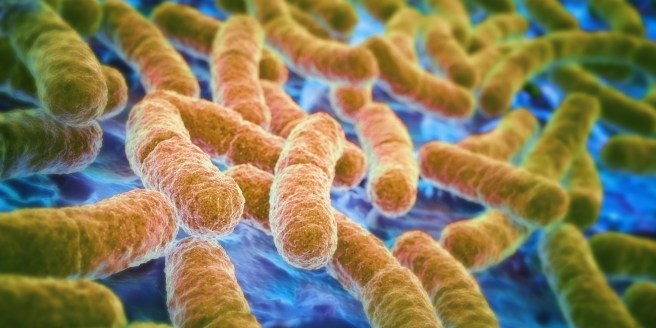
Bacteria in the gut. Source: Huffington Post

Before you go to bed tonight, contemplate this:

The human gut hosts tens of trillions of microorganisms, including at least 1000 species of bacteria (which is a guess-timate as we are not really sure how many species there are).

And whenever you feel like you are all alone, know that you are not.

You are never alone: tens of trillions of microorganisms are with you!

And there is sooooooo many of these microorganisms, that they can make up as much as 2 kg of your total weight.

What do the microorganisms do?

Ours bodies are made up of microbiota – that is,  collections of microbes or microorganisms inhabiting particular environments (or region of our body) and creating “mini-ecosystems”. And whether you like this idea or not, you need them.

The microorganisms in the human gut, for example, perform all manner of tasks for you to make your life easier. From helping to break down food, to aiding with the production of some vitamins (in particular B and K).

That’s great, but what does the bacteria in our gut have to do with Parkinson’s disease?

People with Parkinson’s disease quite often have issues associated with the gastrointestinal tract (or the gut), such as constipation for example. Some people believe that some of these gut related symptoms may actually pre-date a diagnosis of Parkinson’s disease, which has led many researchers to speculate as to whether the gut could be a starting point for the condition.

We have previously discussed the gut and Parkinson’s disease in several posts (click here, here and here to read them).

Today we re-address this topic because a group of scientists from the USA have determined that the populations of bacteria in the guts of people with Parkinson’s disease are different to those of healthy individuals.

Sounds interesting. What exactly is the difference?

Well, before we discuss that, we need a little bit of background.

In 2015, a group of scientists from Finland, published this research paper:

biota-title

Title: Gut microbiota are related to Parkinson’s disease and clinical phenotype.
Authors: Scheperjans F, Aho V, Pereira PA, Koskinen K, Paulin L, Pekkonen E, Haapaniemi E, Kaakkola S, Eerola-Rautio J, Pohja M, Kinnunen E, Murros K, Auvinen P.
Journal: Mov Disord. 2015 Mar;30(3):350-8.
PMID: 25476529

In this study the researchers compared the fecal microbiomes of 72 people with Parkinson’s disease and 72 control subjects by sequencing the V1-V3 regions of the bacterial 16S ribosomal RNA gene.

Hang on a minute. What does… any of that mean?

Yeah. Ok, that was a bit technical.

The microbiome refers to the genetics of the microorganisms – that is their genomes (or DNA). When researchers want to look at the microbiome of your gut, they do so by collecting fecal samples (delightful job, huh?).

Interesting facts: Fresh feces is made up of approx. 75% water. Of the remaining solid fraction, 84–93% is organic solids. These organic solids consist of: 25–55% gut bacterial matter, 2–25% protein, 25% carbohydrates, and 2–15% fat (Source: Wikipedia).

Still with me?

After collecting the fecal samples, researchers will extract the DNA from the gut bacterial material, which they can then analyse.

And what are the V1-V3 regions of the bacterial 16S ribosomal RNA gene?

The 16S ribosomal RNA gene is universal in bacteria – it is present in all of their genomes/DNA. The genetic sequence of this particular gene is approximately 1,550 base pairs long, and contains regions that are highly conserved (that is they are shared between species) and highly variable (very different between species).

M_00-06 DNA based kuva 3.jpg

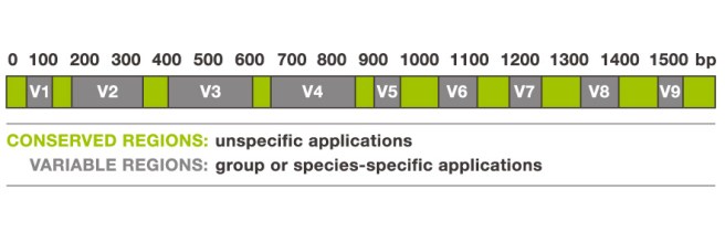
The 16S ribosomal RNA gene. Source: Alimetrics

The gene contains nine of these highly variable regions (called V1 – V9) that display considerable differences in the genetic sequence between different groupings of bacteria. The V2 and V3 regions are considered the most suitable for distinguishing all bacterial species to the genus level (‘genus‘ being a method of classification).

Now scientist can amplify the 16S ribosomal RNA gene by making lots of copies of the highly conserved regions (using PCR) which are shared between bacteria, but then they will genetic sequence the variable sections in between (in this case V2 & V3), which will allow them to discriminate and quantify the different species of microorganisms (such as bacteria) within a particular sample.

16s-ribosomal-dna-sequence-analysis-13-638

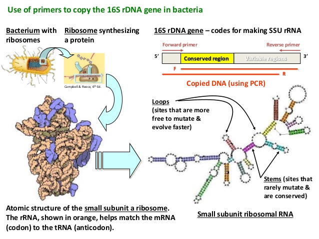
16S rRNA gene analysis – looks complicated. Source: Slideshare

And this is what the scientists in this study did.

They took fecal samples of 72 people with Parkinson’s disease and 72 control subjects, amplified the V1-V3 regions of the bacterial 16S ribosomal RNA gene, and then sequenced the variables regions in between to determine what sorts of bacteria were present (and/or different) in the guts of people with Parkinson’s disease.

The researchers found that there was a reduced abundance of Prevotellaceae in the guts of people with Parkinson’s disease (Prevotellaceae are commonly found in the gastric system of people who maintain a diet low in animal fats and high in carbohydrates, for example vegetarians).

216-5198-1-pb

Prevotella multisaccharivorax which belongs to the Prevotellaceae family. Source: MindsofMalady

In addition, the investigators also reported a positive association between the abundance of Enterobacteriaceae and postural instability and gait difficulty symptoms – that is to say, people with Parkinson’s disease who also had postural instability and gait difficulties had significantly more Enterobacteriaceae in their guts than people with Parkinson’s disease who were more tremor dominant.

Due to the design of the study, the researchers were not able to make conclusions about causality from their study. Neither could they tell whether the microbiome changes were present before the onset of Parkinson’s disease or whether they simply developed afterwards. All they could really say was at the time of analysis, they did see a difference in the gut microbiota between people with and without Parkinson’s disease.

And while these same researchers are currently conducting a two year follow up study to determine the stability of these differences over time in the same subjects, they admit that much larger prospective studies are required to address such issues as causality.

Which brings us to the new research published last week:

gut-title

Title: Parkinson’s disease and Parkinson’s disease medications have distinct signatures of the gut microbiome.
Authors: Hill-Burns EM, Debelius JW, Morton JT, Wissemann WT, Lewis MR, Wallen ZD, Peddada SD, Factor SA, Molho E, Zabetian CP, Knight R, Payami H.
Journal: Mov Disord. 2017 Feb 14. [Epub ahead of print]
PMID: 28195358

The researchers in this study (completely independent from the previous study) applied the same study design as the previous study, but on a much larger scale:

They took samples from a total of 197 people with Parkinson’s disease and 130 healthy controls. And importantly, none of the individual subjects in the study were related (this was an attempt to reduce the effect of shared microbiota between people who live together). Participants were enrolled from the NeuroGenetics Research Consortium in the cities of Seattle (Washington), Atlanta (Georgia) and Albany (New York).

So what did they find?

The researcher’s data revealed alterations in at least 7 families of bacteria: Bifidobacteriaceae, Christensenellaceae, Tissierellaceae, Lachnospiraceae, Lactobacillaceae, Pasteurellaceae, and Verrucomicrobiaceae families

Of particular interest was their observation of reduced levels of Lachnospiraceae in Parkinson’s disease subjects. Lachnospiraceae is involved with the production of short chain fatty acids (SCFA) in the gut. Depletion of SCFA has been implicated in the pathogenesis of Parkinson’s disease (Click here for more on this), and it could potentially explain the inflammation and microglial cell activation observed in the brain (Click here for more on this).

Importantly, they did not replicate the association of Parkinson’s disease with Prevotellaceae (see the previous study above).

The investigators also looked at the medication that the subjects were taking and they found a significant difference in the gut microbiome in relation to treatment with COMT inhibitors and anticholinergics. The effects of COMT inhibitors and anticholinergics on hte microbiome was independent of the effect that Parkinson’s disease was having.

The investigators concluded that Parkinson’s disease is accompanied by ‘dysbiosis of gut microbiome’ (that is, microbial imbalance). Again they could not determine whether the ‘chicken came before the egg’ so to speak, but it will be interesting to see what follow up work in this study highlights.

What does it all mean?

The studies that we have reviewed today provide us with evidence that the bacteria in the guts of people with Parkinson’s disease are different to that of healthy control subjects. Whether the differences between the studies results are due to regional effects (Finland vs USA) will require further investigation. But given that so much attention is now focused on the role of the gut in Parkinson’s disease, it is interesting that there are differences in the gut microbiome between people with and without Parkinson’s disease.

One issue that both studies do not address is whether this difference is specific to Parkinson’s disease and not other neurodegenerative conditions. That is to say, it would have been very interesting if the investigators had included a small set of samples from people with Alzheimer’s disease, for example. This would indicate which differences are specific to Parkinson’s disease as opposed to differences that a general to individuals who have a neurodegenerative condition. If they can tease out medication-related differences (in the second study), then this should be a do-able addition to any future studies.

One would also hope that the researchers will go back and dig a little deeper with future analyses. Using 16S ribosomal RNA gene analysis to determine and quantify the different families of bacteria is analogous to dividing people according to hair and eye colour. The bacteria of our gut is a lot more complicated than this review has suggested. For example, future studies and follow up research could include some genetic techniques that go beyond simply sequencing the 16S ribosomal RNA gene. The investigators could sequence the entire genomes of these species of bacteria to see if genetic mutations within a particular family of bacteria is present in people with Parkinson’s disease.

Easy to say of course. A lot of work, in practise.

There is most likely going to be more of a focus on the gastrointestinal tract in Parkinson’s disease research as a result of these studies. It will be interesting to see where this research leads.


The banner for today’s post was sourced from Youtube

Pink flies in Leicester at it again

mito-em-web-1-1024x256

Imagine discovering a protein that could make the power supply of your cells healthier AND perhaps provide a new therapeutic target for Parkinson’s disease.

That would be a pretty big deal right?

Well, this week, researchers may have found just such a protein. In today’s post we will review their finding and discuss what it means for Parkinson’s disease.


This is Dr Miguel Martins:

miguel_martins

Source: Tox.mrc.ac.uk

He’s a dude.

Dr Martins is a group leader at the MRC toxicology unit in Leicester – a city in the East Midlands of England.

leicester-town-hall-squareLeicester. Source: Keithvazmp

You may have heard of Leicester. Last year their football team had a dream season, miraculously winning the Premier league title despite starting with odds of 5000:1.

hd-leicester-city-champions_1d6y6oasvbk3n1q8iqxzkguv82

Last season’s winners. Source: Goal.com

This season, however,….well, uh…

Let’s move on, shall we.

Recently we reviewed Dr Martins research group’s work on ‘Pink flies’ and how they survive longer on Niacin rich diets (Click here for that post). He and his group were again publishing research this week, involving new a new study highlighting a protein that may help with keeping mitochondria healthy.

What are mitochondria?

Good question.

Mitochondria are the power house of each cell. They keep the lights on. Without them, the lights go out and the cell dies.

Mitochondria

Mitochondria and their location in the cell. Source: NCBI

You may remember from high school biology class that mitochondria are bean-shaped objects within the cell. They convert energy from food into Adenosine Triphosphate (or ATP). ATP is the fuel which cells run on. Given their critical role in energy supply, mitochondria are plentiful and highly organised within the cell, being moved around to wherever they are needed.

So what has Dr Martins group found?

This week they published this study:

atf4

Title: dATF4 regulation of mitochondrial folate-mediated one-carbon metabolism is neuroprotective.
Authors: Celardo I, Lehmann S, Costa AC, Loh SH, Miguel Martins L.
Journal: Cell Death Differ. 2017 Feb 17. [Epub ahead of print]
PMID: 28211874       (This article is OPEN ACCESS if you would like to read it)

In the study, the researchers were interested in determining what changes occur in the flies that are missing the Parkinson’s disease associated genes PINK1 or PARKIN, particularly which transcription factors are affected.

What is a transcription factor?

Another good question.

Ok, so you remember your high school science class when the adult at the front of the class was explaining biology 101? And they were saying that DNA gives rise to RNA, RNA gives rise to protein? The central dogma of biology. Remember this?

maxresdefault

The basic of biology. Source: Youtube

Ultimately this DNA-RNA-Protein mechanism is a circular cycle, because the protein that is produced using RNA is required at all levels of this process. Some of the protein is required for making RNA from DNA, while other proteins are required for making protein from the RNA instructions.

A transcription factor is a protein that is involved in the process of converting (or transcribing) DNA into RNA.

Importantly, a transcription factor can be an ‘activator’ of transcription – that is initiating or helping the process of generating RNA from DNA.

6567f50d30ad3ac65aff1e815caf202b3abd7111

An example of a transcriptional activator. Source: Khan Academy

Or it can be a repressor of transcription – blocking the machinery (required for generating RNA) from doing it’s work.

6286f2dbd5e353145bef785aecb273d25176ff23

An example of a transcriptional repressor. Source: Khan Academy

In their study, Dr Martins and colleagues were looking for changes in the levels of proteins that either initiate or repress transcription, as these are the proteins that are ultimately at the start of the process of making things happen.

And what do Parkin and Pink1 actually do?

About 10% of cases of Parkinson’s disease can be attributed to genetic mutations in particular genes. PINK1 and PARKIN are two of those genes.

People with particular mutations in the PINK1 or PARKIN gene are vulnerable to developing an early onset form of Parkinson’s disease.

As to what the protein that is generated from PINK1 or PARKIN DNA & RNA, well in normal, healthy cells, the PINK1 protein is absorbed by mitochondria and eventually degraded. In unhealthy cells, however, this process is inhibited and PINK1 starts to accumulate on the outer surface of the mitochondria. There, it starts grabbing the PARKIN protein. This pairing is a signal to the cell that this particular mitochondria is not healthy and needs to be removed.

601587-fig-003

Pink1 and Parkin in normal (right) and unhealthy (left) situations. Source: Hindawi

The process by which mitochondria are removed is called autophagy. Autophagy is an absolutely essential function in a cell. Without it, old proteins will pile up making the cell sick and eventually it dies. Through the process of autophagy, the cell can break down the old protein, clearing the way for fresh new proteins to do their job.

Think of autophagy as the waste disposal process of the cell.

In the absence of PINK1 and PARKIN – as is the case in some people with Parkinson’s disease who have genetic mutations in these genes – we believe that sick/damaged mitochondria start to pile up and are not disposed of appropriately. This results in the cell dying.

Ok, so the researchers were looking for transcription factors that change in the absence of PINK1 and PARKIN. How did they do this experiment?

They used flies.

pink_fly-1410843

PINK flies. Source: Wallpapersinhq

The researchers took the heads (yes, I know, delightful stuff) of ‘young’ 3-day-old Pink1 and Parkin mutant flies and compared them to ‘aged’ heads from 21- and 30-day-old Parkin and Pink1 mutant flies, respectively. The comparison was specifically looking at transcription factors that change over time.

This analysis revealed a protein called activating transcription factor 4 (or ATF4).

The researchers found that ATF4 levels were higher in both Pink1 and Parkin mutants than levels in control flies. Importantly, the researchers next looked at the genes that this transcription factor (ATF4) was regulating, and they found that ATF4 was encouraging the production of proteins that protect mitochondria. The researchers noticed that when they reduced ATF4 in flies, the levels of these critical mitochondrial proteins dropped as well.

When the researchers reduced the levels of each of these critical mitochondrial proteins in flies, it resulted in impaired climbing ability (suggesting a locomotor deficit) and decreased lifespan. Interestingly, these protective mitochondrial proteins are increased in the Pink1 and Parkin flies, suggesting that efforts to keep the mitochondria healthy are active inside the cells.

Finally, the researchers increased the levels of these protective mitochondrial proteins in the Pink1 and Parkin mutants and they found that the mitochondrial function was improved, and neuronal cell loss was avoided. They concluded that their findings demonstrate a central role for ATF4 signalling in Parkinson’s disease and that this protein may represent a target for new therapeutic strategy.

So what does it all mean?

The researchers behind this study were looking for biological pathways that are altered in genetic forms of Parkinson’s disease and they have identified a protein that is involved with keeping mitochondria healthy. This pathway could represent a new therapeutic target for future treatments, and also opens a new door in our understanding of Parkinson’s disease.

ATF4 is currently not directly targeted by any medications (that we are aware of), but there are drugs in clinical trials that target proteins that subsequently activate ATF4. For example, Oncoceutics Inc. have a drug candidate called ONC201 (currently in phase II trials for brain cancer) which kills solid tumor cells by triggering an stress response which is dependent on ATF4 activation.

moa-diagram-5-31-16

Source: Oncoceutics Inc

We are not for a second suggesting that this is a viable drug for Parkinson’s disease (so PLEASE DON’T rush out and besiege the company for all of their stocks!) – ATF4 should be considered a very experimental target until these results are replicated by independent research groups. We are mentioning ONC201 here simply to indicate that there is a field of research surrounding this potential target (ATF4) and it may be worthwhile for the Parkinson’s community to follow up this line of investigation.

We are assuming that while Leicester football club is struggling, the Martins lab are currently investigating compounds that activate ATF4 (and the other critical mitochondrial proteins), and we will report any follow up work as it comes to hand.

Watch this space.


And if nothing we’ve written here makes any sense, the good folks at Leicester University have kindly provided a short video explaining the research:


Postscript (March 2017):

matters_journal

The Martins lab have done it again!

This time in the OPEN ACCESS online journal Science Matters, they have published this article:

Matters

Title: Folinic acid is neuroprotective in a fly model of Parkinson’s disease associated with pink1 mutations
Authors: Lehmann S , Jardine J, Garrido – Maraver J, Loh SH, & Martins LM
Journal: Science Matters

In this study, the researchers demonstrated that a folinic acid-enriched diet might delay or prevent the neuronal loss in people with PINK1 associated Parkinson’s disease. They present data suggesting that beginning an intake of Folinic acid in early to middle stages of adulthood prevents the degeneration of dopamine neurons in pink1 mutant flies.

Folinic acid (also known as leucovorin) is a medication used to decrease the toxic effects of chemotherapy drugs. The pharmacokinetics of leucovorin suggests that it readily crosses the blood-brain-barrier (Source), so it would be possible for a clinical trial to be set up in human. Before taking that path, however, more testing is required (ideally in a mammalian model of Parkinson’s disease).

Amazing that all these results are coming from silly old flies though, huh?


The banner for today’s post was sourced from Tox.mrc.ac.uk

HIV and Parkinson’s disease

hiv-aids-definition2


I was recently made aware of an interesting fact:

Approximately 5% of people with Human immunodeficiency virus (HIV) infections develop Parkinson’s disease-like features.

Why is this?

In today’s post we will try to understand what is going on, and what it may mean for Parkinson’s disease.


hiv-budding-colo2r

HIV (in green) budding (being released) from a blood cell (lymphocyte). Source: Wikipedia

Ok, let’s start at the beginning:

What is HIV?

Human immunodeficiency virus (or HIV) – as the name suggests – is the virus.

It causes the infection which gives rise to Acquired Immune Deficiency Syndrome (or AIDS). AIDS is a progressive failure of the immune system – the body loses its ability to fight infections. Without treatment, average survival period after infection with HIV is between 9 – 12 years.

HIV can be spread by the transfer of bodily fluids, such as blood and semen. The World Health Organisation (WHO) has estimated that approximately 36.9 million people worldwide were living with HIV/AIDS at the end of 2014 (that is equivalent to the entire population of Canada!).

hi-virion-structure_en-svg

The structure of the HIV virus. Source: Wikipedia

Does HIV affect the brain?

Yes.

At postmortem examinations, less than 10% of the brains from HIV infected individuals are histologically normal (Source).

HIV is a member of the lentivirus family of viruses, which readily infect immune cells (such as blood cells). HIV can also infect other types of cells though, including those in the brain. HIV will usually enter the central nervous system within the first month following infection. It enters the brain via infected blood cells which come into contact with brain ‘immune system/helper’ cells such as microglia and macrophages at the blood-brain-barrier.

f1-large

How HIV enters the brain. Source: Disease Models and Mechanisms

HIV can also infect astrocytes (albeit at a lower frequency than microglia and macrophages), by direct cell-cell contact with infected T cells (blood cells) at the blood-brain-barrier (No. 1 in the image above). After infecting astrocytes, there is dysfunction in the astrocyte and it will no longer be so supportive to the local neurons (No. 2 in the image above). Once inside the brain, HIV-infected macrophages will allow for infection of other macrophages and microglia (No. 3 in the image above), and all together these HIV-infected astrocytes and microglia will cause damage to neurons by releasing viral proteins (two in particular, called Tat and gp120) and additional nasty chemicals which are bad for the neurons (No. 4 in the image above). Finally, as the disease progresses, the protective layer of the blood-brain-barrier becomes compromised and HIV-infected T cells eventually enter the brain and they cause damage to neurons by releasing pro-inflammatory chemicals (making the environment harsh for neurons).

There is remarkably little evidence of HIV actually infecting neurons (Click here for a review on this), so any cell loss in the brain that is associated with HIV does not result from neurons themselves being infected. This may be due to the fact that neurons do not have the HIV receptors (such as CD4) on their cell membrane. Similarly, oligodendrocytes (a supporting cell) does not appear to be easily infected by HIV. The bulk of the infected cells in the brain appear to be of the microglial, macrophage and astrocytes. And without these supporting cells doing their jobs in a normal fashion, it is easy to see how neurons can start dying off.

The severity, characteristics and distribution of HIV-induced injury in the brain varies greatly between affected individuals. It is most likely associated with the viral load (or the number of viral particles) in the brain, which can vary from a few thousand to more than a million copies per mL.

Do HIV-infected people show any signs of the virus entering the brain?

For the majority of people infected with HIV, this entry of the virus into the nervous system is neurologically asymptomatic (meaning they will not notice it), except for the occasional mild headache (for more on this read this review). As a result of the HIV virus entering the brain, however, many infected individuals will suffer from a specific set of neurological disorders, collectively called the AIDS dementia complex (ADC) (also known as HIV-associated cognitive/motor complex, or simply HIV dementia).

So how does HIV infection result in Parkinson’s disease-like features?

As we have suggested in the introduction to this post, on rare occasions (approximately 5% of cases), HIV-infected patients may present an illness virtually identical to Parkinson’s disease. More commonly, people with HIV will exhibit an increased sensitivity to dopamine receptor-blocking agents, such as drugs with a low potential for inducing Parkinsonism, (for example prochlorperazine and metoclopropamide).

The exact mechanism by which HIV infection results in Parkinson’s disease-like features is the subject of debate, but what is clear is that the basal ganglia (a structure involved in Parkinson’s disease) faces the brunt of the HIV infection in the brain. HIV-infected microglia and macrophage are most prominent in the basal ganglia when compared to other brain regions (Click here and here for more on this), and the basal ganglia is where the chemical dopamine from the midbrain is being released.

In addition, there are other changes in the brains of HIV infected people which may aid in the appearance of Parkinsonian features:


viraltitle

Title: Increased frequency of alpha-synuclein in the substantia nigra in human immunodeficiency virusinfection.
Authors: Khanlou N, Moore DJ, Chana G, Cherner M, Lazzaretto D, Dawes S, Grant I, Masliah E, Everall IP; HNRC Group.
Journal: J Neurovirol. 2009 Apr;15(2):131-8.
PMID: 19115126       (This article is OPEN ACCESS if you would like to read it)

The researchers in this study used staining techniques to look at the amount of alpha synuclein – the Parkinson’s associated protein – in slices of brain tissue taken from postmortem autopsies of 73 HIV+ individuals aged between 50 and 76 years of age.

The presence of alpha synuclein in the substantia nigra (an area of the brain affected by Parkinson’s disease) was a lot higher in the HIV+ brains when compared with healthy control samples (16% of the HIV+ brains had high levels of alpha synclein vs 0% for the healthy brains).

Interestingly, nearly all of the brains analysed (35 out of 36 HIV+ brains) had high levels of the Alzheimer’s disease associated protein, beta amyloid (which again raises the question of whether beta amyloid could be playing a defensive role in infections – see our previous post on this). Also interesting, was that there was no correlation between these proteins being present and the age of the person at death – that is to say, older brains did not have more of these proteins when compared with younger brains.

There are also additional ways in which HIV could be causing Parkinson’s-like features, such as:

  • HIV has been shown to affect the protein levels of Parkinson’s disease associated proteins, such as DJ1 and Lrrk2 (Click here and here to read more on this).
  • HIV can, in some cases, increase the level of Dopamine transporter, which would reduce the levels of free floating dopamine in the brain (Click here to read more about this).

How is HIV treated?

aidspills

Treating HIV. Source: NPR

There is currently no cure for HIV infection.

There are, however, treatments which help to slow the virus down. These are called Anti-retroviral drugs (HIV is a retrovirus). There are different kinds of anti-retroviral drugs, which act at different stages of the HIV life cycle. Combinations of several anti-retroviral drugs (generally three or four) is known as ‘Highly Active Anti-Retroviral Therapy'(or HAART).

hiv-drug-classes-svg

Mechanism by which four classes of anti-retroviral drugs work against HIV. Source: Wikipedia

As the schematic image above highlights, there are many ways to slow down the HIV virus. For example, you can prevent it from attaching to a cell and fusing with the cell membrane (fusion inhibitors). By treating HIV infected people with multiple medications attacking different parts of the HIV life cycle, the virus has been slowed down.

Does HAART treatments for HIV help with these Parkinson’s-like features?

In some cases, the answer appears to be yes.

There are numerous case studies in the literature which demonstrate the alleviation of HIV-associated Parkinsonian symptoms with HAART, such as this report:

hersh

Title: Parkinsonism as the presenting manifestation of HIV infection: improvement on HAART.
Authors: Hersh BP, Rajendran PR, Battinelli D.
Journal: Neurology. 2001 Jan 23;56(2):278-9.
PMID: 11160977

In this study the researchers described the case of a 37 year old man who developed Parkinson’s like features in the setting of an HIV infection, which were resolved after 1 year of HAART.

Over a period of 4 months, the man developed co-ordination issue, clumsiness and an irregular tremor in his right hand (there was, however, no resting tremor). He noted a generalised slowness and exhibited a tendency towards decreased right arm swinging. He also developed dystonia in the right hand/arm. Following L-dopa treatment (25/100; one tablet 3x per day) there was improvement in balance & co-ordination, speech, facial expression, and the tremor (L-dopa does appear to improve most cases of HIV-associated Parkinson’s-like features).

Six months after first displaying these Parkinsonian features (and two month after initiating L-dopa treatment), the subject was placed on HAART treatment. Four months later, he discontinued L-dopa treatment and 12 months after starting the HAART regime his Parkinsonian features were largely resolved.

More case studies of HAART alleviating HIV-associated Parkinsonisms can be found by clicking here and here.

What does this mean for Parkinson’s disease?

This post was written for the research community rather than people with Parkinson’s disease. I thought the fact that some people with HIV can start to have Parkinson’s like features was an interesting curiosity and wanted to share/spread the information.

Having said that, this post raises some really interesting questions, such as if a virus like HIV can have this effect on the brain, could other viruses be having similar effects? Could some cases of Parkinson’s disease simply be the result of a viral infection? Either multiple hits from a particular virus or different viruses each taking a varying toll over the course of a life time.

This idea would explain many of the curious features of Parkinson’s disease, such as:

  • the asymmetry of the symptoms (people with Parkinson’s usually have the disease starting on one side of the body.
  • the fact that some cells in the brain are more vulnerable to the disease than others (perhaps they are more receptive to a particular virus).
  • the protein clusterings in the cells (Lewy bodies may be defensive efforts against viral infections).

As we have previous mentioned, theories of viral causes for Parkinson’s have been circulating ever since the 1918 flu pandemic (Click here to read our previous post on this topic). About the same time as the influenza virus was causing havoc around the world, another condition began to appear called ‘encephalitis lethargica‘. This disease left many of the victims in a statue-like condition, both motionless and speechless – similar to Parkinson’s disease. Initially, it was assumed that the influenza virus was the causal factor, but more recent research has left us not so sure anymore.

The point is, however, perhaps it is time for us to re-examine the possibility of a viral agent being involved in the development of Parkinson’s disease.

There is new technology that allows us to determine the viral history of each individual from a simple blood test (Click here for more on this), so it would be interesting to compare blood samples from people with Parkinson’s disease with healthy controls to determine any differences.

In addition to the overall question of a viral role in Parkinson’s disease, there also remains the question of why only a small fraction of people with HIV are affected by Parkinsonisms. It could be interesting to genetically screen those people with HIV that exhibit Parkinsonisms and compare them with people with HIV that do not. Do those affected individuals have recognised Parkinson’s related genetic mutations? Or do they have novel genetic variations that could tell us more about Parkinson’s disease?

Food for thought. Would be happy to hear others thoughts.


The banner for today’s post was sourced from AidsServices

Busy day for Parkinson’s – 9/2/2017


o-busy-facebook

Today there was a lot of Parkinson’s related activity in the news… well, more than usual at least.

Overnight there was the publication of a blood test for Parkinson’s disease, which looks very sensitive. And this afternoon, Acorda Therapeutics announced positive data for their phase three trial.

In this post, we’ll look at what it all means.


happens-many-red-blood-cells_891c9a08c6bfe4aa

Blood cells. Source: Reference.com

Today we found out about an interesting new study from scientists at Lund University (Sweden), where they are developing a test that can differentiate between different types of Parkinsonisms (See our last post about this) using a simple blood test.

We have previously reported about an Australian research group working on a blood test for Parkinson’s disease, but they had not determined whether their test could differentiate between different kinds of neurodegenerative conditions (such as Alzheimer’s disease). And this is where the Swedish study has gone one step further…

blood
Title: Blood-based NfL: A biomarker for differential diagnosis of parkinsonian disorder
Authors: Hansson O, Janelidze S, Hall S, Magdalinou N,  Lees AJ, Andreasson U, Norgren N, Linder J, Forsgren L, Constantinescu R, Zetterberg H, Blennow K, & For the Swedish BioFINDER study
Journal: Neurology, Published online before print February 8, 2017
PMID: N/A       (This article is OPEN ACCESS if you would like to read it)

The research group in Lund had previously demonstrated that they could differentiate between people with Parkinson’s disease and other types of Parkinsonism to an accuracy of 93% (Click here to read more on this). That is a pretty impressive success rate – equal to basic clinical diagnostic success rates (click here for more on this).

The difference was demonstrated in the levels of a particular protein, neurofilament light chain (or Nfl). NfL is a scaffolding protein, important to the cytoskeleton of neurons. Thus when cells die and break up, Nfl could be released. This would explain the rise in Nfl following injury to the brain. Other groups (in Germany and Switzerland) have  also recently published data suggesting that Nfl could be a good biomarker of disease progression (Click here to read more on this).

There was just one problem: that success rate we were talking about above, it required cerebrospinal fluid. That’s the liquid surrounding your brain and spinal cord, which can only be accessed via a lumbar puncture – a painful and difficult to perform procedure.

lumbar-puncture-cropped

Lumbar puncture. Source: Lymphomas Assoc.

Not a popular idea.

This led the Swedish researchers to test a more user friendly approach: blood.

In the current study, the researchers took blood samples from three sets of subjects:

  • A Lund set (278 people, including 171 people with Parkinson’s disease (PD), 30 people with Multiple system atrophy (MSA), 19 people with Progressive Supranuclear Palsy (PSP), 5 people with corticobasal syndrome (CBS), and 53 people who were neurologically healthy (controls).
  • A London set (117 people, including 20 people with PD, 30 people with MSA, 29 people with PSP, 12 people with CBS, and 26 neurologically healthy controls
  • An early disease set (109 people, including 53 people with PD, 28 people with MSA, 22 people with PSP, 6 people with CBS). All of the early disease set had a disease duration less than 3 years.

When the researchers looked at the levels of NfL in blood, they found that they could distinguish between people with PD and people with PSP, MSA, and CBS with an accuracy of 80-90% – again a very impressive number!

One curious aspect of this finding, however, is that the levels of Nfl in people with PD are very similar to controls. So while this protein could be used to differentiate between PD and other Parkinsonisms, it may not be a great diagnostic aid for determining PD verses non-PD/healthy control.

In addition, what could the difference in levels of Nfl between PD and other Parkinsonisms tell us about the diseases themselves? Does PD have less cell death, or a more controlled and orderly cell death (such as apoptosis) than the other Parkinsonisms? These are questions that can be examined in follow up work.


but-wait

Source: 3rd-Solutions

Like we said at the top, it’s been a busy day for Parkinson’s disease: Good news today for Acorda Therapeutics, Inc.

acordatherapeuticslogo_calogo2306

Source: Acorda

They announced positive Phase 3 clinical trial results for their inhalable L-dopa treatment, called CVT-301, which demonstrated a statistically significant improvement in motor function in people with Parkinson’s disease experiencing OFF periods.

We have previously discussed the technology and the idea behind this approach to treating Parkinson’s disease (Click here for that post).

levodopa-inhaler-lead-658x394

The ARCUS inhalation technology. Source: ParkinsonsLife

Basically, the inhaler contains capsules of L-dopa, which are designed to break open so that the powder can escape. By sucking on the inhaler (see image below), the open capsule starts spinning, releasing the levodopa into the air and subsequently into the lungs. The lungs allow for quicker access to the blood system and thus, the L-dopa can get to the brain faster. This approach will be particularly useful for people with Parkinson’s disease who have trouble swallowing pills/tablets – a common issue.

The Phase 3, double-blind, placebo-controlled clinical trial evaluated the efficacy and safety of CVT-301 when compared with a placebo in people with Parkinson’s disease who experience motor fluctuations (OFF periods). There were a total of 339 study participants, who were randomised and received either CVT-301 or placebo. Participants self-administered the treatment (up to five times daily) for 12 weeks.

The results were determined by assessment of motor score, as measured by the unified Parkinson’s disease rating scale III (UPDRS III) which measures Parkinson’s motor impairment. The primary endpoint of the study was the amount of change in UPDRS motor score at Week 12 at 30 minutes post-treatment. The change in score for CVT-301 was -9.83 compared to -5.91 for placebo (p=0.009). A negative score indicates an improvement in overall motor ability, suggesting that CVT-301 significantly improved motor score.

The company will next release 12-month data from these studies in the next few months, and then plans to file a New Drug Application (NDA) with the Food and Drug Administration (FDA) in the United States by the middle of the year and file a Marketing Authorization Application (MAA) in Europe by the end of 2017. This timeline will depend on some long-term safety studies – the amount of L-dopa used in these inhalers is very high and the company needs to be sure that this is not having any adverse effects.

All going well we will see the L-dopa inhaler reaching the clinic soon.


The banner for today’s post was sourced from the Huffington Post

George H and Vascular Parkinsonism

george-hw-bush

During Super Bowl 51, ex-president George HW Bush was visibly wheel chair bound. He has in fact been using motorised scooters and wheelchairs since 2012.

His doctors have indicated that he suffers from Vascular Parkinsonism.

In today’s post we will discuss what Vascular Parkinsonism is and how it differs from Parkinson’s disease.


gettyimages-173500627

During a visit to the White house. Source: Heavy

An important concept to understand about the subject matter here:

Parkinsonism is a syndrome, while Parkinson’s is a disease.

A syndrome is a collection of symptoms that characterise a particular condition, while a disease is a pathophysiological response to internal or external factors. The term ‘Parkinsonism’ is an umbrella term that encompasses many conditions which share some of the symptoms of Parkinson’s disease.

There are many different types of Parkinsonism, such as:

  • Idiopathic Parkinson’s disease (the most common type of parkinsonism)
  • Progressive Supranuclear Palsy (PSP)
  • Corticobasal Degeneration (CBD)
  • Multiple System Atrophy (MSA)
  • Essential tremor
  • Vascular Parkinsonism
  • Drug-induced Parkinsonism
  • Dementia with Lewy bodies
  • Inherited Parkinson’s disease
  • Juvenile Parkinson’s disease

All of these conditions fall under the syndrome title of ‘Parkinsonism’, but are all considered distinct/separate diseases in themselves.

So what is Vascular Parkinsonism?

Vascular Parkinsonism was first described in 1929 by Dr Macdonald Critchley (King’s College Hospital, London).

409633

Macdonald Critchley. Source: Npgprints

vascularpd

Title: Arteriosclerotic Parkinsonism.
Author: Critchley, M.
Journal: Brain (1929) 52, 23–83
PMID: N/A                                (this article is accessible by clicking here)

It is estimated that approximately 3% to 6% of all cases of Parkinsonism may have a vascular cause. Vascular (or Arteriosclerotic) Parkinsonism is results from a series of small strokes in the basal ganglia area of the brain and can lead to the appearance of symptoms that look like Parkinson’s disease: slow movements, tremors, difficulty walking, and rigidity.

Walking problems are particularly prominent with Vascular Parkinsonism, as the lower half of the body is usually more affected than the upper half. Another sign of Vascular Parkinsonism can be a poor or no response to L-dopa treatment, as production of dopamine is not the problem. Using brain scanning techniques we can see that some people with Vascular Parkinsonism will have a normal Dopamine transporter (DAT) scan – which demonstrates appropriate levels of dopamine being released and reabsorbed in the striatum (the red-white areas in the image below).

f1-large

DAT-scan and MR images of 62-y-old male  with Vascular Parkinsonism (A) and 62-y-old male with Parkinson’s disease (B). Source: JNM

The brain scans above are from a person with Vascular Parkinsonism (A) and another person with Parkinson’s disease (B). Firstly, note the reduced levels of red-white areas in the image (B) – this reduction is due to less dopamine is being released and reabsorbed in the striatum in Parkinson’s disease (as there are less dopamine fibres present). Compare that with the relatively normal levels of red-white areas in the image (A), indicating normal levels of dopamine turnover (suggesting dopamine fibres are still present). Next, look at the black and white image in panel (A) and you will see a red arrow pointing at damaged areas (darkened regions) of the striatum – indicative of mini strokes. A dopamine receptor scan may show a reduction in the levels of dopamine receptors as a result of the strokes, meaning that the released dopamine will not be having much effect.

Do we know what can cause the strokes associated with Vascular Parkinsonism?

The symptoms of Vascular Parkinsonism tend to appear suddenly and generally do not progress, unlike Parkinson’s disease. We don’t know for sure what causes the mini strokes associated with Vascular Parkinsonism, and it probably varies from person to person, In general, however, doctors believe that high blood pressure and diabetes are the most likely causal factors (heart disease may also play a role).

What does it all mean?

Some people of Parkinson’s disease may actually have Vascular Parkinsonism, which can result from mini strokes in the basal ganglia region of the brain. They will usually be unresponsive to L-dopa and have more motor issues with their lower half of the body.

While Ex-President George HW Bush’s situation is extremely unfortunate, it reminds us that not all forms of Parkinsonism are Parkinson’s disease – an important factor to keep in mind when considering treatment regimes. We have posted this information here to make the Parkinson’s community more aware of this form of Parkinsonism. Later in the year we will discuss another form of Parkinsonism.


The banner for today’s post was sourced from Ew