2017 – Year in Review: A good vintage

At the end of each year, it is a useful practise to review the triumphs (and failures) of the past 12 months. It is an exercise of putting everything into perspective. 

2017 has been an incredible year for Parkinson’s research.

And while I appreciate that statements like that will not bring much comfort to those living with the condition, it is still important to consider and appreciate what has been achieved over the last 12 months.

In this post, we will try to provide a summary of the Parkinson’s-related research that has taken place in 2017 (Be warned: this is a VERY long post!)


The number of research reports and clinical trial studies per year since 1817

As everyone in the Parkinson’s community is aware, in 2017 we were observing the 200th anniversary of the first description of the condition by James Parkinson (1817). But what a lot of people fail to appreciate is how little research was actually done on the condition during the first 180 years of that period.

The graphs above highlight the number of Parkinson’s-related research reports published (top graph) and the number of clinical study reports published (bottom graph) during each of the last 200 years (according to the online research search engine Pubmed – as determined by searching for the term “Parkinson’s“).

PLEASE NOTE, however, that of the approximately 97,000 “Parkinson’s“-related research reports published during the last 200 years, just under 74,000 of them have been published in the last 20 years.

That means that 3/4 of all the published research on Parkinson’s has been conducted in just the last 2 decades.

And a huge chunk of that (almost 10% – 7321 publications) has been done in 2017 only.

So what happened in 2017? Continue reading “2017 – Year in Review: A good vintage”

Novartis focuses on improving PARKIN control

Last week, as everyone was preparing for Christmas celebrations, researchers at the pharmaceutic company Novartis published new research on a gene that is involved with Parkinson’s, called PARKIN (or PARK2).

They used a new gene editing technology – called CRISPR – to conduct a large screening study to identify proteins that are involved with the activation of PARKIN.

In today’s post we will look at what PARKIN does, review the research report, and discuss how these results could be very beneficial for the Parkinson’s community.


Source: Novartis

As many people within the Parkinson’s community will be aware, 2017 represented the 200th anniversary of the first report of Parkinson’s disease by James Parkinson.

It also the 20th anniversary of the discovery of first genetic mutation (or variant) that increases the risk of developing Parkinson’s. That genetic variation occurs in a region of DNA (a gene) called ‘alpha synuclein’. Yes, that same alpha synuclein that seems to play such a critical role in Parkinson’s (Click here to read more about the 20th anniversary).

In 2018, we will be observing the 20th anniversary of the second genetic variation associated with Parkinson.

That gene is called PARKIN:

Title: Mutations in the parkin gene cause autosomal recessive juvenile parkinsonism.
Authors: Kitada T, Asakawa S, Hattori N, Matsumine H, Yamamura Y, Minoshima S, Yokochi M, Mizuno Y, Shimizu N
Journal: Nature. 1998 Apr 9; 392(6676):605-8
PMID: 9560156

In 1998, Japanese researchers published this report based on 5 individuals from 4 Japanese families who were affected by juvenile-onset Parkinson’s. In family 1, the affected individual was a female, 43 years old, born of first-cousin parents, and her two younger brothers are healthy. Her condition was diagnosed in her teens and it had then progressed very slowly afterwards. Her response to L-dopa was very positive, but L-dopa-induced dyskinesia were frequent. In family 2-4, affected individuals (born to unrelated parents) exhibited very similar clinical features to the subject in family 1. The age of onset was between 18 to 27 years of age.

Using previous research and various techniques the investigators were able to isolate genetic variations that were shared between the 5 affected individuals. They ultimately narrowed down their search to a section of DNA containing 2,960 base pairs, which encoded a protein of 465 amino acids.

They decided to call that protein PARKIN.

PARKIN Protein. Source: Wikipedia

How much of Parkinson’s is genetic?

Continue reading “Novartis focuses on improving PARKIN control”

Non-invasive gene therapy: “You never monkey with the truth”

Gene therapy involves treating medical conditions at the level of DNA – that is, altering or enhancing the genetic code inside cells to provide therapeutic benefits rather than simply administering drugs. Usually this approach utilises specially engineered viruses to deliver the new DNA to particular cells in the body.

For Parkinson’s, gene therapy techniques have all involved direct injections of these engineered viruses into the brain – a procedure that requires brain surgery. This year, however, we have seen the EXTREMELY rapid development of a non-invasive approach to gene therapy for neurological condition, which could ultimately see viruses being injected in the arm and then travelling up to the brain where they will infect just the desired population of cells.

Last week, however, this approach hit a rather significant obstacle.

In today’s post, we will have a look at this gene therapy technology and review the new research that may slow down efforts to use this approach to help to cure Parkinson’s.


Gene therapy. Source: rdmag

When you get sick, the usual solution is to visit your doctor.

They will prescribe a medication for you to take, and then all things going well (fingers crossed/knock on wood) you will start to feel better. It is a rather simple and straight forward process, and it has largely worked well for most of us for quite some time.

As the overall population has started to live longer, however, we have begun to see more and more chronic conditions which require long-term treatment regimes. The “long-term” aspect of this means that some people are regularly taking medication as part of their daily lives. In many cases, these medications are taken multiple times per day.

A good example of this is Levodopa (also known as Sinemet or Madopar) which is the most common treatment for the chronic condition of Parkinson’s disease.

When you swallow your Levodopa pill, it is broken down in the gut, absorbed through the wall of the intestines, transported to the brain via our blood system, where it is converted into the chemical dopamine – the chemical that is lost in Parkinson’s disease. This conversion of Levodopa increases the levels of dopamine in your brain, which helps to alleviate the motor issues associated with Parkinson’s disease.

7001127301-6010801

Levodopa. Source: Drugs

This pill form of treating a disease is only a temporary solution though. People with Parkinson’s – like other chronic conditions – need to take multiple tablets of Levodopa every day to keep their motor features under control. And long term this approach can result in other complications, such as Levodopa-induced dyskinesias in the case of Parkinson’s.

Yeah, but is there a better approach?

Continue reading “Non-invasive gene therapy: “You never monkey with the truth””

CRISPR-Cas9: “New CRISPY Parkinson’s research”

Recently a Parkinson’s-associated research report was published that was the first of many to come.

It involves the use of a genetic screening experiment that incorporates new technology called ‘CRISPR’.

There is an absolute tidal wave of CRISPR-related Parkinson’s disease research coming down the pipe towards us, and it is important that the Parkinson’s community understands how this powerful technology works.

In today’s post we will look at what the CRISPR technology is, how it works, what the new research report actually reported, and discuss how this technology can be used to tackle a condition like Parkinson’s.


Me and my mother (and yes, the image is to scale). Source: Openclipart

My mother: Simon, what is all this new ‘crispy’ research for Parkinson’s I heard about on the news?

Me: Huh? (I was not really paying attention to the question. Terrible to ignore one’s mother I know, but what can I say – I am the black sheep of the family)

My mother: Yes, something about ‘crispy’ and Parkinson’s.

Me: Oh! You mean CRISPR. Yeah, it’s really cool stuff.

My mother: Ok, well, can you explain it all to me please, this ‘Crisper’ stuff?

Me: Absolutely.

CRISPR.101 (or CRISPR for beginners)

In almost every cell of your body, there is a nucleus.

It is the command centre for the cell – issuing orders and receiving information concerning everything going on inside and around the cell. The nucleus is also a storage bank for the genetic blueprint that provides most of the instructions for making a physical copy of you. Those grand plans are kept bundled up in 23 pairs of chromosomes, which are densely coiled strings of a molecule called Deoxyribonucleic acid (or DNA).

DNA’s place inside the cell. Source: Kids.Britannica

Continue reading “CRISPR-Cas9: “New CRISPY Parkinson’s research””

AAV-PHP.B: The future is apparently now

In addition to looking at current Parkinson’s disease research on this website, I like to look at where technological advances are taking us with regards to future therapies.

In July of this year, I wrote about a new class of engineered viruses that could potentially allow us to treat conditions like Parkinson’s disease using a non-invasive, gene therapy approach (Click here to read that post). At the time I considered this technology way off at some point in the distant future. Blue sky research. “Let’s wait and see” – sort of thing.

So imagine my surprise when an Italian research group last weekend published a new research report in which they used this futurist technology to correct a mouse model of Parkinson’s disease. Suddenly the distant future is feeling not so ‘distant’.

In today’s post we will review and discuss the results, and look at what happens next.


Technological progress – looking inside the brain. Source: Digitial Trends

I have said several times in the past that the pace of Parkinson’s disease research at the moment is overwhelming.

So much is happening so quickly that it is quite simply difficult to keep up. Not just here on the blog, but also with regards to the ever increasing number of research articles in the “need to read” pile on my desk. It’s mad. It’s crazy. Just as I manage to digest something new from one area of research, two or three other publications pop up in different areas.

But it is the shear speed with which things are moving now in the field of Parkinson’s research that is really mind boggling!

Source: Pinterest

Take for example the case of Squalamine.

In February of this year, researchers published an article outlining how a drug derived from the spiny dogfish could completely suppress the toxic effect of the Parkinson’s associated protein Alpha Synuclein (Click here to read that post).

The humble dogfish. Source: Discovery

And then in May (JUST 3 MONTHS LATER!!!), a biotech company called Enterin Inc. announced that they had just enrolled their first patient in the RASMET study: a Phase 1/2a randomised, controlled, multi-center clinical study evaluating a synthetic version of squalamine (called MSI-1436) in people with Parkinson’s disease. The study will enrol 50 patients over a 9-to-12-month period (Click here for the press release).

Source: Onemednews

Wow! That is fast.

Yeah, I thought so too, but then this last weekend a group in Italy published new research that completely changed my ideas on the meaning of the word ‘fast’. Regular readers will recall that in July I discussed amazing new technology that may one day allow us to inject a virus into a person’s arm and then that virus will make it’s way up to the brain and only infect the cells that we want to have a treatment delivered to. This represents non-invasive (as no surgery is required), gene therapy (correcting a medical condition with the delivery of DNA rather than medication). This new study used the same virus we discussed in July.

Continue reading “AAV-PHP.B: The future is apparently now”

Voyager Therapeutics: phase Ib clinical trial results


This week a biotech company called Voyager Therapeutics announced the results of their ongoing phase Ib clinical trial. The trial is investigating a gene therapy approach for people with severe Parkinson’s disease.

Gene therapy is a technique that involves inserting new DNA into a cell using a virus. The DNA can help the cell to produce beneficial proteins that go on help to alleviate the motor features of Parkinson’s disease.

In today’s post we will discuss gene therapy, review the new results and consider what they mean for the Parkinson’s community.


Source: Joshworth

On 25th August 2012, the Voyager 1 space craft became the first human-made object to exit our solar system.

After 35 years and 11 billion miles of travel, this explorer has finally left the heliosphere (which encompasses our solar system) and it has crossed into the a region of space called the heliosheath – the boundary area that separates our solar system from interstellar space. Next stop on the journey of Voyager 1 will be the Oort cloud, which it will reach in approximately 300 years and it will take the tiny craft about 30,000 years to pass through it.

Where is Voyager 1? Source: Tampabay

Where is Voyager actually going? Well, eventually it will pass within 1 light year of a star called AC +79 3888 (also known as Gliese 445), which lies 17.6 light-years from Earth. It will achieve this goal on a Tuesday afternoon in 40,000 years time.

Gliese 445 (circled). Source: Wikipedia

Remarkably, the Gliese 445 star itself is actually coming towards us. Rather rapidly as well. It is approaching with a current velocity of 119 km/sec – nearly 7 times as fast as Voyager 1 is travelling towards it (the current speed of the craft is 38,000 mph (61,000 km/h).

Interesting, but what does any of that have to do with Parkinson’s disease?

Well closer to home, another ‘Voyager’ is also ‘going boldly where no man has gone before’ (sort of).

Continue reading “Voyager Therapeutics: phase Ib clinical trial results”

Future of gene therapy: hAAVing amazing new tools

image-20151106-16253-1rzjd0s

In this post I review recently published research describing interesting new gene therapy tools.

“Gene therapy” involved using genetics, rather than medication to treat conditions like Parkinson’s disease. By replacing faulty sections of DNA (or genes) or providing supportive genes, doctors hope to better treat certain diseases.

While we have ample knowledge regarding how to correct or insert genes effectively, the problem has always been delivery: getting the new DNA into the right types of cells while avoiding all of the other cells.

Now, researchers at the California Institute of Technology may be on the verge of solving this issue with specially engineered viruses.


gene_therapy_augmentation_yourgenome

Gene therapy. Source: yourgenome

When you get sick, the usual solution is to visit your doctor. They will prescribe a medication for you to take, and then all things going well (fingers crossed/knock on wood) you will start to feel better. It is a rather simple and straight forward process, and it has largely worked well for most of us for quite some time.

As the overall population has started to live longer, however, we have become more and more exposed to chronic conditions which require long-term treatment regimes. The “long-term” aspect of this means that some people are regularly taking medication as part of their daily lives. In many cases, these medications are taken multiple times per day.

An example of this is Levodopa (also known as Sinemet or Madopar) which is the most common treatment for the chronic condition of Parkinson’s disease. When you swallow your Levodopa pill, it is broken down in the gut, absorbed through the wall of the intestines, transported to the brain via our blood system, where it is converted into the chemical dopamine – the chemical that is lost in Parkinson’s disease. This conversion of Levodopa increases the levels of dopamine in your brain, which helps to alleviate the motor issues associated with Parkinson’s disease.

7001127301-6010801

Levodopa. Source: Drugs

This pill form of treating a disease is only a temporary solution though. People with Parkinson’s disease – like other chronic conditions – need to take multiple tablets of Levodopa every day to keep their motor features under control. And long term this approach can result in other complications, such as Levodopa-induced dyskinesias in the case of Parkinson’s.

Yeah, but is there a better approach?

Some researchers believe there is. But we are not quite there yet with the application of that approach. Let me explain:

Continue reading “Future of gene therapy: hAAVing amazing new tools”

Flu jabs and Parkinson’s disease

o-FLU-JAB-facebook

Our apologies to anyone who is squeamish about needles, but this is generally how most people get their seasonal flu vaccination.

Why are we talking about flu vaccines?

Because new research, published last week, suggests everyone should be going out and getting them in the hope of reducing our risk of Parkinson’s disease.

In today’s post we will review the research, exactly what a flu vaccine is, and how it relates to Parkinson’s disease.


influenza-virus-electron-micrograph1

Electron micro photograph of Influenza viruses. Source: Neuro-hemin

Long time readers of the SoPD blog will know that I have a particular fascination with theories regarding a viral or microbial role in the development of Parkinson’s disease (the ‘idiopathic’ – or arising spontaneously – variety at least).

Why?

Numerous reasons. For example:

  • The targeted nature of the condition (why are only selective groups of cells are lost in the brain during the early stages of the condition?)
  • The unexplained protein aggregation (eg. Lewy bodies; could they be a cellular defensive mechanism against viruses/microbes – Click here to read more on this idea)
  • The asymmetry of the onset (why do tremors start on only one side of the body in most cases?)

And we have previously discussed research here on the website regarding possible associations between Parkinson’s disease and and various types of viruses (including Hepatitis C, Herpes Simplex, and Influenza).

Today we re-visit influenza as new research has been published on this topic.

What is influenza?

Influenza is a single-stranded, RNA virus of the orthomyxovirus family of viruses.

3D_Influenza_transparent_key_pieslice_lrg

A schematic of the influenza virus. Source: CDC

It is the virus that causes ‘the flu’ – (runny nose, sore throat, coughing, and fatigue) – with the symptom arising two days after exposure and lasting for about a week. In humans, there are three types of influenza viruses, called Type A, Type B, and Type C. Type A are the most virulent in humans. The influenza virus behind both of the outbreaks in the 1918 pandemic was a Type A.

influenzaha-na

Schematic of Influenza virus. Source: Bcm

As the image above indicates, the influenza virus has a rounded shape, with “HA” (hemagglutinin) and “NA” (neuraminidases) proteins on the outer surface of the virus. The HA protein allows the virus to stick to the outer membrane of a cell. The virus can then infect the host cell and start the process of reproduction – making more copies of itself. The NA protein is required for the virus to exit the host cell and go on to infect other cells. Different influenza viruses have different combinations of hemagglutinin and neuraminidase proteins, hence the numbering. For example, the Type A virus that caused the outbreaks in the 1918 pandemic was called H1N1.

Inside the influenza virus, there are there are eight pieces (segments) of RNA, hence the fact that influenza is an RNA virus. Some viruses have DNA while others have RNA. The 8 segments of RNA provide the information that is required for making new copies of the virus. Each of these segments provides the instructions for making one or more proteins of the virus (eg. segment 4 contains the instructions to make the HA protein).

martinez-influenza-virus

The 8 segments of RNA in influenza. Source: URMC

The Influenza virus is one of the most changeable viruses we are aware of, which makes it such a tricky beast to deal with. Influenza uses two techniques to change over time. They are called shift and drift.

Shifting is an sudden change in the virus, which produces a completely new combination of the HA and NA proteins. Virus shift can take place when a person or animal is infected with two different subtypes of influenza. When new viral particles are generated inside the cell, there is a mix of both subtypes of virus which gives rise to an all new type of virus.

flu-reassortment-320-240-20131210133600

An example of viral shift. Source: Bcm

Drifting is the process of random genetic mutation. Gradual, continuous, spontaneous changes that occur when the virus makes small “mistakes” during the replication of its RNA. These mistakes can results in a slight difference in the HA or NA proteins, and although those changes are small, they can be significant enough that the human immune system will no longer recognise and attack the virus. This is why you can repeatedly get the flu and why flu vaccines must be administered each year to combat new forms of circulating influenza virus.

What is a flu jab exactly?

Seasonal flu vaccination is a treatment that is given each year to minimise the risk of being infected by an influenza virus.

The ‘seasonal’ part of the label refers to the fact that the flu vaccine changes each year. Most flu vaccines target three strains of the viruses (and are thus called ‘Trivalent flu vaccines’) which are selected each year based on data collected by various health organisations around the world.

The three chosen viruses for a particular year are traditionally injected into and grown in hens’ eggs, then harvested and purified before the viral particles are chemically deactivated. The three dead viruses are then pooled together and packaged as a vaccine. As you can see in the image below, the process of vaccine production is laborious and takes a full year:

35619a7

The process of vaccine production. Source: Linkedin

By injecting people with the dead viruses from three different strains of the influenza virus, however, the immune system has the chance to build up a defence against those viruses without the risk of the individual becoming infected (the dead viruses in the vaccine can not infect cells).

Flu vaccines cause the immune system to produce antibodies which are used by the immune system to help defend the body against future attacks from viruses. These antibodies generally take about two weeks to develop in the body after vaccination.

As we have said most injected flu vaccines protect against three types of flu virus. Generally each of the three viruses is taken from the following strains:

  • Influenza A (H1N1) – the strain of flu that caused the swine flu pandemic in 2009.
  • Influenza A (H3N2) – a strain of flu that mainly affects the elderly and people at risk with long term health conditions. In 2016/17 the vaccine contains an A/Hong Kong/4801/2014 H3N2-like virus.
  • Influenza B – a strain of flu that particularly affects children. In 2016/17 the vaccine contains B/Brisbane/60/2008-like virus.

How effective are the vaccines?

Well, it really depends on which strains of influenza are going to affect the most people each year, and this can vary greatly. Overall, however, research from the Centers for Disease Control and Prevention (or CDC) suggests that the seasonal flu vaccine reduces the chance of getting sick by approximately 50% (Source). Not bad when you think about it.

Ok, so are there actually any connections between influenza and Parkinson’s disease?

This question is up for debate.

There are certainly some tentative associations between influenza and Parkinson’s disease. Early on, those connections were coincidental, but more recently research is suggesting that there could be a closer relationship.

Coincidental?

Between January 1918 and December 1920 there were two outbreaks of an influenza virus during an event that became known as the 1918 flu pandemic. Approximately 500 million people across the globe were infected by the H1N1 influenza virus, and this resulted in 50 to 100 million deaths (basically 3-5% of the world’s population). Given that is occurred during World War 1, censors limited the media coverage of the pandemic in many countries in order to maintain morale. The Spanish media were not censored, however, and this is why the 1918 pandemic is often referred to as the ‘Spanish flu’.

photo_66943_landscape_650x433

1918 Spanish flu. Source: Chronicle

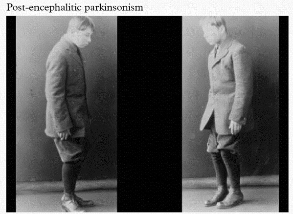
At the same time that H1N1 was causing havoc, a Romanian born neurologist named Constantin von Economo reported a number of unusual symptoms which were referred to as encephalitis lethargica (EL). This disease left victims in a statue-like condition, speechless and motionless.

Economo

Constantin von Economo. Source: Wikipedia

By 1926, EL had spread around the world, with nearly five million people being affected. Many of those who survived never returned to their pre-existing state of health. They were left frozen in an immobile state.

vonecomo-parkinson

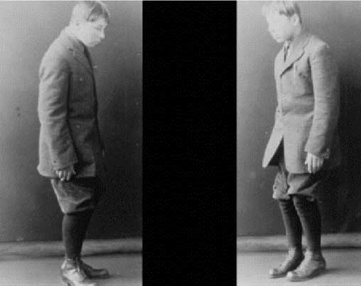
An individual with encephalitis lethargica. Source: Baillement

Historically, it was believed that EL was caused by the influenza virus from the 1918 Spanish influenza pandemic. This was largely due to a temporal association (things happening at approximately the same time) and the finding of influenza antigens in some of the suffers of EL (Click here to read more about this).

And then there were also the observations of Dr Oliver Sacks:

Oliver-Sacks-1933-2015-1

Amazing guy! Dr Oliver Sacks. Source: Pensologosou

During the late 1960s, while employed as a neurologist at Beth Abraham Hospital’s chronic-care facility in New York, Dr Sacks began working with a group of survivors of EL, who had been left immobile by the condition. He treated these individuals with L-dopa (the standard treatment for Parkinson’s disease now, but it was still experimental at the time) and he observed them become miraculously reanimated. The sufferers went from being completely motionless to suddenly active and mobile. Unfortunately the beneficial effects were very short lived.

You may be familiar with Dr Sack’s book about his experience of treating these patients. It is called ‘Awakenings’ and it was turned into a film starring actors Robin Williams and Robert De Niro.

robin_williams_con_robert_de_niro_en_1990

Robin Williams and Robert De Niro in Awakenings. Source: Pinterest

More recent, postmortem analysis of the brains of EL patients found an absence of influenza RNA – click here for more on this), which has led many researchers to simply reject the association between influenza and EL. The evidence supporting this rejection, however, has also been questioned (click here to read more on this), leaving the question of an association between influenza and EL still open for debate.

I think it’s fair to say that we genuinely do not know what caused EL. Whether it was influenza or not is still be undecided.

Ok, so that was the coincidental evidence. Has there been a more direct connection between influenza and Parkinson’s disease?

This is Dr Richard J Smeyne:

Richard_Smeyne

Source: Researchgate

Nice guy.

He is a research faculty member in the Department of Developmental Neurobiology at St. Jude Children’s Research Hospital (Memphis, Tennessee).

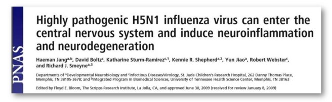
He has had a strong interest in what role viruses like influenza could be playing in the development of Parkinson’s disease, and his research group has published several interesting research reports on this topic, including:

PNAS

Title: Highly pathogenic H5N1 influenza virus can enter the central nervous system and induce neuroinflammation and neurodegeneration.
Author: Jang H, Boltz D, Sturm-Ramirez K, Shepherd KR, Jiao Y, Webster R, Smeyne RJ.
Journal: Proc Natl Acad Sci U S A. 2009 Aug 18;106(33):14063-8.
PMID: 19667183                 (This article is OPEN ACCESS if you would like to read it)

Dr Smeyne and his colleagues found in this study that when they injected the highly infectious A/Vietnam/1203/04 (H5N1) influenza virus into mice, the virus progressed from the periphery (outside the brain) into the brain itself, where it induced Parkinson’s disease-like symptoms.

The virus also caused a significant increase in the accumulation of the Parkinson’s disease-associated protein Alpha Synuclein. In addition, they witnessed the loss of dopamine neurons in the midbrain of the mice at 60 days after the infection – that cell loss resembling what is observed in the brains of people with Parkinson’s disease.

Naturally this got the researchers rather excited!

In a follow up study on H5N1, however, these same researchers found that the Parkinson’s disease-like symptoms that they observed were actually only temporary:

JNS

Title: Inflammatory effects of highly pathogenic H5N1 influenza virus infection in the CNS of mice.
Authors: Jang H, Boltz D, McClaren J, Pani AK, Smeyne M, Korff A, Webster R, Smeyne RJ.
Journal: Journal for Neuroscience, 2012 Feb 1;32(5):1545-59.
PMID: 22302798                   (This article is OPEN ACCESS if you would like to read it)

Dr Smeyne and colleagues repeated the 2009 study and had a closer look at what was happening to the dopamine neurons that were disappearing at 60 days post infection with the virus. When they looked at mice at 90 days post infection, they found that the number of dopamine neurons had returned to their normal number. This pattern was also observed in a region of the brain called the striatum, where the dopamine neurons release their dopamine. The levels of dopamine dropped soon after infection, but rose back to normal by 90 days post infection.

How does that work?

The results suggest that rather than developing new dopamine neurons in some kind of miraculous regenerative process, the dopamine neurons that were infected by the virus simply stopped producing dopamine while they dealt with the viral infection. Once the crisis was over, the dopamine neurons went back to life as normal. And because the researcher use chemicals in the production of dopamine to identify the dopamine neurons, they mistakenly thought that the cells had died when they couldn’t see those chemicals.

One interesting observation from the study was that H5N1 infection in mice induced a long-lasting inflammatory response in brain. The resident helper cells, called microglia, became activated by the infection, but remained active long after the dopamine neurons returned to normal service. The investigators speculated as to whether this activation may be a contributing factor in the development of neurodegenerative disorders.

And this is an interesting idea.

In a follow up study, they investigated this further by looking another influenza viruse that doesn’t actually infect cells in the brain:

PLOS

Title: Induction of microglia activation after infection with the non-neurotropic A/CA/04/2009 H1N1 influenza virus.
Author: Sadasivan S, Zanin M, O’Brien K, Schultz-Cherry S, Smeyne RJ.
Journal: PLoS One. 2015 Apr 10;10(4):e0124047.
PMID: 25861024                (This article is OPEN ACCESS if you would like to read it)

In this study, a different type of influenza (H1N1) was tested, and while it did not infect the brain, it did cause the microglia cells to flare up and become activated. And again, this activation was sustained for a long period after the infection (at least 90 days).

This is a really interesting finding and relates to the idea of a “double hit” theory of Parkinson’s disease, in which the virus doesn’t necessarily cause Parkinson’s disease but may play a supplemental or distractionary role, grabbing the attention of the immune system while some other toxic agent is also attacking the body. Or perhaps simply weakening the immune system by forcing it to fight on multiple fronts. Alone the two would not cause as much damage, but in combination they could deal a terrible blow.

So what was the flu vaccine research published last week?

Again, from Dr Smeyne’s research group, this report looked whether the combination of an influenza virus infection plus a toxic agent gave a worse outcome than just the toxic agent by itself. An interesting idea for a study, but then the investigators threw in another component: what effect would a influenza vaccine have in such an experiment. And the results are interesting:

Flu

Title: Synergistic effects of influenza and 1-methyl-4-phenyl-1,2,3,6-tetrahydropyridine (MPTP) can be eliminated by the use of influenza therapeutics: experimental evidence for the multi-hit hypothesis
Authors: Sadasivan S, Sharp B, Schultz-Cherry S, & Smeyne RJ
Journal: npj Parkinson’s Disease 3, 18
PMID: N/A                    (This article is OPEN ACCESS if you would like to read it)

What the researchers found was that H1N1-infected mice that were treated with a neurotoxin (called MPTP – a toxin that specifically kills dopamine neurons) exhibit a 20% greater loss of dopamine neurons than mice that were treated with MPTP alone.

And this increase in dopamine neuron loss was completely eliminated by giving the mice the influenza vaccination. The researchers concluded that the results demonstrate that multiple insults (such as a viral infection and a toxin) can enhance the impact, and may even be significant in allowing an individual to cross a particular threshold for developing a disease.

It’s an intriguing idea.

Have epidemiologists (population data researchers) ever investigated a connection between Parkinson’s disease and influenza?

Good question.

And yes they have:

flu1
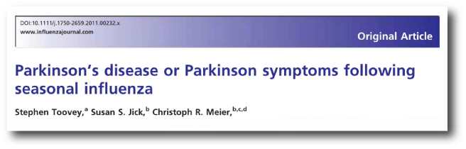
Title: Parkinson’s disease or Parkinson symptoms following seasonal influenza.
Authors: Toovey S, Jick SS, Meier CR.
Journal: Influenza Other Respir Viruses. 2011 Sep;5(5):328-33.
PMID: 21668692            (This article is OPEN ACCESS if you would like to read it)

In this first study, the researcher used the UK‐based General Practice Research Database to perform a case–control analysis (that means they compare an affected population with an unaffected ‘control’ population. They identified individual cases who had developed an ‘incident diagnosis’ of Parkinson’s disease or Parkinson’s like symptoms between 1994 and March 2007. For each of those case files identified, they matched them with at least four age matched control case files for comparative sake.

Their analysis found that the risk of developing Parkinson’s disease was not associated with previous influenza infections. BUT, they did find that Influenza was associated with Parkinson’s‐like symptoms such as tremor, particularly in the month after an infection. One can’t help but wonder if the dopamine neurons stopped producing dopamine during that period while they dealt with the viral infection.

But of course, I’m only speculating here… and it’s not like there was a second study suggesting that there is actually an association between Parkinson’s disease and influenza.

A year after that first study, a second study was published:

occupation
Journal: Association of Parkinson’s disease with infections and occupational exposure to possible vectors.
Authors: Harris MA, Tsui JK, Marion SA, Shen H, Teschke K.
Journal: Movement Disorder. 2012 Aug;27(9):1111-7.
PMID: 22753266

This second study reported that there is actually an association between Parkinson’s disease and influenza.

This investigation was also a case-control study, but it was based in British Columbia, Canada. The researchers recruited 403 individuals detected by their use of antiparkinsonian medications and matched them with 405 control subjects selected from the universal health insurance plan. Severe influenza was associated with Parkinson’s disease at an odds ratio of 2.01 (1 being no difference) and the range of the odds was 1.16-3.48. That’s pretty significant.

Interestingly, the effect is reduced when the reports of infection were restricted to those occurring within 10 years before diagnosis. This observation would suggest that early life infections may have more impact than previously thought.

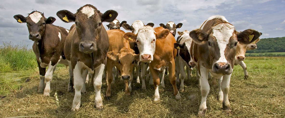
Curiously, the researchers also found that exposure to certain animals (cats odds ration of 2.06; range 1.09-3.92) and cattle (2.23; range 1.22-4.09) was also associated with developing Parkinson’s disease.

Time to get rid of the pet cow.

1016238_tcm9-156853

Source: RSPB

Do any other neurodegenerative condition have associations with influenza?

In the limited literature search that we conducted, we only found reports dealing with influenza and Alzheimer’s disease.

Large studies suggest that Alzheimer’s is not associated with influenza (click here to read more on this). Interestingly, the Alzheimer’s associated protein beta amyloid has been shown to inhibit influenza A viruses (Click here to read that report), which may partly explain the lack of any association.

Influenza does have a mild association, however, with depression (Click here to see that report).

So what does it all mean?

A viral theory for Parkinson’s disease has existed since the great epidemic of 1918. Recent evidence points towards several viruses potentially having some involvement in the development of this neurodegenerative condition. And recent evidence suggests that influenza in particular could be particularly influential.

In 1938, Jonas Salk and Thomas Francis developed the first vaccine against flu viruses. It could be interesting for epidemiologists to go back and see if regular flu vaccination usage (if such data exists) reduces the risk of developing Parkinson’s disease.

But until such data is published, however, perhaps it would be wise to go and get a flu vaccine shot.


The banner for today’s post was sourced from the HuntingtonPost

Hepatitis – Parkinson’s goes viral?

maxresdefault

Last week a new piece of Parkinson’s disease research has been widely discussed in the media.

It involves Hepatitis – the viral version of it at least.

In today’s post we will review the research and discuss what it may mean for Parkinson’s disease.


Fig2_v1c

A lewy body (brown with a black arrow) inside a cell. Source: Cure Dementia

A definitive diagnosis of Parkinson’s disease can only be made at the postmortem stage with an examination of the brain. Until that moment, all cases of Parkinson’s disease are ‘suspected’.

Critical to that postmortem diagnosis is the presence of circular shaped, dense clusters of proteins, called Lewy bodies (see the image above for a good example).

What causes Lewy bodies? We don’t know, but many people have theories.

This is Friedrich Heinrich Lewy (1885-1950).

DrLewy

Friedrich Lewy. Source: Lewy Body Society

As you can probably guess, Friedrich was the first to discover the ‘Lewy body’. His finding came by examining the brains of 85 people who died with Parkinson’s disease between 1908 – 1923.

In 1931, Friedrich Lewy read a paper at the International Congress of Neurology in Bern. During that talk he noted the similarities between the circular inclusions (called ‘negri bodies’) in the brains of people who suffered from rabies and his own Lewy bodies (observed in Parkinson’s disease).

rabies

A Negri body in a cell affected by rabies (arrow). Source: Nethealthbook

Given the similarities, Lewy proposed a viral cause for Parkinson’s disease.

Now, the idea that Parkinson’s disease could have a viral component has existed for a long time – even before Lewy made his conclusion. As we have previous mentioned, theories of viral causes for Parkinson’s have been circulating ever since the 1918 flu pandemic (Click here to read our post on this topic).

vonecomo-parkinson

An example of post-encephalitic Parkinsonism. Source: Baillement

About the same time as the influenza virus was causing havoc around the world, another condition began to appear called ‘encephalitis lethargica‘ (also known as post-encephalitic Parkinsonism). This disease left many of the victims in a statue-like condition, both motionless and speechless – similar to Parkinson’s disease. Initially, it was assumed that the influenza virus was the causal factor, but more recent research has left us not so sure anymore.

Since then there, however, has been additional bits of evidence suggesting a viral role in Parkinson’s disease. Such as this report:

H1N1

Title: Highly pathogenic H5N1 influenza virus can enter the central nervous system and induce neuroinflammation and neurodegeneration.
Author: Jang H, Boltz D, Sturm-Ramirez K, Shepherd KR, Jiao Y, Webster R, Smeyne RJ.
Journal: Proc Natl Acad Sci U S A. 2009 Aug 18;106(33):14063-8.
PMID: 19667183

The researchers in this study found that when they injected the highly infectious H5N1 influenza virus into mice, the virus progressed from the periphery (outside the brain) into the brain itself, where it induced Parkinson’s disease-like symptoms. The virus also caused a significant increase in the accumulation of the Parkinson’s associated protein Alpha Synuclein. Importantly, they witnessed the loss of dopamine neurons in the midbrain of the mice 60 days after resolution of the infection – that cell loss resembling what is observed in the brains of people with Parkinson’s disease.

The Parkinson’s associated protein alpha synuclein has also recently demonstrated anti-viral properties:

Beckham

Title: Alpha-Synuclein Expression Restricts RNA Viral Infections in the Brain.
Authors: Beatman EL, Massey A, Shives KD, Burrack KS, Chamanian M, Morrison TE, Beckham JD.
Journal: J Virol. 2015 Dec 30;90(6):2767-82. doi: 10.1128/JVI.02949-15.
PMID: 26719256               (This article is OPEN ACCESS if you would like to read it)

David Beckham (not the football player) and his research colleagues introduced West nile virus to brain cells grown in cell culture and they observed an increase in alpha synuclein production. They also found that the brains of people with West nile infections had increased levels of alpha synuclein.

The researchers then injected West Nile virus into both normal mice and genetically engineered mice (which produced no alpha synuclein) and they found that the genetically engineered mice which produced no alpha synuclein died quicker than the normal mice. They reported that there was an almost 10x increase in viral production in the genetically engineered mice. This suggested to them that alpha synuclein may be playing a role in protecting cells from viral infections.

Interesting, but what about this new data involving Hepatitis?

Yes, indeed. Let’s move on.

Wait a minute, what is Hepatitis exactly?

The name Hepatitis comes from the Greek: Hepat – liver; and itis – inflammation, burning sensation. Thus – as the label suggests – Hepatitis is inflammation of liver tissue.

Progress-of-Liver-Damage

Hepatitis and the liver. Source: HealthandLovepage

It can be caused by infectious agents (such as viruses, bacteria, and parasites), metabolic changes (induced by drugs and alcohol), or autoimmune/genetic causes (involving a genetic predisposition).

The most common cause of hepatitis is viral.

There are five main types of viral hepatitis (labelled A, B, C, D, and E). Hepatitis A and E are mainly spread by contaminated food and water. Both hepatitis B and hepatitis C are commonly spread through infected blood (though Hepatitis B is mainly sexually transmitted). Curiously, Hepatitis D can only infect people already infected with hepatitis B.

Hepatitis A, B, and D are preventable via the use of immunisation. A vaccine for hepatitis E has been developed and is licensed in China, but is not yet available elsewhere

Hepatitis C, however, is different.

There is currently no vaccine for it, mainly because the virus is highly variable between strains and the virus mutates very quickly, making an effective vaccine a difficult task. A number of vaccines under development (Click here for more on this).

What is known about Hepatitis C and the brain?

Quite a bit.

Similar to HIV (which we discussed in a previous post), the hepatitis C virus (HCV) enters the brain via infected blood-derived macrophage cells. In the brain, it is hosted by microglial cells, which results in altered functioning of those microglial cells. This causes problems for neuronal cells – including dopamine neurons. For example, people infected with HCV have reduced dopamine transmission, based on brain imaging studies (Click here and here for more on this result).

Have there been connections between hepatitis C virus and Parkinson’s disease before?

Yes.

Dopatitle


Title: Hepatitis C virus infection: a risk factor for Parkinson’s disease.
Authors: Wu WY, Kang KH, Chen SL, Chiu SY, Yen AM, Fann JC, Su CW, Liu HC, Lee CZ, Fu WM, Chen HH, Liou HH.
Journal: J Viral Hepat. 2015 Oct;22(10):784-91.
PMID: 25608223

The researchers in this study used data collected from a community-based screening program in north Taiwan which involved 62,276 people. The World Health Organisation (WHO) estimates that the prevalence of hepatitis C viral infection worldwide is approximately 2.2–3%, representing 130–170 million people. Taiwan is a high risk area for hepatitis, with antibodies for hepatitis viruses in Taiwan present in 4.4% in the general population (Source).

The researchers found that the significant association between hepatitis C viral infections and Parkinson’s disease – that is to say, a previous infection of hepatitis C increased the risk of developing Parkinson’s disease (by 40%). The researchers then looked at what the hepatitis C and B viral infections do to dopamine neurons growing in cell culture. They found that hepatitis C virus induced 60% dopaminergic cell death, while hepatitis B had no effect.

This study was followed up a few months later, by a second study suggesting an association between Hepatitis C virus and Parkinson’s disease:

Hep title

Title: Hepatitis C virus infection as a risk factor for Parkinson disease: A nationwide cohort study.
Authors: Tsai HH, Liou HH, Muo CH, Lee CZ, Yen RF, Kao CH.
Journal: Neurology. 2016 Mar 1;86(9):840-6.
PMID: 26701382

The researchers in this study wanted to investigate whether hepatitis C could be a risk factor for Parkinson’s disease. They did this by analyzing data from 2000-2010 drawn again from the Taiwan National Health Insurance Research Database.

The database included 49,967 people with either hepatitis B, hepatitis C or both, in addition to 199,868 people without hepatitis. During the 12 year period, 270 participants who had a history of hepatitis developed Parkinson’s disease (120 still had hepatitis C). This compared with 1,060 participants who were free of hepatitis, but went on to develop Parkinson’s disease.

When the researchers controlled for potentially confounding factors (such as age, sex, etc), the researchers found participants with hepatitis C had a 30% greater risk of developing Parkinson’s disease than the controls.

So if this has been demonstrated, why is this new study last week so important?

Good question.

The answer is very simple: This study is not based on statistics from Taiwan – this new study has found the same result from a new population.

HEP TITLE

Title: Viral hepatitis and Parkinson disease: A national record-linkage study.
Authors: Pakpoor J, Noyce A, Goldacre R, Selkihova M, Mullin S, Schrag A, Lees A, Goldacre M.
Journal: Neurology. 2017 Mar 29. [Epub ahead of print]
PMID: 28356465

These researchers used the English National Hospital Episode Statistics database and linked it to mortality data collected from 1999 till 2011. They too have found a strong association between hepatitis C and Parkinson’s disease (standardized rate ratio 1.51, 95% CI 1.18–1.9).

Curiously (and different from the previous studies), the researchers in this study also found a strong association for hepatitis B and Parkinson’s disease (standardized rate ratio 1.76, 95% CI 1.28–2.37). And these associations appear to be specific to Hepatitis B and C, as the investigators did not find any association between autoimmune hepatitis, chronic hepatitis, or HIV.

One important caveat with this new study, however, is that the authors could not
control for lifestyle factors (such as smoking or alcohol consumption). In addition, their system of linking medical records may underestimate the numbers of patients with
Parkinson’s disease as it would not take into account people with Parkinson’s disease who do not seek medical advice or those who are misdiagnosed (given a wrong diagnosis – it does happen!).

Regardless of these cautionary notes, the results still add to the accumulating evidence of an association between the virus that causes Hepatitis and the neurodegenerative condition of Parkinson’s disease.

But what about those people with Parkinson’s disease who have never had Hepatitis?

Yeah, this is a good question.

But there is a rather uncomfortable answer to it.

Here’s the rub: “Approximately 70%–80% of people with acute Hepatitis C do not have any symptoms” (Source: Centre for Disease Control). That is to say, the majority of people infected with the Hepatitis C virus will not be aware that they are infected. Some of those people who are infected may think that they have a case of the flu (HCV symptoms include fever, fatigue, loss of appetite,…), while others will simply not display any symptoms at all.

So many people with Parkinson’s disease may have had HCV, but never been aware of it.

And this is the really difficult part of researching the causal elements of Parkinson’s disease.

The responsible agent may actually leave little or no sign that they were ever present. For a long time, people have suggested that Parkinson’s disease is caused by a thief in the night – some agent that comes in, causes a problem and disappears without detection.

Perhaps Hepatitis is that thief.

But hang on a second, 60–70% of HCV infected people will go on to develop chronic liver disease (Source). Do people with Parkinson’s disease have liver issue?

Umm, well actually, in some cases: yes.

There have been studies of liver function in Parkinson’s disease where abnormalities have been found (Click here for more on this). And dopamine cell dysfunction has been seen in people with cirrhosis issues (Click here for more on this). In fact, the prevalence of Parkinsonism in people with cirrhosis has been estimated to be as high as 20% (and Click here for more on that).

So what are we saying? Hepatitis causes Parkinson’s disease???

No, we are not saying that.

Proving causality is the hardest task in science.

In addition, there have been a few studies in the past that have looked at viral infections as the cause of Parkinson’s disease that found strong associations with other viruses. For example this study:

Title: Infections as a risk factor for Parkinson’s disease: a case-control study.
Authors: Vlajinac H, Dzoljic E, Maksimovic J, Marinkovic J, Sipetic S, Kostic V.
Journal: Int J Neurosci. 2013 May;123(5):329-32.
PMID: 23270425

In this study, the researchers found that Parkinson’s Disease was also significantly associated to mumps, scarlet fever, influenza, and whooping cough as well as herpes simplex 1 infections. They found no association between Parkinson’s disease and Tuberculosis, measles or chickenpox though.

This result raises the tantalizing possibility that other viruses may also be involved with the onset of Parkinson’s disease (it should be added though that this study was based on only 110 people with Parkinson’s (compared with 220 controls) in one particular geographical location (Belgrade, Serbia)).

So different viruses may cause Parkinson’s disease?

We are not saying that either, but we would like to see more research on this topic.

And the situation may actually be more complicated than we think.

Recently, it has been reported that previous infection with flaviviruses (such as dengue) actually enhances the effect of Zika virus infect (Click here to read more on this). That is to say, a prior infection by one particular virus may exacerbate the infection of another virus. It could be that a previous infection by one virus increases that chance that a later infection by another virus – a particular combination of viral infections – may result in Parkinsonian symptoms (we are simply speculating here). 

Add to this complicated situation, the sheer number of unknown viruses. It is estimated that there are a minimum of 320,000 mammalian viruses still awaiting discovery (Click here for the source of this statistic), thus it is possible that additional unknown viruses may be involved with disease initiation for conditions like Parkinson’s disease.

A gang of unknown thieves in the night perhaps?

So what does it all mean?

Summing up: last week a new study was published that supported previous results that Hepatitis C viral infections could increase the risk of developing Parkinson’s disease. The results are important because they replicate previous findings from a different population of people.

The findings do not immediately mean that people with Hepatitis C are going to develop Parkinson’s disease, but it does suggest that they may be more vulnerable. The findings also suggest that more research is needed on the role of viral/infectious agents in the development of Parkinson’s disease.

We would certainly like to see more research in this area.


The banner for today’s post was sourced from Youtube

HIV and Parkinson’s disease

hiv-aids-definition2


I was recently made aware of an interesting fact:

Approximately 5% of people with Human immunodeficiency virus (HIV) infections develop Parkinson’s disease-like features.

Why is this?

In today’s post we will try to understand what is going on, and what it may mean for Parkinson’s disease.


hiv-budding-colo2r

HIV (in green) budding (being released) from a blood cell (lymphocyte). Source: Wikipedia

Ok, let’s start at the beginning:

What is HIV?

Human immunodeficiency virus (or HIV) – as the name suggests – is the virus.

It causes the infection which gives rise to Acquired Immune Deficiency Syndrome (or AIDS). AIDS is a progressive failure of the immune system – the body loses its ability to fight infections. Without treatment, average survival period after infection with HIV is between 9 – 12 years.

HIV can be spread by the transfer of bodily fluids, such as blood and semen. The World Health Organisation (WHO) has estimated that approximately 36.9 million people worldwide were living with HIV/AIDS at the end of 2014 (that is equivalent to the entire population of Canada!).

hi-virion-structure_en-svg

The structure of the HIV virus. Source: Wikipedia

Does HIV affect the brain?

Yes.

At postmortem examinations, less than 10% of the brains from HIV infected individuals are histologically normal (Source).

HIV is a member of the lentivirus family of viruses, which readily infect immune cells (such as blood cells). HIV can also infect other types of cells though, including those in the brain. HIV will usually enter the central nervous system within the first month following infection. It enters the brain via infected blood cells which come into contact with brain ‘immune system/helper’ cells such as microglia and macrophages at the blood-brain-barrier.

f1-large

How HIV enters the brain. Source: Disease Models and Mechanisms

HIV can also infect astrocytes (albeit at a lower frequency than microglia and macrophages), by direct cell-cell contact with infected T cells (blood cells) at the blood-brain-barrier (No. 1 in the image above). After infecting astrocytes, there is dysfunction in the astrocyte and it will no longer be so supportive to the local neurons (No. 2 in the image above). Once inside the brain, HIV-infected macrophages will allow for infection of other macrophages and microglia (No. 3 in the image above), and all together these HIV-infected astrocytes and microglia will cause damage to neurons by releasing viral proteins (two in particular, called Tat and gp120) and additional nasty chemicals which are bad for the neurons (No. 4 in the image above). Finally, as the disease progresses, the protective layer of the blood-brain-barrier becomes compromised and HIV-infected T cells eventually enter the brain and they cause damage to neurons by releasing pro-inflammatory chemicals (making the environment harsh for neurons).

There is remarkably little evidence of HIV actually infecting neurons (Click here for a review on this), so any cell loss in the brain that is associated with HIV does not result from neurons themselves being infected. This may be due to the fact that neurons do not have the HIV receptors (such as CD4) on their cell membrane. Similarly, oligodendrocytes (a supporting cell) does not appear to be easily infected by HIV. The bulk of the infected cells in the brain appear to be of the microglial, macrophage and astrocytes. And without these supporting cells doing their jobs in a normal fashion, it is easy to see how neurons can start dying off.

The severity, characteristics and distribution of HIV-induced injury in the brain varies greatly between affected individuals. It is most likely associated with the viral load (or the number of viral particles) in the brain, which can vary from a few thousand to more than a million copies per mL.

Do HIV-infected people show any signs of the virus entering the brain?

For the majority of people infected with HIV, this entry of the virus into the nervous system is neurologically asymptomatic (meaning they will not notice it), except for the occasional mild headache (for more on this read this review). As a result of the HIV virus entering the brain, however, many infected individuals will suffer from a specific set of neurological disorders, collectively called the AIDS dementia complex (ADC) (also known as HIV-associated cognitive/motor complex, or simply HIV dementia).

So how does HIV infection result in Parkinson’s disease-like features?

As we have suggested in the introduction to this post, on rare occasions (approximately 5% of cases), HIV-infected patients may present an illness virtually identical to Parkinson’s disease. More commonly, people with HIV will exhibit an increased sensitivity to dopamine receptor-blocking agents, such as drugs with a low potential for inducing Parkinsonism, (for example prochlorperazine and metoclopropamide).

The exact mechanism by which HIV infection results in Parkinson’s disease-like features is the subject of debate, but what is clear is that the basal ganglia (a structure involved in Parkinson’s disease) faces the brunt of the HIV infection in the brain. HIV-infected microglia and macrophage are most prominent in the basal ganglia when compared to other brain regions (Click here and here for more on this), and the basal ganglia is where the chemical dopamine from the midbrain is being released.

In addition, there are other changes in the brains of HIV infected people which may aid in the appearance of Parkinsonian features:


viraltitle

Title: Increased frequency of alpha-synuclein in the substantia nigra in human immunodeficiency virusinfection.
Authors: Khanlou N, Moore DJ, Chana G, Cherner M, Lazzaretto D, Dawes S, Grant I, Masliah E, Everall IP; HNRC Group.
Journal: J Neurovirol. 2009 Apr;15(2):131-8.
PMID: 19115126       (This article is OPEN ACCESS if you would like to read it)

The researchers in this study used staining techniques to look at the amount of alpha synuclein – the Parkinson’s associated protein – in slices of brain tissue taken from postmortem autopsies of 73 HIV+ individuals aged between 50 and 76 years of age.

The presence of alpha synuclein in the substantia nigra (an area of the brain affected by Parkinson’s disease) was a lot higher in the HIV+ brains when compared with healthy control samples (16% of the HIV+ brains had high levels of alpha synclein vs 0% for the healthy brains).

Interestingly, nearly all of the brains analysed (35 out of 36 HIV+ brains) had high levels of the Alzheimer’s disease associated protein, beta amyloid (which again raises the question of whether beta amyloid could be playing a defensive role in infections – see our previous post on this). Also interesting, was that there was no correlation between these proteins being present and the age of the person at death – that is to say, older brains did not have more of these proteins when compared with younger brains.

There are also additional ways in which HIV could be causing Parkinson’s-like features, such as:

  • HIV has been shown to affect the protein levels of Parkinson’s disease associated proteins, such as DJ1 and Lrrk2 (Click here and here to read more on this).
  • HIV can, in some cases, increase the level of Dopamine transporter, which would reduce the levels of free floating dopamine in the brain (Click here to read more about this).

How is HIV treated?

aidspills

Treating HIV. Source: NPR

There is currently no cure for HIV infection.

There are, however, treatments which help to slow the virus down. These are called Anti-retroviral drugs (HIV is a retrovirus). There are different kinds of anti-retroviral drugs, which act at different stages of the HIV life cycle. Combinations of several anti-retroviral drugs (generally three or four) is known as ‘Highly Active Anti-Retroviral Therapy'(or HAART).

hiv-drug-classes-svg

Mechanism by which four classes of anti-retroviral drugs work against HIV. Source: Wikipedia

As the schematic image above highlights, there are many ways to slow down the HIV virus. For example, you can prevent it from attaching to a cell and fusing with the cell membrane (fusion inhibitors). By treating HIV infected people with multiple medications attacking different parts of the HIV life cycle, the virus has been slowed down.

Does HAART treatments for HIV help with these Parkinson’s-like features?

In some cases, the answer appears to be yes.

There are numerous case studies in the literature which demonstrate the alleviation of HIV-associated Parkinsonian symptoms with HAART, such as this report:

hersh

Title: Parkinsonism as the presenting manifestation of HIV infection: improvement on HAART.
Authors: Hersh BP, Rajendran PR, Battinelli D.
Journal: Neurology. 2001 Jan 23;56(2):278-9.
PMID: 11160977

In this study the researchers described the case of a 37 year old man who developed Parkinson’s like features in the setting of an HIV infection, which were resolved after 1 year of HAART.

Over a period of 4 months, the man developed co-ordination issue, clumsiness and an irregular tremor in his right hand (there was, however, no resting tremor). He noted a generalised slowness and exhibited a tendency towards decreased right arm swinging. He also developed dystonia in the right hand/arm. Following L-dopa treatment (25/100; one tablet 3x per day) there was improvement in balance & co-ordination, speech, facial expression, and the tremor (L-dopa does appear to improve most cases of HIV-associated Parkinson’s-like features).

Six months after first displaying these Parkinsonian features (and two month after initiating L-dopa treatment), the subject was placed on HAART treatment. Four months later, he discontinued L-dopa treatment and 12 months after starting the HAART regime his Parkinsonian features were largely resolved.

More case studies of HAART alleviating HIV-associated Parkinsonisms can be found by clicking here and here.

What does this mean for Parkinson’s disease?

This post was written for the research community rather than people with Parkinson’s disease. I thought the fact that some people with HIV can start to have Parkinson’s like features was an interesting curiosity and wanted to share/spread the information.

Having said that, this post raises some really interesting questions, such as if a virus like HIV can have this effect on the brain, could other viruses be having similar effects? Could some cases of Parkinson’s disease simply be the result of a viral infection? Either multiple hits from a particular virus or different viruses each taking a varying toll over the course of a life time.

This idea would explain many of the curious features of Parkinson’s disease, such as:

  • the asymmetry of the symptoms (people with Parkinson’s usually have the disease starting on one side of the body.
  • the fact that some cells in the brain are more vulnerable to the disease than others (perhaps they are more receptive to a particular virus).
  • the protein clusterings in the cells (Lewy bodies may be defensive efforts against viral infections).

As we have previous mentioned, theories of viral causes for Parkinson’s have been circulating ever since the 1918 flu pandemic (Click here to read our previous post on this topic). About the same time as the influenza virus was causing havoc around the world, another condition began to appear called ‘encephalitis lethargica‘. This disease left many of the victims in a statue-like condition, both motionless and speechless – similar to Parkinson’s disease. Initially, it was assumed that the influenza virus was the causal factor, but more recent research has left us not so sure anymore.

The point is, however, perhaps it is time for us to re-examine the possibility of a viral agent being involved in the development of Parkinson’s disease.

There is new technology that allows us to determine the viral history of each individual from a simple blood test (Click here for more on this), so it would be interesting to compare blood samples from people with Parkinson’s disease with healthy controls to determine any differences.

In addition to the overall question of a viral role in Parkinson’s disease, there also remains the question of why only a small fraction of people with HIV are affected by Parkinsonisms. It could be interesting to genetically screen those people with HIV that exhibit Parkinsonisms and compare them with people with HIV that do not. Do those affected individuals have recognised Parkinson’s related genetic mutations? Or do they have novel genetic variations that could tell us more about Parkinson’s disease?

Food for thought. Would be happy to hear others thoughts.


The banner for today’s post was sourced from AidsServices