
|
# # # # In 2015, a woman with dementia was sitting in hospice care, wheelchair-bound and largely non-communicative. As part of a research project, she was given repeated computed tomography (CT) scans of her brain. The effect of this seemingly harmless treatment was rather astonishing, and it has led to more research exploring the effect of low dose ionizing radiation on neurodegenerative conditions. Including Parkinson’s. In today’s post, we will discuss what low dose ionizing radiation is, what research has been conducted thus far, and what could be happening in the brain to explain the effect. # # # # |
 Source: Linkedin
During the lunch break of a recent research conference and desiring some ‘alone time’, I approached a table toward the back of the conference center.
There was only one other inhabitant of this oasis of solitude and after asking if they minded if I sat there (“Please” they gestured), I sat down. A moment or two later, a polite conversation was started. It was innocent enough (“Enjoying the conference?“) but this quickly shifted to specifics (Me asking “So what area of the research is of most interest to you?“).
And this was where the rabbit hole began.
You see, my table companion explained that they had trained as a radiologist – this is a group of doctors that specialise in diagnosing and treating injuries/diseases using medical imaging.
And upon being told their occupation, I asked if they were interested in the research exploring diagnostic imaging for Parkinson’s.
Their answer was: “Well, yes, but I’m more interested in using the imaging therapeutically“.
To which, my facial expression must have shifted towards something like:

My table companion was amused by the expression on my face and added that they lived locally and they were curious about the research being done on Parkinson’s. But more specifically, they were interested to know if anyone was investigating low-dose ionizing radiation as a potential therapeutic treatment.
My facial confusion must have become somewhat more exaggerated at that point, and so they gave me the background history and context to their reason for interest.
It was a fascinating story.
What did they say?
In 2015, an 81 year old female with a 10 year history of dementia was sitting in hospice care in the state of Michigan (USA).
Her neuropsychologist reported that she was non-responsive, non-communicative and largely refined to her wheelchair. She “would only rarely utter a single word” and “had not attempted to rise from her wheelchair in months” (Source).
The lady was taking part in some clinical studies and over the period of three months she received five computed tomography (CT) scans of the brain. The first two scans were given in July, 2015.
Two days after the second scan, her caregiver (who was unaware of this radiation treatment) noticed significant changes: “She is doing so well that it is amazing. I have never seen someone improve this much. She wanted to get up and walk. She was talking some, with more sense, and she was feeding herself again” (Source).
The lady received a third and fourth CT scans in August 2015, and her condition continued to improve. So much so in fact that by November 2015, she was deemed no longer eligible for hospice care due to the improvements in both her cognitive and physical condition.
A write up of this case study has been published:
 Title: Treatment of Alzheimer Disease With CT Scans: A Case Report.
Authors: Cuttler JM, Moore ER, Hosfeld VD, Nadolski DL.
Journal: Dose Response. 2016 Apr 1;14(2):1559325816640073.
PMID: 27103883 (This report is OPEN ACCESS if you would like to read it)
The patient’s family were so amazed by her improvements that they requested for her to receive ongoing “booster” CT scans every four to five months, and the clinicians agreed as they were rather intrigued by the woman’s response as well.
From February 2016 to December 2016, the woman received four additional CT scans, and her slow but continuous improvement were also documented in this second report:
 Title: Update on a Patient With Alzheimer Disease Treated With CT Scans.
Authors: Cuttler JM, Moore ER, Hosfeld VD, Nadolski DL.
Journal: Dose Response. 2017 Feb 17;15(1):1559325817693167.
PMID: 28321176 (This report is OPEN ACCESS if you would like to read it)
The lady received an additional two CT scans in January 2017 but sadly began to decline from that point and on March 6th, 2017 she returned to hospice care, and she died on May 18th, 2018.
How sad. But what a fascinating story. This is just one case study though, right? So there could be a placebo effect occurring?
This is true, and the woman could have been experiencing some kind of placebo response (that is the sensing of benefits to a treatment for which there are no biological basis or explanation).
But there has also been a second case study reported: A 73-year-old gentleman in Massachusetts with early-stage Alzheimer’s requested CT scans of the brain after reading about the lady in Michigan, and he too experienced some positive benefits (Click here to read about this individual’s experience).
Oh wow! Really? Has there been a clinical trial of this CT scan treatment?
Yes. Based on these anecdotal observations a pilot study was conducted between 2017 and 2020, and the results were published in 2021.
This is the report:
 Title: Low Doses of Ionizing Radiation as a Treatment for Alzheimer’s Disease: A Pilot Study.
Authors: Cuttler JM, Abdellah E, Goldberg Y, Al-Shamaa S, Symons SP, Black SE, Freedman M.
Journal: J Alzheimers Dis. 2021;80(3):1119-1128.
PMID: 33646146 (This report is OPEN ACCESS if you would like to read it)
In this study, four dementia patients (82-90 years of age) volunteered to undergo three consecutive treatments of low-dose ionizing radiation, with each scan being two weeks apart. Clinical measures of cognition and behavior were taken both pre- and post- scans. Each scan delivered a dose of 40 mGy (we’ll discuss what this means in a moment) within 10 seconds to the head (Click here to read more about the details of this study)
The key detail to note here was that this was a single arm, open label study – this means that everyone was receiving the treatment and everyone knew that they were receiving the treatment.
Minor and variable improvements were observed on quantitative clinical measures in three of the four participants at the end of the study. But the investigators reported qualitative observations from immediate relatives (descriptions, photos, and videos) suggested beneficial improvements, including greater overall alertness.
Interesting. Are there plans to do a larger study?
I have no idea if this is planned but I am assuming so and will post an addendum at the bottom of this post if/when I found out.
Before we go any further: What exactly is low-dose ionizing radiation?
Radiation is energy that comes from a source and travels through space at the speed of light. This energy itself has an electric field and a magnetic field associated with it, and it has wave-like properties.
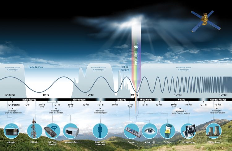 Source: NASA
Importantly, it is very natural and it is all around us. We are all exposed to radiation from outer space, in the air we breathe, and from the earth on which we tread. We even have naturally occurring radioactive elements in our bones which continually irradiate us.
This video provides an overview of radiation:
Radiation comes in two forms: ionising and non-ionising.
Ionising radiation is a type of high-energy radiation that has enough energy to remove an electron (negative particle) from an atom or molecule, causing it to become ionised. Non-ionising is a low energy form of radiation that does not remove electrons
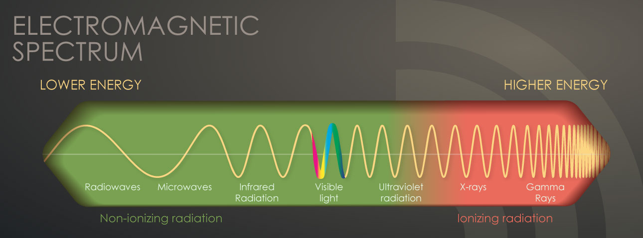 Source: CDC
As a result of this electron-removing property, ionising radiation can cause chemical changes in cells and damage our DNA. And to be clear, this is not good. The risk of health issues (like cancer) later in life is are small for low levels of exposure, but this increases quickly with exposure to high levels of ionising radiation.
This danger was observed in the survivors of the Hiroshima atomic blast at the end of World War 2. These individuals were exposed to very high levels of ionising radiation when the bomb detonated in 1945. In the map below, of the survivors who sheltered from the heat and force of the blast within 1 mile of ground zone (the gray dots in the centre), very few survived to 1950 and beyond:
 Rings represent 2- and 3-km distances. Source: PMC
As you assess survival beyond the 1 mile radius (the red, orange, yellow and green dots), the number of survivors living beyond the 1950s increases, providing researchers with a (very unfortunate) dataset for investigating the effects of lower doses of ionizing radiation.
Low-dose ionizing radiation is considered as radiation dosages that range from values near 0 up to about 100 mSV (100 mGy).
What are mSV and mGy?
A millisievert (or mSv) is “the average accumulated background radiation dose to an individual for 1 year, exclusive of radon, in the United States” (Source). And 1 mSv is the dose produced by exposure to 1 milligray (mGy) of radiation.
Humans are regularly exposed to low-dose ionizing radiation. For wandering around in the modern world, the average annual background radiation is 3 mGy. Additionally, people are exposed to radioactive materials that are widely used in industrial applications and to that of diagnostic equipment, such as CT scans, in which the radiation dose ranges between 3 to 20 mGy
And for the record, cell phones emit low levels of non-ionising radiation when in use (Source).
Across our planet, the radiation level varies from very low levels in underground spaces (20 nGy/hr) to ambient levels (60–100 nGy/hr). These levels can get very high levels in nuclear disaster zones such as Chernobyl (Ukraine) where average levels of radiation is about 1.2 mSv per hour (Source).
Ok. And what is a computed tomography (CT) scan? And how does it involve low-dose ionizing radiation?
A computed tomography (CT) scan uses X-rays and a computer to create detailed images of the inside of the body. While traditional x-rays provide a flat 2 dimensional image of a body part (where different organs can be seen to overlapping), CT scans involve lots of images being taken of a region of the body and then computers can generate a robust 3-dimensional image of the internal organs.
 Source: Omegapds
This video provides a useful overview of CT technology:
CT scans use X-ray to generate their images. X-ray is one of the three main forms of ionizing radiation. The radiation dose from a CT scan ranges between 3 to 20 mGy (Source).
 Source: Quora
Understood. So CT scans use x-ray as ionizing radiation to build 3-dimensional images. But how could this procedure be having a beneficial effect in people with dementia?
So that is the $60,000 question.
And researchers have got some ideas about how the effect might be working. They think that there might be a ‘Goldilocks-like’ aspect to low dose ionizing radiation that causes key cellular processes to kick into action. If you turn up the volume too much on the dose, then you cross a threshold that puts you into ‘high dose’ range where the radiation starts to have detrimental results.
 Source: PMC
What are the “key cellular processes” you mention?
So when mice are exposed to low dose ionising radiation, and researchers then look at their brains, the scientists find increasing in neuroprotective functions. Most notably they see an increase in cellular components involved in DNA repair, cell-cycle control, lipid metabolism, and stress response.
Interestingly, when the researchers wait some time after the radiation exposure they see additional changes in cellular activity which are associated with metabolic function, myelin and protein synthesis, as well as increases in levels of antioxidative enzymes (Click here and here to read more about this).
Has anyone tested low dose ionizing radiation in preclinical models of dementia?
Yes, there have been multiple studies demonstrating beneficial outcomes – in fly models of dementia (Click here to read an example) and mice models (Click here for an example).
And most recently in this report:
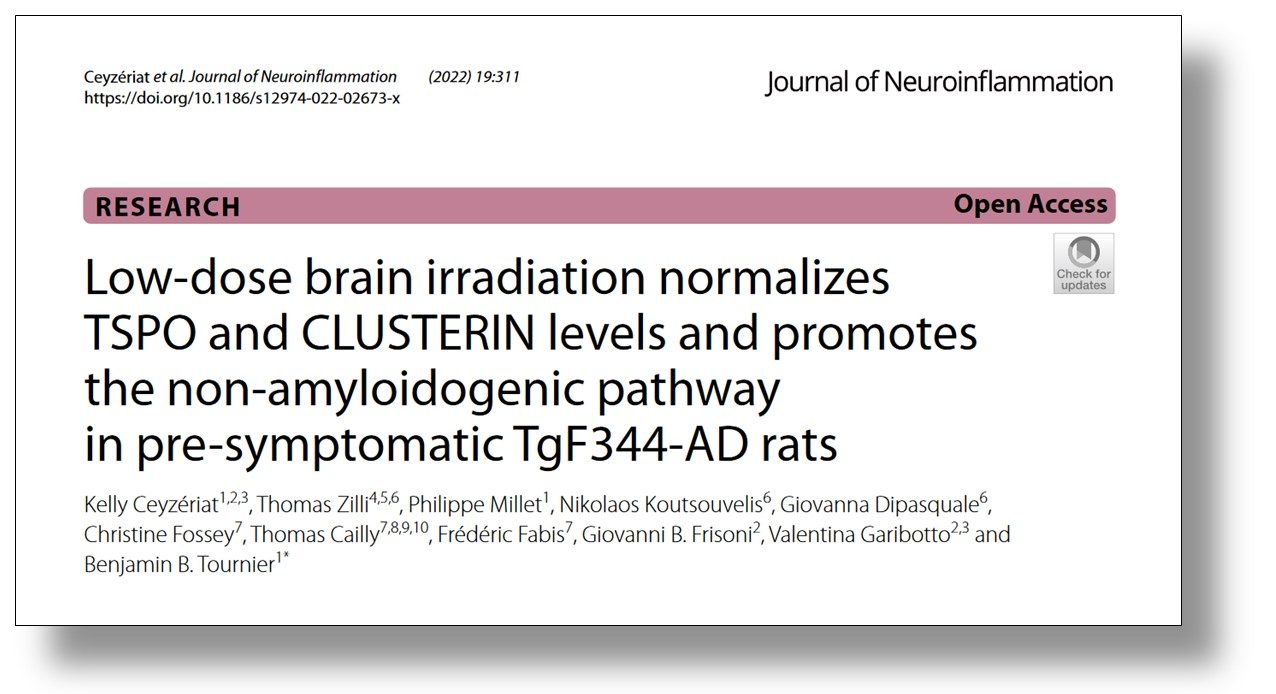 Title: Low-dose brain irradiation normalizes TSPO and CLUSTERIN levels and promotes the non-amyloidogenic pathway in pre-symptomatic TgF344-AD rats.
Authors: Ceyzériat K, Zilli T, Millet P, Koutsouvelis N, Dipasquale G, Fossey C, Cailly T, Fabis F, Frisoni GB, Garibotto V, Tournier BB.
Journal: J Neuroinflammation. 2022 Dec 22;19(1):311.
PMID: 36550510 (This report is OPEN ACCESS if you would like to read it)
In this study, the researchers exposed a genetically engineered rat model of Alzheimer’s (TgF344-AD rats, which produce high levels of human amyloid precursor protein and presenilin 1 protein) to low dose ionising radiation over 5 days. The procedure resulted in levels of neuroinflammation being reduced to normal ranges in these rodents. It also reduced the levels of aggregated protein in the animals brains by approximately 60 to − 80%.
And it should be noted all of the researchers involved with these preclinical studies cited here are completely independent from those investigators that reported the case studies and pilot clinical trial research mentioned further above.
Has low-dose radiation ever been tested in a model of Parkinson’s?
This was the first preclinical study exploring low-dose radiation in a model of Parkinson’s:
 Title: Elevation of antioxidant potency in the brain of mice by low-dose gamma-ray irradiation and its effect on 1-methyl-4-phenyl-1,2,3,6-tetrahydropyridine (MPTP)-induced brain damage.
Authors: Kojima S, Matsuki O, Nomura T, Yamaoka K, Takahashi M, Niki E.
Journal: Free Radic Biol Med. 1999 Feb;26(3-4):388-95.
PMID: 9895231
Published way back in 1999, the researchers who conducted this study exposed a neurotoxin (MPTP) mouse model of Parkinson’s to low dose ionising radiation, and they reported that low dose ionising radiation induced antioxidative mechanisms in the brain that reduced the impact of the neurotoxin. The procedure increased levels of antioxidants like glutathione and thioredoxin.
And other research groups have suggested similar results (Click here and here to read more about this).
Has anyone with Parkinson’s ever tried this type of treatment?
So this is where there is a really good twist in this tale.
You remember the lady with dementia in Michigan? The first case study we discussed above? Well, she had a husband and he had been diagnosed with Parkinson’s.
Naturally, after observing his wife’s improvements from the CT scans, he immediately requested the same treatment for himself.
And what happened?
This situation was discussed in a second update on that particular lady’s case study:
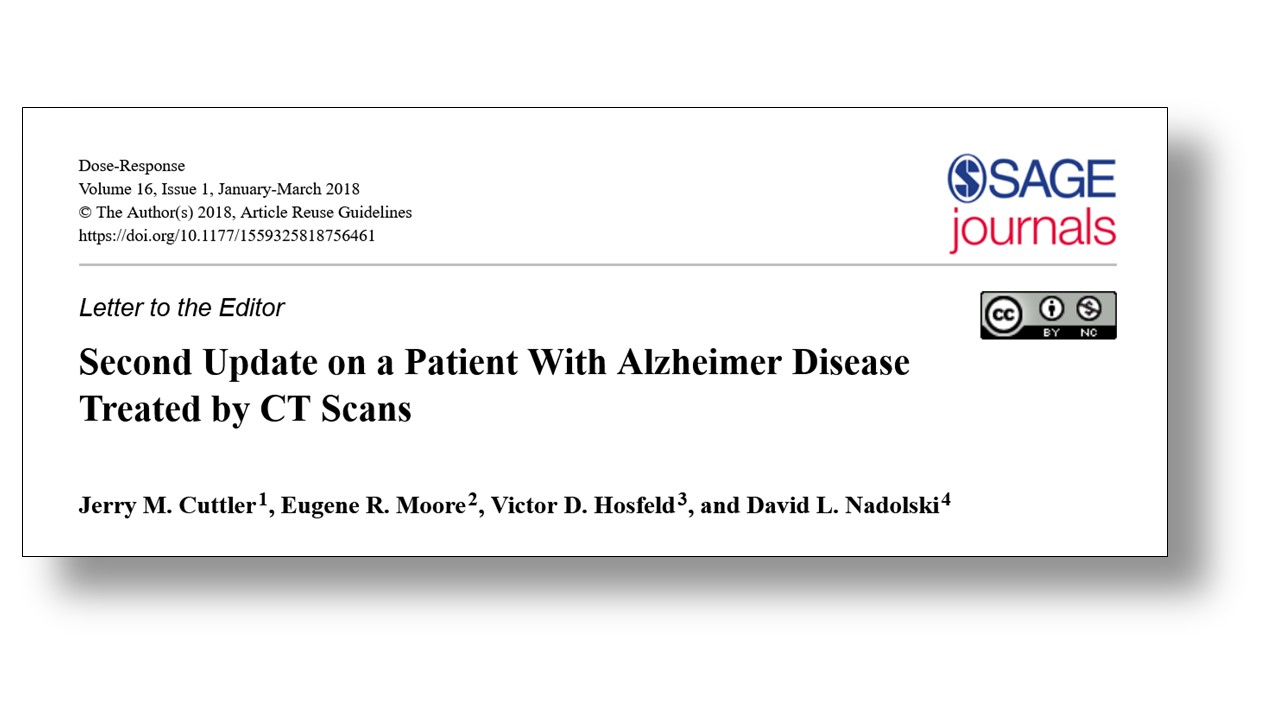 Title: Second Update on a Patient With Alzheimer Disease Treated by CT Scans.
Authors: Cuttler JM, Moore ER, Hosfeld VD, Nadolski DL.
Journal: Dose Response. 2018 Feb 14;16(1):1559325818756461.
PMID: 29479296 (This report is OPEN ACCESS if you would like to read it)
The husband started receiving CT scans in October, 2015. The night after his first scan, he reported a significant reduction in his tremor. Short after this, his medication was reduced from 6 to just 2 or 3 pills per day. By December 2016, continued improvement in tremor was reported and his constipation was also better. “The patient consistently observed a decrease in tremor shortly after each low dose of ionizing radiation” (Source).
Again, this is just a single case study and in this particular case the individual was potentially prone to a placebo response in that he knew what he was being treated with and had seen the effect of it on his wife. But the account still provides some preliminary data supporting further research into this area.
But do you think low dose ionising radiation is safe?
The researchers who conducted the pilot research on low dose ionising radiation wrote in their discussion of the results that a certain level of caution is required. “Low dose ionizing radiation therapy is controversial because it is generally accepted that ionizing radiation is a significant cause of DNA mutations and increases cancer risk” (Source).
That said, if a particular dose of ionising radiation can be identified as having beneficial effects on cellular function in people with neurodegenerative conditions, it might still provide a means of providing some alleviation of the symptoms and restore some quality of life.
If nothing else it certainly sounds like something worth exploring.
So what does it all mean?
In April 1973, the US Food and Drug Administration approved the use of the influenza treatment amantadine for alleviating the symptoms of Parkinson’s (Click here to read more about this). It is a classic example of drug repurposing that began with a single anecdotal patient-doctor interaction. A woman with Parkinson’s was administered amantadine to treat an influenza infection that she was struggling with. She reported that her Parkinson’s symptoms improved significantly while on amantadine, and that they worsened after she finished the treatment. This observation resulted in clinical trials, which ultimately led to a new treatment option for the Parkinson’s community.
Much of medical history is based on anecdotal discoveries. Some play out and become game changing events (like amantadine). In other cases, efforts are made to replicate the findings of the original observations and they do not see the same outcome. The case of low dose ionising radiation is intriguing because in the original case study – the lady with dementia in Michigan – the individuals may not have been aware of the impact of the CT scans. It was probably just a routine imaging procedure, and this would indicate that a placebo response is less likely. But it leaves one questioning how many other individuals with dementia or Parkinson’s have received CT scans? And did they observe noticeable effects?
Regardless, I was pleased to have struck up a conversation with my fellow conference attendee during the lunch break and I left the table with a facial expression resembling this:

I will follow the area of low dose of ionising radiation research in neurodegeneration with interest. It will be very interesting to see if it plays out.
 All of the material on this website is licensed under a
Creative Commons Attribution 4.0 International License
You can do whatever you like with it!
The banner for today’s post was sourced from ans

Antioxidant pathways are promoted by 1,25(OH)D signalling, including Nrf2, and glutathione synthesis and utilisation. I shall not be at all surprised if D3 deficiency -which is widespread – explains the sensitivty to Xrays. Sporadic PD is absent in elderly rural Kenyans, with a lifestyle exposed to the sun similar to the Masai who are regarded as having physiological D3 status.
LikeLike
I have to say that I worry that some “biohacker” type folks living with PD or other neurodegenerative conditions will try to jump ahead of your interesting blogpost because case reports and rodent studies suggest that low dose ionizing radiation exposure might be beneficial for PwP — Please strongly discourage folks from purposefully exposing themselves to additional ionizing radiation by requesting CT scans or other higher levels of exposure that could have major NEGATIVE effects on health. A 2020 meta-analytic review (https://www.ncbi.nlm.nih.gov/pmc/articles/PMC9710150/) notes that CT scans increase risk of cancer significantly (“increase of radiation dose (OR, 33.31 [95% CI, 21.33 to 52.02]), and multiple CT scan sites (OR, 14.08 [95% CI, 6.60 to 30.05]).”
LikeLike
Fascinating article – thanks for posting. I seem to remember a BBC documentary looking at whether there were any positive health effects from the radiation exposure in regions around Chernobyl (think it was http://news.bbc.co.uk/1/hi/sci/tech/5173310.stm) Presumably there are some good datasets on health outcomes for these people, and radiation workers. Interesting paper here: https://www.sciencedirect.com/science/article/pii/S016041202032167X
LikeLike
Yes, I also recall the BBC feature from over a decade ago. Thanks. This was during a time I was considering electrical stimulation & DBS for my wife (YOPD) and had been researching ANY treatment that could help reduce the severity of her systems and the unrelenting impact on her daily activities (& family life).
As this interesting post brings up, much of medical history is based on anecdotal discoveries. I look forward to responsible study and to learning more about low dose ionizing radiation.
LikeLiked by 1 person