New stem cell transplantation trial for Parkinson’s proposed in China

7661_Screen-Shot-2013-11-08-at-11.37.38-AM

We have been contacted by some readers asking about a new stem cell transplantation clinical trial for Parkinson’s disease about to start in China (see the Nature journal editorial regarding this new trial by clicking here).

While this is an exciting development, there have been some concerns raised in the research community regarding this trial.

In today’s post, we will discuss what is planned and what it will mean for stem cell transplantation research.


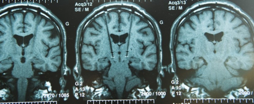
Deep-Brain-Stimulation-60pghfsukanm4j4bljb8mbq9hyafm3pj0e6t4iuyndm

Brain surgery. Source Bionews-tx

Parkinson’s disease is a progressive neurodegenerative condition.

This means that cells in the brain are slowly being lost over time. What makes the condition particularly interesting is that certain types of brain cells are more affected than others. The classic example of this is the dopamine neurons in an area of the brain called the substantia nigra, which resides in the midbrain.

d1ea3d21c36935b85043b3b53f2edb1f87ab7fa6

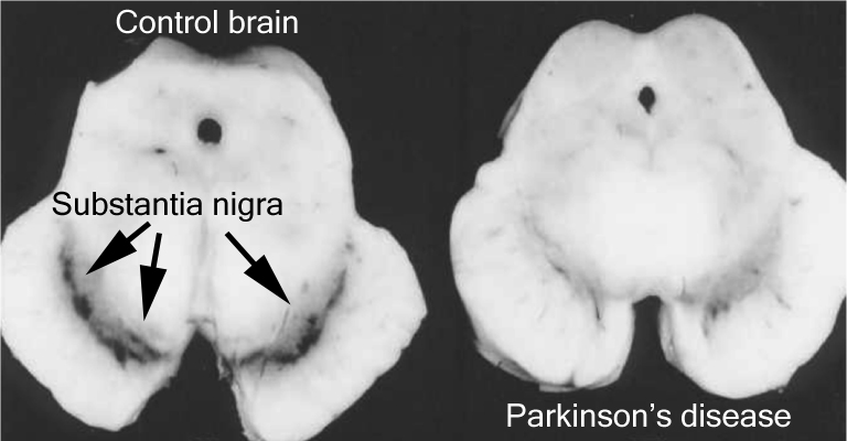
The number of dark pigmented dopamine cells in the substantia nigra are reduced in the Parkinson’s disease brain (right). Source: Adapted from Memorangapp

Approximately 50% of the dopamine neurons in the midbrain have been lost by the time a person is diagnosed with Parkinson’s disease (note the lack of dark colouration in the substantia nigra of the Parkinsonian brain in the image above), and as the condition progresses the motor features – associated with the loss of dopamine neurons – gradually get worse. This is why dopamine replacement treatments (like L-dopa) are used for controlling the motor symptoms of Parkinson’s disease.

A lot of research effort is being spent on finding disease slowing/halting treatments, but these will leave many people who have already been diagnosed with Parkinson’s disease still dealing with the condition. What those individuals will require is a therapy that will be able to replace the lost cells (particularly the dopamine neurons). And researchers are also spending a great deal of time and effort on findings ways to do this. One of the most viable approaches at present is cell transplantation therapy. This approach involves actually injecting cells back into the brain to adopt the functions of the lost cells.

How does cell transplantation work?

We have discussed the history of cell transplantation in a previous post (Click here to read that post), and today we are simply going to focus on the ways this experimental treatment is being taken forward in the clinic.

Many different types of cells have been tested in cell transplantation experiments for Parkinson’s disease (Click here for a review of this topic), but to date the cells that have given the best results have been those dissected from the developing midbrain of aborted embryos.

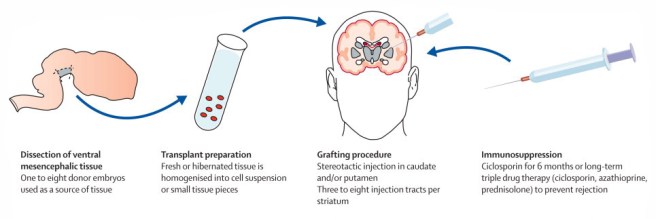
This now old fashioned approach to cell transplantation involved dissecting out the region of the developing dopamine neurons from a donor embryo, breaking up the tissue into small pieces that could be passed through a tiny syringe, and then injecting those cells into the brain of a person with Parkinson’s disease.

gr3

The old cell transplantation process for Parkinson’s disease. Source: The Lancet

Critically, the people receiving this sort of transplant would require ‘immunosuppression treatment’ for long periods of time after the surgery. This additional treatment involves taking drugs that suppress the immune system’s ability to defend the body from foreign agents. This step is necessary, however, in order to stop the body’s immune system from attacking the transplanted cells (which would not be considered ‘self’ by the immune system), allowing those cells to have time to mature, integrate into the brain and produce dopamine.

The transplanted cells are injected into an area of the brain called the putamen. This is one of the main regions of the brain where the dopamine neurons of the substantia nigra release their dopamine. The image below demonstrates the loss of dopamine (the dark staining) over time as a result of Parkinson’s disease (PD):

m_awt192f1p

The loss of dopamine in the putamen as Parkinson’s disease progresses. Source: Brain

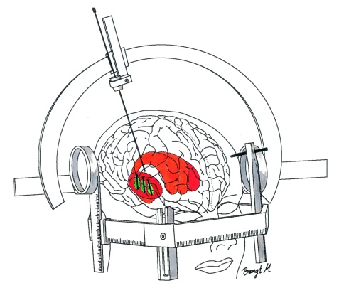
In cell transplant procedures for Parkinson’s disease, multiple injections are usually made in the putamen, allowing for deposits in different areas of the structure. These multiple sites allow for the transplanted cells to produce dopamine in the entire extent of the putamen. And ideally, the cells should remain localised to the putamen, so that they are not producing dopamine in areas of the brain where it is not desired (possibly leading to side effects).

image2

Targeting transplants into the putamen. Source: Intechopen

Postmortem analysis – of the brains of individuals who have previously received transplants of dopamine neurons and then subsequently died from natural causes – has revealed that the transplanted cells can survive the surgical procedure and integrate into the host brain. In the image below, you can see rich brown areas of the putamen in panel A. These brown areas are the dopamine producing cells (stained in brown). A magnified image of individual dopamine producing neurons can be seen in panel B:

Microsoft Word - Li W-Revision-Final.docx

Transplanted dopamine neurons. Source: Sciencedirect

The transplanted cells take several years to develop into mature neurons after the transplantation surgery, and the benefits of the transplantation technique may not be apparent for some time (2-3 years on average). Once mature, however, it has also been demonstrated (using brain imaging techniques) that these transplanted cells can produce dopamine. As you can see in the images below, there is less dopamine being processed (indicated in red) in the putamen of the Parkinsonian brain on the left than the brain on the right (several years after bi-lateral – both sides of the brain – transplants):

3_2

Brain imaging of dopamine processing before and after transplantation. Source: NIH

Sounds like a great therapy for Parkinson’s disease right?

So why aren’t we doing it???

Two reasons:

1. The tissue used in the old approach for cell transplantation in Parkinson’s disease was dissected from embryonic brains. Obviously there are serious ethical and moral problems with using this kind of tissue. There is also a difficult problem of supply: tissue from at least 3 embryos is required for transplanting each side of the brain (6 embryos in total). Given these issues, researchers have focused their attention on a less controversial and more abundant supply of cells: brain cells derived from embryonic stem cells (the new approach to cell transplantation).

1024px-Humanstemcell

Human embryonic stem cells. Source: Wikipedia

2. The second reason why cell transplantation is not more widely available is that in the mid 1990’s, the US National Institutes of Health (NIH) provided funding for the two placebo-controlled, double blind studies to be conducted to test the efficacy of the approach. Unfortunately, both studies failed to demonstrate any beneficial effects on Parkinson’s disease features.

In addition, many (15% – 50%) of transplanted subjects developed what are called ‘graft-induced dyskinesias’. This involves the subjects display uncontrollable/erratic movement (or dyskinesias) as a result of the transplanted cells. Interestingly, patients under 60 years of age did show signs of improvement on when assessed both clinically (using the UPDRS-III) and when assessed using brain imaging techniques (increased F-dopa uptake on PET).

Both of the NIH trials have been criticised by experts in the field for various procedural failings that could have contributed to the failures. But the overall negative results left a dark shadow over the technique for the better part of a decade. Researchers struggled to get funding for their research.

And this is the reason why many researchers are now urging caution with any new attempts at cell transplantation clinical trials in Parkinson’s disease – any further failures will really harm the field, if not kill if off completely.

Are there any clinical trials for cell transplantation in Parkinson’s disease currently being conducted?

Yes, there are currently two:

Firstly there is the Transeuro being conducted in Europe.

Transeuro_Logo_100

The Transeuro trial. Source: Transeuro

The Transeuro trial is an open label study, involving 40 subjects, transplanted in different sites across Europe. They will receive immunosuppression for at least 12 months post surgery, and the end point of the study will be 3 years post surgery, with success being based on brain imaging of dopamine release from the transplanted cells (PET scans). Based on the results of the previous NIH funding double blind clinical studies discussed above, only subject under 65 years of age have been enrolled in the study.

transeuro

The European consortium behind the Transeuro trial. Source: Transeuro

In addition to testing the efficacy of the cell transplantation approach for Parkinson’s disease, another goal of the Transeuro trial is to optimise the surgical procedures with the aim of ultimately shifting over to an embryonic stem cells oriented technique in the near future with the proposed G-Force embryonic stem cell trials planned for 2018 (the Transeuro is testing the old approach to cell transplantation).

The second clinical study of cell transplantation for Parkinson’s disease is being conducted in Melbourne (Australia), by an American company called International Stem Cell Corporation.

logo

This study is taking the new approach to cell transplantation, but the company is using a different type of stem cell to produce dopamine neurons in the Parkinsonian brain.

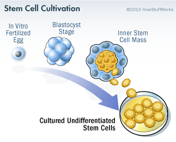
Specifically, the researchers will be transplanting human parthenogenetic stem cells-derived neural stem cells (hpNSC). These hpNSCs come from an unfertilized egg – that is to say, no sperm cell is involved. The female egg cell is chemically encouraged to start dividing and then it becoming a collection of cells that is called a blastocyst, which ultimately go on to contain embryonic stem cell-like cells.

stem-cell-cultivation-3

The process of attaining embryonic stem cells. Source: Howstuffworks

This process is called ‘Parthenogenesis’, and it’s not actually as crazy as it sounds as it occurs naturally in some plants and animals (Click here to read more about this). Proponents of the parthenogenic approach suggest that this is a more ethical way of generating ES cells as it does not result in the destruction of a viable organism.

Regular readers of this blog will be aware that we are extremely concerned about this particular trial (Click here and here to read previous posts about this). Specifically, we worry that there is limited preclinical data from the company supporting the efficacy of these hpNSC cells being used in the clinical study (for example, researchers from the company report that the hpNSC cells they inject spread well beyond the region of interest in the company’s own published preclinical research – not an appropriate property for any cells being taken to the clinic). We have also expressed concerns regarding the researchers leading the study making completely inappropriate disclosures about the study while the study is ongoing (Click here to read more about this). Such comments only serve the interests of the company behind the study. And this last concern has been raised again with a quote in the Nature editorial about the Chinese trial:

“Russell Kern, chief scientific officer of the International Stem Cell Corporation in Carlsbad, California, which is providing the cells for and managing the Australian trial, says that in preclinical work, 97% of them became dopamine-releasing cells” (Source)

We are unaware of any preclinical data produced by Dr Kern and International Stem Cell Corporation…or ANY other research lab in the world that has achieved 97% dopamine-releasing cells. We (and others) would be interested in learning more about Dr Kerns amazing claim.

The International Stem Cell Corporation clinical trial is ongoing. For more details about this second ongoing clinical trial, please click here.

So what do we know about the new clinical study?

The clinical trial (Titled: A Phase I/II, Open-Label Study to Assess the Safety and Efficacy of Striatum Transplantation of Human Embryonic Stem Cells-derived Neural Precursor Cells in Patients With Parkinson’s Disease) will take place at the First Affiliated Hospital of Zhengzhou University in Henan province.

The researchers are planning to inject neuronal-precursor cells derived from embryonic stem cell into the brains of individuals with Parkinson’s disease. They have 10 subjects that they have found to be well matched to the cells that they will be injecting, which will help to limit the chance of the cells being rejected by the body.

In testing the safety and efficacy of these cells, the trial will have two primary outcome measures:
  1. Incidence of treatment-emergent adverse events, as assessed by brain imaging and blood examination at 6 months post transplant.
  2. Number of subjects with adverse events (such as the evidence of transplant failure or rejection)

In addition to these, there will also be a series of secondary outcome measures, which will include:

  1. Change in Unified Parkinson’s Disease Rating Scale (UPDRS) score at 12 months post surgery, when compared to baseline scores. Each subject was independently rated by two observers at each study visit and a mean score was calculated for analysis.
  2. Change in DATscan brain imaging at 12 months when compared to a baseline brain scan taken before surgery. DATscan imaging provides an indication of dopamine processing.
  3. Change in Hoehn and Yahr Stage at 12 months, compared to baseline scores. The Hoehn and Yahr scale is a commonly used system for Parkinson’s disease.

The trial will be a single group, non-randomized analysis of the safety and efficacy of the cells. The estimated date of completion is December 2020.

Why are some researchers concerned about the study?

Professor Qi Zhou, a stem-cell specialist at the Chinese Academy of Sciences Institute of Zoology will be leading the study and he has a REALLY impressive track record in the field of stem cell biology. His team undertaking this study have a great deal of experience working with embryonic stem cells, having published some extremely impressive research on this topic. But, (and it’s a big but) they have published a limited amount of research in peer-reviewed journals on cell transplantation in models of Parkinson’s disease. Lorenz Studer is one of the leading scientists in this field, was quoted in an editorial in the journal Nature this week:

“Lorenz Studer, a stem-cell biologist at the Memorial Sloan Kettering Cancer Center in New York City who has spent years characterizing such neurons ahead of his own planned clinical trials, says that “support is not very strong” for the use of precursor cells. “I am somewhat surprised and concerned, as I have not seen any peer-reviewed preclinical data on this approach,” he says.” (Source)

In addition to the lack of published research by the team undertaking the trial, the research community is also worried about the type of cells that are going to be transplanted in this clinical trial. Most of the research groups heading towards clinical trials in this area are all pushing embryonic stem cells towards a semi-differentiated state. That is, they are working on recipes that help the embryonic stem cells grow to the point that they have almost become dopamine neurons. Prof Zhou and his colleagues, however, are planning to transplant a much less differentiated type of cell called a neural-precursor cell in their transplants.

Pan

Neuronal-precursor cells. Source: Wired

Neuronal-precursors are very early stage brain cells. They are most likely being used in the study because they will survive the transplantation procedure better than a more mature neurons which would be more sensitive to the process – thus hopefully increasing the yield of surviving cells. But we are not sure how the investigators are planning to orient the cells towards becoming dopamine neurons at such an early stage of their development. Neuronal-precursors could basically become any kind of brain cell. How are the researchers committing them to become dopamine neurons?

Are these concerns justified?

We feel that there are justified reasons for concern.

While Prof Zhou and his colleagues have a great deal of experience with embryonic stem cells and have published very impressive research on that topic, the preclinical data for this trial is limited. In 2015, the research group published this report:

zhou
Title: Lmx1a enhances the effect of iNSCs in a PD model
Authors: Wu J, Sheng C, Liu Z, Jia W, Wang B, Li M, Fu L, Ren Z, An J, Sang L, Song G, Wu Y, Xu Y, Wang S, Chen Z, Zhou Q, Zhang YA.
Journal: Stem Cell Res. 2015 Jan;14(1):1-9.
PMID: 25460246              (This article is OPEN ACCESS if you would like to read it)

In this study, the researchers engineered embryonic stem cells to over-produce a protein called LMX1A to help produce dopamine neurons. LMX1A is required for the development of dopamine neurons (Click here to read more about this). The investigators then grew these cells in cell culture and compared their ability to develop into dopamine neurons against embryonic stem cells with normal levels of LMX1A. After 14 days in cell culture, 16% of the LMX1A cells were dopamine neurons, compared to only 5% of the control cells.

When the investigators transplanted these cells into a mouse model of Parkinson’s disease, they found that the behavioural recovery in the mice did not differ from the control injected mice, and when they looked at the brains of the mice 11 weeks after transplantation “very few engrafted cells had survived”.

In addition to this previously published work, the Chinese team do have unpublished research on 15 monkeys that have undergone the neuronal-precursor cell transplantation procedure having had Parkinson’s disease induced using a neurotoxin. The researchers have admitted that they initially did not see any improvements in movement (which is expected given the slow maturation of the cells). At the end of the first year, however, they examined the brains of some of the monkeys and they found that the transplanted stem cells had turned into dopamine-releasing cells (exactly what percentage of the cells were dopamine neurons is yet to be announced). The monkey study has been running for several years now and they have seen a 50% improvement in the motor ability of the remaining monkeys, supported by brain imaging data. The publication of this research is in preparation, but it probably won’t be available until after the trial has started.

So yes, there is a limited amount of preclinical research supporting the clinical trial.

As for concerns regarding the type of cells that are going to be transplanted:

Embryonic stem cells have robust tumour forming potential. If you inject them into the brain of mice, there is the potential for them to develop into dopamine neurons, but also tumours:

Ole
Title: Embryonic stem cells develop into functional dopaminergic neurons after transplantation in a Parkinson rat model
Authors: Bjorklund LM, Sánchez-Pernaute R, Chung S, Andersson T, Chen IY, McNaught KS, Brownell AL, Jenkins BG, Wahlestedt C, Kim KS, Isacson O.
Journal: Proc Natl Acad Sci U S A. 2002 Feb 19;99(4):2344-9.
PMID: 11782534               (This article is OPEN ACCESS if you want to read it)

In this study, the researchers found that of the twenty-five rats that received embryonic stem cell injections into their brains to correct the modelled Parkinson’s disease, five rats died before completed behavioural assessment and the investigators found teratoma-like tumours in their brains – less than 16 weeks after the cells had been transplanted.

eb7315426f2171c7cb96dc4d980686_big_gallery

A teratoma (white spot) inside a human brain. Source: Radiopaedia

Given this risk of tumour formation, research groups in the cell transplantation field have been trying to push the embryonic stem cells as far away from their original pluripotent state and as close to a dopamine fate as possible without producing mature dopamine neurons which will not survive the transplantation procedure very well.

Prof Zhou’s less mature neuronal-precursor cells are closer to embryonic stem cells than dopamine neurons on this spectrum than the kinds of cells other research groups are testing in cell transplantation experiments. As a result, we are curious to know what precautions the investigators are taking to limit the possibility of an undifferentiated, still pluripotent embryonic stem cell from slipping into this study (the consequences could be disastrous). And given their results from the LMX1A study described above, we are wondering how they are planning to push the cells towards a dopamine fate. If they do not have answers to this issues, they should not be rushing to the clinic with these cells.

So yes, there are reasons for concern regarding the cells that the researchers plan to use in this clinical trial.

And, as with the International Stem Cell Corporation stem cell trial in Australia, we also worry that the follow up-period (or endpoint in the study) of 12 months is not long enough to determine the efficacy of these cells in improving Parkinson’s rating scores and brain imaging results. All of the previous clinical research in this field indicates that the transplanted cells require years of maturation before their dopamine production has an observable impact on the participant. Using 12 months as an end point for this study is tempting a negative result when the long term outcome could be positive.

As we mentioned above, any negative outcomes for these studies could have dire consequences for the field as a whole.

So what does it all mean?

Embryonic stem cells hold huge potential in the field of regenerative medicine. Their ability to become any cell type in the body means that if we can learn how to control them correctly, these cells could represent a fantastic new tool for future cell replacement therapies in conditions like Parkinson’s disease.

Strong demand for such therapies from groups like the Parkinsonian community, has resulted in research groups rushing to the clinic with different approaches using these cells. Concerns as to whether such approaches are ready for the clinic are warranted, if only because mistakes by individual research groups/consortiums in the past have caused delays for everyone in the field.

While China is very keen (and should be encouraged) to take bold steps in its ambition to be a world leader in this field, open and transparent access to extensive preclinical research would help assuage concerns within the research community that prudent care is being taken heading forward.

We’ll keep you aware of developments in this clinical trial.


EDITORIAL NOTE No.1 – It is important for all readers of this post to appreciate that cell transplantation for Parkinson’s disease is still experimental. Anyone declaring otherwise (or selling a procedure based on this approach) should not be trusted. While we appreciate the desperate desire of the Parkinson’s community to treat the disease ‘by any means possible’, bad or poor outcomes at the clinical trial stage for this technology could have serious consequences for the individuals receiving the procedure and negative ramifications for all future research in the stem cell transplantation area. 

EDITORIAL NOTE No.2 – the author of this blog is associated with research groups conducting the current Transeuro transplantation trials and the proposed G-Force embryonic stem cell trials planned for 2018. He has endeavoured to present an unbiased coverage of the news surrounding the current clinical trials, though he shares the concerns of the Parkinson’s scientific community that the research supporting the current Australian trial is lacking in its thoroughness and will potentially jeopardise future work in this area. He is also concerned by the lack of peer-reviewed published research on cell transplantation in models of Parkinson’s disease for the proposed clinical studies in China. 


The banner for today’s post was sourced from Ozy

Oleuropein – “surely the richest gift of heaven?”

thomas-jefferson

The title of this post is a play on a Thomas Jefferson quote (“the olive tree is surely the richest gift of heaven“). Jefferson, the third President of the United States (1801 to 1809), was apparently quite the lover of food. During the Revolutionary War, while he was a U.S. envoy to France, Jefferson travelled the country. In Aix-en-Provence, he developed an admiration for olive trees, calling them “the most interesting plant in existence”.

Being huge food lovers ourselves, we here at the SoPD wholeheartedly agree with Jefferson. But we also think that olives are interesting for another reason:

They contain a chemical called Oleuropein.

In today’s post we’ll explore what is known about this chemical and discuss what it could mean for Parkinson’s disease.


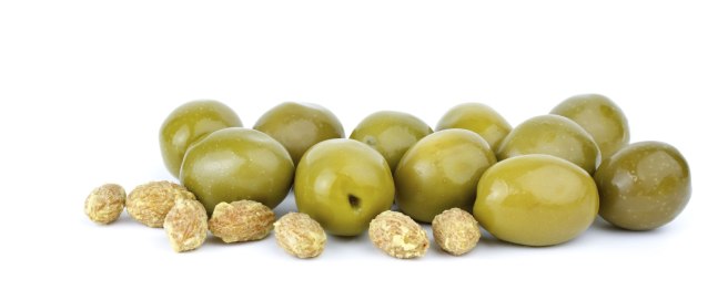
olivve-pits

Olives. Source: Gardeningknowhow

The olive, also known by the botanical name ‘Olea europaea,’ is an evergreen tree that is native to the Mediterranean, Asia and Africa, but now found around the world. It has a rich history of economic and symbolic importance within western civilisation. And the fruit of the tree also tastes good, either by themselves or in a salad or pasta dish.

Traditional diets of people living around the Mediterranean sea are very rich in extra-virgin olive oil. Olives are an excellent source of ‘good’ fatty acids (monounsaturated and di-unsaturated), antioxidants and vitamins. Indeed, research has shown that the traditional Mediterranean diet reduces the risk of heart disease (Click here to read more on this).

preview

Olive oil. Source: Bonzonosvilla

There are also chemicals within the olive fruit that may have very positive benefits for Parkinson’s disease.

But before you rush out and gorge yourself on olives, we have one small piece of advice:

The chemical is called Oleuropein, and it is usually removed from olives due to its bitterness.

What is Oleuropein?

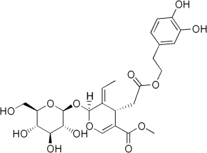
Oleuropein is a ‘phenylethanoid’ – a type of phenolic compound that is found in the leaf and the fruit of the olive. Phenolic compounds are produced by plants as a protective measure against different kinds of stress.

Oleuropein

Oleuropein. Source: Wikipedia

The main phenolic compounds found in olives are hydroxytyrosol and oleuropein – both of which give extra-virgin olive oil its bitter taste and both have demonstrated neuroprotective effects.

More research has been done on oleuropein so we will focus on it here (for more on hydroxytyrosol – please click here).

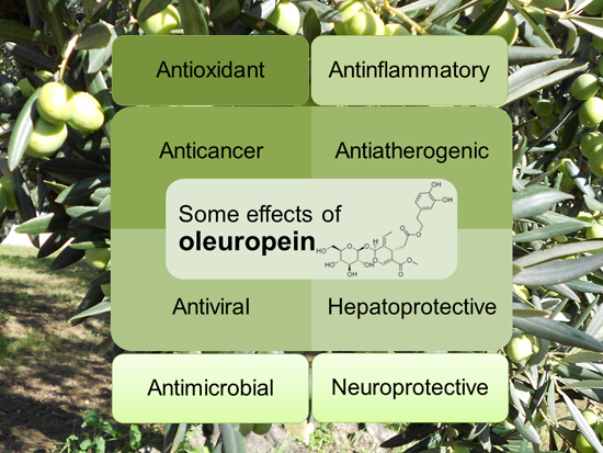
Oleuropein has been found to have many interesting properties, such as:

ijms-15-18508-ag

The many properties of oleuropein. Source: Mdpi

What neuroprotective research has been done on Oleuropein?

Thus far, most of the research addressing this question has been conducted on models of Alzheimer’s disease. The first study

PLos1

Title: Oleuropein aglycone protects transgenic C. elegans strains expressing Aβ42 by reducing plaque load and motor deficit.
Authors: Diomede L, Rigacci S, Romeo M, Stefani M, Salmona M.
Journal: PLoS One. 2013;8(3):e58893.
PMID: 23520540                 (This article is OPEN ACCESS if you would like to read it)

The Italian researchers who conducted this study treated a microscopic worm model of Alzheimer’s disease with oleuropein aglycone. We should not that oleuropein aglycone is a hydrolysis product of oleuropein (a hydrolysis product is a chemical compound that is broken apart by the addition of water). The microscopic worm used in the study are called Caenorhabditis elegans:

c_elegans

Caenorhabditis elegans – cute huh? Source: Nematode

Caenorhabditis elegans (or simply C. Elegans) are tiny creatures that are widely used in biology because they can be easily genetically manipulated and their nervous system is very simple and well mapped out (they have just 302 neurons and 56 glial cells!). The particular strain of C. elegans used in this first study produced enormous amounts of a protein called Aβ42.

Amyloid beta (or Aβ) is the bad boy/trouble maker of Alzheimer’s disease; considered to be critically involved in the condition. A fragment of this protein (called Aβ42) begins clustering in the brains of people with Alzheimer’s disease. This clustering of Aβ42 goes on to form the plaques that are so characteristic of the Alzheimer’s affected brain.

The Italian researchers conducting this study had previously shown that oleuropein can inhibit the ability of Aβ42 to aggregate in cells growing in culture dishes (Click here to read more about that study), and they wanted to see if oleuropein had the same properties in actual live animals. So they chose the C. Elegans that had been genetically engineered to produce a lot of Aβ42 to test this idea.

In the C. Elegans that produce a lot of Aβ42 gradually become paralysed and their lives are shortened. By treating these worms with oleuropein, however, the Italian researchers found that there was less aggregation of Aβ42 (though the levels of the protein stayed the same), resulting in less plaque formation, and improved mobility (>50% reduction in paralysis) and survival compared to untreated Aβ42 producing C. Elegans.

Encouraged by this result, the researchers next moved on to studies in mice:

Plos2

Title: The polyphenol oleuropein aglycone protects TgCRND8 mice against Aß plaque pathology.
Authors: Grossi C, Rigacci S, Ambrosini S, Ed Dami T, Luccarini I, Traini C, Failli P, Berti A, Casamenti F, Stefani M.
Journal: PLoS One. 2013 Aug 8;8(8):e71702.
PMID: 23951225                   (This article is OPEN ACCESS if you would like to read it)

For this study, the Italian researchers used the genetically engineered TgCRND8 mice. These mice have a mutant form of amyloid precursor protein (which, similar to Aβ42, is associated with Alzheimer’s disease). In the brains of these mice, amyloid clustering begins at 3 months of age, and dense plaques are evident from 5 months of age. The mice also exhibit a clear learning impairment from 3 months of age.

By treating these mice with oleuropein aglycone, the researchers observed a remarkable reduction in plaques in the brain, and those that were present appeared less compact and “fluffy” (their very technical description, not ours). In addition, there was a reduction in the activation of astrocytes and microglia (the helper cells in the brain), indicating a healthier environment.

These same researchers have observed the same results in a rat model of Alzheimer’s disease in a report published the next year (Click here to read more about this).

Interestingly, the oleuropein treated TgCRND8 mice also displayed a major increase in autophagy activity. As we discussed in our previous post (Click here to read that post), autophagy is the rubbish disposal/recycling system of each cell, and increasing the activity of this system can help to keep cells health (particularly if there is a lot of a genetically engineered protein present!).

The Italian researchers repeated this study, and published the results this year, with an interesting twist:

JCBP

Title: Oleuropein aglycone and polyphenols from olive mill waste water ameliorate cognitive deficits and neuropathology.
Authors: Pantano D, Luccarini I, Nardiello P, Servili M, Stefani M, Casamenti F.
Journal: Br J Clin Pharmacol. 2017 Jan;83(1):54-62.
PMID: 27131215

In this study, the researchers tested the same genetically engineered mice, but with two different treatments:

  1.  Two much lower doses of oleuropein (4 and 100 times lower)
  2.  A mixture of polyphenols from olive mill concentrated waste water

The lowest dose of oleuropein (100 times less oleuropein than the previous study) did not provide any significant improvements for the mice, but the intermediate dose (only 4 times less oleuropein than the previous study) did provide significant benefits. These result indicate that there is a dose-dependent range to the beneficial properties of oleuropein.

The researchers also observed very similar beneficial effects from the mice drinking a mixture of polyphenols from olive mill concentrated waste water. Given these results, the investigators are now seeking to design appropriate conditions to perform a clinical trial to assess better the possible use of oleuropein (or a mix of olive polyphenols) against Alzheimer’s disease.

Ok, but what research has been done with oleuropein and Parkinson’s disease?

Unfortunately, not much.

A research group in Iran has looked at the effect of oleuropein in aged rodents and found an interesting result:

Iran
Title: Antioxidant role of oleuropein on midbrain and dopaminergic neurons of substantia nigra in aged rats.
Authors: Sarbishegi M, Mehraein F, Soleimani M.
Journal: Iran Biomed J. 2014;18(1):16-22.
PMID: 24375158                 (This article is OPEN ACCESS if you would like to read it)

In this study, the investigators took twenty aged rats (18-month-old) and randomly assigned them to two groups: a treatment group (which received a daily dose of 50 mg/kg of oleuropein for 6 months) and a control group (which received just water). Following these treatments, the investigators found an increase in the activity of anti-oxidant agents (such as superoxide dismutase, catalase and glutathione) in the treatment group compared to control group. The treated rats also had significantly more dopamine neurons in the region of the brain affected by Parkinson’s disease (the substantia nigra). The investigators concluded that oleuropein consumption in a daily diet may be useful in reducing oxidative stress damage by increasing the antioxidant activity in the brain.

This first study was followed more recently by a report from a group in Quebec (Canada) who investigated oleuropein use in a cell culture model of Parkinson’s disease:

Oleu
Title: Oleuropein Prevents Neuronal Death, Mitigates Mitochondrial Superoxide Production and Modulates Autophagy in a Dopaminergic Cellular Model.
Authors: Achour I, Arel-Dubeau AM, Renaud J, Legrand M, Attard E, Germain M, Martinoli MG.
Journal: Int J Mol Sci. 2016 Aug 9;17(8).
PMID: 27517912              (This article is OPEN ACCESS if you would like to read it)

The researcher conducting this study wanted to determine if oleuropein could prevent neuronal degeneration in a cellular model of Parkinson’s disease. They exposed cells to the neurotoxin 6-hydroxydopamine (6-OHDA) and then investigated mitochondrial oxidative stress and autophagy.

What is mitochondrial oxidative stress?

Mitochondria are the power house of each cell. They keep the lights on. Without them, the lights go out and the cell dies.

Mitochondria

Mitochondria and their location in the cell. Source: NCBI

Oxidative stress results from too much oxidation. Oxidation is the loss of electrons from a molecule, which in turn destabilises the molecule. Think of iron rusting. Rust is the oxidation of iron – in the presence of oxygen and water, iron molecules will lose electrons over time. Given enough time, this results in the complete break down of objects made of iron.

1112dp_01rust_bustingrusty_bottom_of_door

Rust, the oxidation of metal. Source: TravelwithKevinandRuth

The exact same thing happens in biology. Molecules in your body go through a similar process of oxidation – losing electrons and becoming unstable. This chemical reaction leads to the production of what we call free radicals, which can then go on to damage cells. A free radical is an unstable molecule – unstable because they are missing electrons.

imgres

How free radicals and antioxidants work. Source: h2miraclewater

In an unstable format, free radicals bounce all over the place, reacting quickly with other molecules, trying to capture the much needed electron to re-gain stability. Free radicals will literally attack the nearest stable molecule, to steal an electron. This leads to the “attacked” molecule becoming a free radical itself, and thus a chain reaction is started. Inside a living cell this can cause terrible damage, ultimately killing the cell.

Now if this oxidative process starts in the mitochondria, it can be very bad for a cell.

And what is autophagy?

Yes, the researchers also looked at autophagy levels in their cells. Autophagy is an absolutely essential function in a cell. Without autophagy, old proteins and mitochondria will pile up making the cell sick and eventually it dies. Through the process of autophagy, the cell can break down the old protein, clearing the way for fresh new proteins to do their job.

Think of autophagy as the waste disposal/recycling process of the cell.

Print

The process of autophagy. Source: Wormbook

Waste material inside a cell is collected in membranes that form sacs (called vesicles). These vesicles then bind to another sac (called a lysosome) which contains enzymes that will breakdown and degrade the waste material. The degraded waste material can then be recycled or disposed of by spitting it out of the cell.

Ok, so what did the researchers find?

Well, by pretreating the their cells with oleuropein 3 hours before exposing them to the neurotoxin, the investigators found a significant neuroprotective effect. There was a significant reduction in mitochondrial production of free radicals, and the investigators found an important role for oleuropein in the regulation of autophagy.

And the good news is that other research groups have observed similar beneficial effects of oleuropein in cell culture models of Parkinson’s disease (Click here to read more about that).

The bad news is: that is all the published research on oleuropein and Parkinson’s disease we could find (and we would be happy to be corrected on this if people are aware of other reports!).

So what does Oleuropein do in the brain?

This is a good question, but with so little research done in this area, it is hard to answer.

We know that oleuropein is well absorbed by the human body and that it is relatively stable (Click here to read more on this). In addition, it can cross the blood-brain-barrier – in rodents at least (Click here to read more on that).

Obviously (based on the research we described above), we know that oleuropein has anti-oxidant promoting activities. In addition, it appears to be doing something with regards to autophagy. And it may be regulating autophagy by acting as an inhibitor of mammalian target of rapamycin (mTOR) activation.

What is mTOR?

mTOR is a protein that binds with other proteins to form the nexus of a signalling pathway which integrates both intracellular and extracellular signals (such asnutrients, growth factors, and cellular energy status) and then serves as one of the central instructors of how the cell should respond.

For example, insulin can signal to mTOR the status of glucose levels in the body. mTOR also deals with infectious or cellular stress-causing agents, thus it could be involved in a cells response to conditions like Parkinson’s disease.

ncb2763-f11

Factors that activate mTOR. Source: Selfhacked

One important property of mTOR is its ability to block autophagy (the recycling process of the cell that we discussed above). Recently, the Italian researchers whose work we reviewed above, found that oleuropein can activate autophagy by blocking the mTOR pathway:

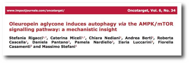
Onco

Title: Oleuropein aglycone induces autophagy via the AMPK/mTOR signalling pathway: a mechanistic insight.
Authors: Rigacci S, Miceli C, Nediani C, Berti A, Cascella R, Pantano D, Nardiello P, Luccarini I, Casamenti F, Stefani M.
Journal: Oncotarget. 2015 Nov 3;6(34):35344-57.
PMID: 26474288                (This article is OPEN ACCESS if you would like to read it)

The researchers conducting this study found that treatment with oleuropein caused an increase in autophagy in both cell culture and in a mouse model of Alzheimer’s disease, and they demonstrated that it achieved this by blocking the mTOR pathway.

Has anyone ever looked at oleuropein in the clinic?

No, not to our knowledge (and we are happy to be corrected on this).

There have been six clinical trials of olive leaf extract (the majority of which is oleuropien), but none of them have been focused on any neurological conditions.


So oleuropein is safe then?

It is a widely available supplement that a lot of people use to help lower bad cholesterol and blood pressure, so yes it can be considered safe. But any decision to experiment with oleuropein should only be made in consultation with your regular medically trained physician.

Why? Because there are always caveats.

Importantly, individuals with low blood pressure and diabetes may suffer even lower blood pressure and blood glucose levels as a result of consumption of oleuropein. Oleuropein may also interact with other pharmaceutical drugs that are designed to lower blood pressure or regulate diabetes. Such interactions could be dangerous.

And this is a particularly important factor for Parkinson’s disease as up to 30% of people with Parkinson’s may be glucose intolerant (Click here to see our post on Parkinson’s & diabetes).

Those who experience symptoms such as headache, nausea, flu-like symptoms, fainting, dizziness, and other life threatening symptoms should medical attention immediately.

What does it all mean?

We are grateful to regular reader (Don) who brought oleuropein to our attention. It is a very interesting chemical and we are definitely intrigued by it. We would certainly like to see more research on oleuropein in models of Parkinson’s disease.

Attentive readers will have noticed that most of the research discussed above have been conducted in the last 5-10 years. This suggests that oleuropein research is still in its infancy, particularly with regards to research on neurological conditions. And we hope that by reporting on it here, we will be bringing it to the attention of researchers.

Oleuropein is extracted from all parts of the olive tree (the leaves, bark, root, and fruit). It forms part of the defence system of the olive tree against stress or infection. Perhaps we could apply some of its interesting properties to Parkinson’s disease.


EDITORIAL NOTE:  Under absolutely no circumstances should anyone reading the material on this website consider it medical advice. The information provided here is for educational purposes only. Before considering or attempting any change in your treatment regime, PLEASE consult with your doctor or neurologist. While some of the drugs and supplements discussed on this website are clinically available, they may have serious side effects. We urge caution and professional consultation before altering any treatment regime. SoPD can not be held responsible for any actions taken based on the information provided here. 


The banner for this post was sourced from jrbenjamin

James: That essay

The-essay

In her excellent book – ‘The Enlightened Mr. Parkinson: The Pioneering Life of a Forgotten English Surgeon’ (Icon Books Ltd) – Dr Cherry Lewis wrote that the earliest reference to Mr James Parkinson’s ‘An Essay on the Shaking Palsy’ was an advert placed in the Morning Chronicle of Saturday 31st May (1817), under a list of books “published this day”.

Given this information, we searched the Britishnewspaperarchive online and captured the image presented above.

Today is the 200th anniversary of the publication of ‘An Essay on the Shaking Palsy’.

In this post, we continue our four part series on the man behind the disease by discussing the ‘Essay’ on the 200th anniversary of its publication.


waterloo_18june1817

The opening of Waterloo Bridge on the 18th of June 1817. Source: Thames

A few weeks before the opening of the Waterloo Bridge, James Parkinson published the booklet that would go on to immortalise him in the annals of medicine. An Essay on the Shaking Palsy, which spans 66 pages, was published by Sherwood, Neely and Jones of London, and printed by Whittingham and Rowland in 1817.

At the date of printing it sold for 3 shillings (approx. £9 or US$12).

Much has been written about the essay, and we here at the SoPD feel that we have little to actually add to the conversation. Thus our post today will simply provide an overview of the book (a highlights package, if you will), summarising it for those who do not have time to read its entirety (a full copy of the essay can be found by clicking here).

Essay

Source: Project Gutenberg

The Essay begins with a preface and is then divided into five chapters, labeled:

1. “DEFINITION—HISTORY—ILLUSTRATIVE CASES”

2. “PATHOGNOMONIC SYMPTOMS EXAMINED—TREMOR COACTUS—SCELOTYRBE FESTINANS”

3. “SHAKING PALSY DISTINGUISHED FROM OTHER DISEASES FROM WHICH IT MAY BE CONFOUNDED”

4. “PROXIMATE CAUSE—REMOTE CAUSES—ILLUSTRATIVE CASES”

5. “CONSIDERATIONS RESPECTING THE MEANS OF CURE”

The preface

In the preface of the book, James gave his reasons for actually writing it. Basically, he wanted to make others aware of what he considered a previously un-described condition.

At the heart of the preface is a paragraph, which reads:

“The disease is of long duration: to connect, therefore, the symptoms which occur in its later stages with those which mark its commencement, requires a continuance of observation of the same case, or at least a correct history of its symptoms, even for several years. Of both these advantages the writer has had the opportunities of availing himself; and has hence been led particularly to observe several other cases in which the disease existed in different stages of its progress. By these repeated observations, he hoped that he had been led to a probable conjecture as to the nature of the malady, and that analogy had suggested such means as might be productive of relief, and perhaps even of cure, if employed before the disease had been too long established. He therefore considered it to be a duty to submit his opinions to the examination of others, even in their present state of immaturity and imperfection.”

At the end of the preface, James hopes that friends to humanity and medical science…might be excited to extend their researches to this malady”. And in that situation James would “think himself fully rewarded by having excited the attention of those, who may point out the most appropriate means of relieving a tedious and most distressing malady”. 

Chapter 1

In the first chapter, James begins with a description of the Shaking Palsy (or ‘Paralysis agitans’ as he called it), that resembles modern Parkinson’s disease almost perfectly:

“Involuntary tremulous motion, with lessened muscular power, in parts not in action and even when supported; with a propensity to bend the trunk forwards, and to pass from a walking to a running pace: the senses and intellects being uninjured.”

He then moves on to provide a breakdown of the features that make up this condition, which includes a history of tremor that takes into account the works of Aelius “Galen” GalenusSylvius de la Boë, and Johann Juncker.

James starts by noting the slow progress of the condition:

“So slight and nearly imperceptible are the first inroads of this malady, and so extremely slow is its progress, that it rarely happens, that the patient can form any recollection of the precise period of its commencement. The first symptoms perceived are, a slight sense of weakness, with a proneness to trembling in some particular part; sometimes in the head, but most commonly in one of the hands and arms.”

How familiar does this sound?

And please remember, James was describing this condition for the first time based only on his own observations of just six individuals (three from a distance). His attention to detail was amazing, taking into account so many different aspects of the condition (from the obvious motor features to issues with bowel movements). And he noted it all down in the essay.

He continues by describing the progress of the condition over time:

“But as the malady proceeds,….The propensity to lean forward becomes invincible, and the patient is thereby forced to step on the toes and fore part of the feet, whilst the upper part of the body is thrown so far forward as to render it difficult to avoid falling on the face.”

His description took into account the entire history of the condition, starting from the appearance of the first features and finishing with the late stages of the disease:

“As the disease proceeds towards its last stage, the trunk is almost permanently bowed, the muscular power is more decidedly diminished, and the tremulous agitation becomes violent….As the debility increases and the influence of the will over the muscles fades away, the tremulous agitation becomes more vehement. It now seldom leaves him for a moment;”

After describing the basic clinical appearance of the condition, James then immediately moves on to each of the six cases he based his description on.

Case I was the first encounter of this condition for James. It was also probably one of the case that James was most familiar with as he wrote “every circumstance occurred which has been mentioned in the preceding history”. In his writing of Case I, however, James was rather brief:

Case I

“The subject of this case was a man rather more than fifty years of age, who had industriously followed the business of a gardener, leading a life of remarkable temperance and sobriety. The commencement of the malady was first manifested by a slight trembling of the left hand and arm, a circumstance which he was disposed to attribute to his having been engaged for several days in a kind of employment requiring considerable exertion of that limb. Although repeatedly questioned, he could recollect no other circumstance which he could consider as having been likely to have occasioned his malady.”

The “next case” (as James wrote it, indicating that the cases are presented in chronological order), Case II, was a man that James casually met with in the street.

Case II

“It was a man sixty-two years of age; the greater part of whose life had been spent as an attendant at a magistrate’s office. He had suffered from the disease about eight or ten years. All the extremities were considerably agitated, the speech was very much interrupted, and the body much bowed and shaken. He walked almost entirely on the fore part of his feet, and would have fallen every step if he had not been supported by his stick. He described the disease as having come on very gradually,…”

Case II attributed his condition to his choice of lifestyle (“irregularities in mode of living and indulgence in spiritous liquors,”), which James did not give any credit. This was probably because much of the rest of the city partook in such a lifestyle without the emergence of the disease. Ever the humanitarian, though, James points towards the unfortunate situation that these individuals found themselves:

“He was the inmate of a poor-house of a distant parish, and being fully assured of the incurable nature of his complaint, declined making any attempts for relief.”

The third case was also “noticed casually in the street“. James did interact with the man though, determining that he had been a sailor who attributed his condition to having been for many months in a Spanish prison:

Case III.

“The subject…was a man of about sixty-five years of age, of a remarkable athletic frame. The agitation of the limbs, and indeed of the head and of the whole body, was too vehement to allow it to be designated as trembling. He was entirely unable to walk; the body being so bowed, and the head thrown so forward, as to oblige him to go on a continued run, and to employ his stick every five or six steps to force him more into an upright posture, by projecting the point of it with great force against the pavement.”

The 4th case was a gentleman (of about fifty-five years of age) who presented himself to James. He claimed that he had first experienced the trembling of the arms about five years before. In this case, we see the nature of the medical treatments during that period (that being a preference for blood letting):

Case IV.

“His application was on account of a considerable degree of inflammation over the lower ribs on the left side, which terminated in the formation of matter beneath the fascia. About a pint was removed on making the necessary opening; and a considerable quantity discharged daily for two or three weeks. On his recovery from this, no change appeared to have taken place in his original complaint; and the opportunity of learning its future progress was lost by his removal to a distant part of the country”

Case V was the subject that James had the least amount of information about and observed the gentleman only from a distance (it is curious to note that all of these cases were males – who have a higher risk of developing Parkinson’s disease – click here for more on this):

Case V.

“…one of the characteristic symptoms of this malady, the inability for motion, except in a running pace, appeared to exist in an extraordinary degree. It seemed to be necessary that the gentleman should be supported by his attendant, standing before him with a hand placed on each shoulder, until, by gently swaying backward and forward, he had placed himself in equipoise; when, giving the word, he would start in a running pace, the attendant sliding from before him and running forward, being ready to receive him and prevent his falling, after his having run about twenty paces”

Case VI may have been the individual that spurred James to write his essay as it was one “which presented itself to observation since those above-mentioned,”. Thus, James had the benefit of hindsight and all the information that he had gained from the previous cases, when he was confronted with Case VI and he could make a thorough study of the individual. In case VI, James also hints at the indiscriminate nature of the condition, afflicting people from all sorts of backgrounds.

Case VI.

“The gentleman who was the subject of it is seventy-two years of age. He has led a life of temperance, and has never been exposed to any particular situation or circumstance which he can conceive likely to have occasioned, or disposed to this complaint; which he rather seems to regard as incidental upon his advanced age, than as an object of medical attention….About eleven or twelve, or perhaps more, years ago, he first perceived weakness in the left hand and arm, and soon after found the trembling commence. In about three years afterwards the right arm became affected in a similar manner: and soon afterwards the convulsive motions affected the whole body, and began to interrupt the speech…Of late years the action of the bowels had been very much retarded;…”

James notes with Case VI that the gentleman had the capacity to temporarily control his situation by his own will:

“…he, being then just come in from a walk, with every limb shaking, threw himself rather violently into a chair, and said, ‘Now I am as well as ever I was in my life.’ The shaking completely stopped; but returned within two minutes”

At the end of the section on CaseVI, James notes some input from the wife of the gentleman:

“…if whilst walking he felt much apprehension from the difficulty of raising his feet, if he saw a rising pebble in his path? he avowed, in a strong manner, his alarm on such occasions; and it was observed by his wife, that she believed, that in walking across the room, he would consider as a difficulty the having to step over a pin”

Having finished reading Chapter 1, it is truly remarkable to recall that James was describing what he thought was a previously unrecognised condition. Remarkable because of the depth and scope he provides. It is difficult to put oneself in his shoes, given that we are now so familiar with the disease. But it does Mr Parkinson great credit both as a surgeon and a writer that what he is describing feels so familiar.

Chapter 2

Here James returns to the cardinal features of the condition as he sees them, starting with the tremor:

1. Involuntary tremulous motion, with lessened voluntary muscular power, in parts, not in action, and even supported.

In this first section, James breaks down the different types of tremor in an effort to better understand this condition he is describing.

“It is necessary that the peculiar nature of this tremulous motion should be ascertained, as well for the sake of giving to it its proper designation, as for assisting in forming probable conjectures, as to the nature of the malady, which it helps to characterise”

And again, James cites the works of Galen and Sylvius de la Boë.
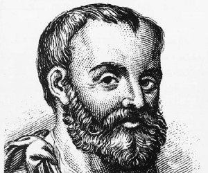
galen-1

Galen. Source: thefamouspeople

“The separation of palpitation of the limbs (Palmos of Galen, Tremor Coactus of de la Boë) from tremor, is the more necessary to be insisted on, since the distinction may assist in leading to a knowledge of the seat of the disease.”
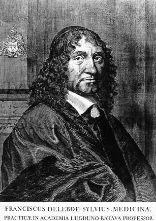
Sylviusf

de la Boë. Source: Wikipedia

James concludes that the tremor associated with his new condition is distinct given that the tremor is nearly constant or “induced immediately on bringing the parts into action”

The second characteristic feature of this newly described condition, according to James, is the gait and posture:

2. A propensity to bend the trunk forwards, and to pass from a walking to a running pace.

Here James discusses the works of François Boissier de Sauvages de Lacroix (1706 – 1767), a French physician and botanist who is credited with establishing a methodical nosology for diseases (a classification system).

boissier01

de Sauvage. Source: Homeoint

“Mons. de Sauvages attributes this complaint to a want of flexibility in the muscular fibres. Hence, he supposes, that the patients make shorter steps, and strive with a more than common exertion or impetus to overcome the resistance; walking with a quick and hastened step, as if hurried along against their will”

It is a demonstration of Mr Parkinson’s studious nature and high general level of intelligence that he was so familiar with the works of de Sauvage – it is a simple task for us modern folk to simply ‘google’ anything we don’t know or are curious about. Where did James go to find his background research for his Esssay?

Having clearly outlined the features of the condition, James next moves to Chapter 3 where he attempts to differentiate this condition from other maladies.

Chapter 3

James did not want to have this new condition he was describing confused with other diseases, hence the meticulous description of the symptoms/features.

“…it is necessary to show that it is a disease which does not accord with any which are marked in the systematic arrangements of nosologists; and that the name by which it is here distinguished has been hitherto vaguely applied to diseases very different from each other, as well as from that to which it is now appropriated”

James’ choice of name for the new condition was ‘Shaking palsy’, but he noted that this label had been used several times before. For example, one Dr. Charlton had used the label in describing a particular case:

“Another case, which the Doctor designates as ‘A Shaking Palsy,’ apparently from worms, he describes thus, “A poor boy, about twelve or thirteen years of age, was seized with a Shaking Palsy. His legs became useless, and together with his head and hands, were in continual agitation; after many weeks trial of various remedies, my assistance was desired…His bowels being cleared, I ordered him a grain of Opium a day in the gum pill; and in three or four days the shaking had nearly left him.” By pursuing this plan, the medicine proving a vermifuge, he could soon walk, and was restored to perfect health”

Given the level of detail that James goes into in other chapters, it is fair to say that chapter 3 is light reading. But it finishes strong as James describes the truly distinguishing feature of his version of Shaking palsy – that being the resting state nature of the tremor:

“If the trembling limb be supported, and none of its muscles be called into action, the trembling will cease. In the real Shaking Palsy the reverse of this takes place, the agitation continues in full force whilst the limb is at rest and unemployed;”

And it was this feature for James that could be used to distinguish it from other conditions.

Chapter 4

In Chapter 4, James tries to understand the cause of the condition, but right up front he acknowledges that this is a rather difficult task:

“Unaided by previous inquiries immediately directed to this disease, and not having had the advantage, in a single case, of that light which anatomical examination yields, opinions and not facts can only be offered”

In addition, James notes that “Cases illustrative of the nature and cause of this malady are very rare”

He does an admirable job in his endeavour here, however, by looking at previously reported cases of other diseases that share some similarities with this new condition. And James actually describes cases that he himself has dealt with (albeit by informally), but he takes pains to point out that these cases are different to the new conditions that he is describing in this essay. For example:

“…the unhappy subject of this malady was casually met in the street, shifting himself along, seated in a chair; the convulsive motions having ceased, and the limbs having become totally inert, and insensible to any impulse of the will”

In this case, the man had been treated with mercury for a venereal infection (click here to read more about early mercury treatments) many years before, which had left him with convulsive movements restricted to the legs.

Using this case study approach, however, James proposes that the disease is targeting or affecting an area of the brain stem called the medulla oblongata (which is affected in Parkinson’s disease, and is actually not too far from the midbrain where the significant loss of the dopamine neurons gives rise to the motor features of Parkinson’s disease).

1311_Brain_Stem

Location of the midbrain and medulla in the human brain. Source: Wikipedia

Chapter 5

In chapter 5, James expresses hope that a successful treatment is almost at hand:

“…there appears to be sufficient reason for hoping that some remedial process may ere long be discovered, by which, at least, the progress of the disease may be stopped”

Exactly 200 years on, I think it is fair to say that James was a bit too optimistic in nature, but we are certainly a lot closer now to stopping the disease than he was then.

James was instructive in how he thought it was best to attack the condition. He divides the condition into two halves, early and late, based on the spread of the motor features from individual limbs to other areas of the body. And he is rather certain that early diagnosis was essential if there was to be any chance of cure.

He also thought that the condition simply required some reverse engineering:

“…it seems as if we were able to trace the order and mode in which the morbid changes may proceed in this disease”

But his thoughts on how to treat the disease were largely based on the medical practises of the time (as they are today):

“…blood should be first taken from the upper part of the neck,…After which vesicatories should be applied to the same part, and a purulent discharge obtained by appropriate use of the Sabine Liniment; having recourse to the application of a fresh blister, when from the diminution of the discharging surface, pus is not secreted in a sufficient quantity”

He provides further thoughts on this treatment, but then offers the caveat that this is merely an opinion:

“Until we are better informed respecting the nature of this disease, the employment of internal medicines is scarcely warrantable;”

James also then comments on the insidious nature and the slow progress of the disease, as it:

“Seldom occurring before the age of fifty, and frequently yielding but little inconvenience for several months, it is generally considered as the irremediable diminution of the nervous influence, naturally resulting from declining life; and remedies therefore are seldom sought for”

And this leaves the sufferer focusing on:

“The weakened powers of the muscles in the affected parts is so prominent a symptom, as to be very liable to mislead the inattentive, who may regard the disease as a mere consequence of constitutional debility. If this notion be pursued, and tonic medicines, and highly nutritious diet be directed, no benefit is likely to be thus obtained; since the disease depends not on general weakness, but merely on the interruption of the flow of the nervous influence to the affected parts”

This is very insightful of James. He understood that it was not the weakness felt in the muscles that was paramount in this condition, but rather a dysfunction in the brain.

He concludes the essay with the following:

“To such researches the healing art is already much indebted for the enlargement of its powers of lessening the evils of suffering humanity. Little is the public aware of the obligations it owes to those who, led by professional ardour, and the dictates of duty, have devoted themselves to these pursuits, under circumstances most unpleasant and forbidding. Every person of consideration and feeling, may judge of the advantages yielded by the philanthropic exertions of a Howard; but how few can estimate the benefits bestowed on mankind, by the labours of a Morgagni, Hunter, or Baillie.

FINIS.”

Regarding the last line, I may be displaying my ignorance here with regards to ‘a Howard’, but I suspect James is referring to John Howard (1726 – 1790), an English philanthropist of James’ era:

800px-John_Howard_by_Mather_Brown

John Howard. Source: Wikipedia

Although, “a Howard” is also an old slang term used to describe a man (any man) of great character!

Giambattista_morgagni

Giovanni Battista MorgagniSource: Wikipedia

Giovanni Battista Morgagni (1682 – 1771) was an Italian anatomist, who is generally regarded as the father of modern anatomical pathology.

280px-John_Hunter_by_John_Jackson

John Hunter. Source: Wikipedia

John Hunter (1728 – 1793) was a Scottish surgeon – one of the most distinguished scientists/surgeons of his day. He was an early advocate of careful observation and scientific method in medicine, and James personally learned a great deal from him. Between October 1785 and April 1786, James attended the evening lectures provided by Hunter. James wrote down the lectures verbatim (in shorthand) and his notes were later published by his son John (“Hunterian Reminiscences, Being The Substance Of A Course Of Lectures On The Principles And Practice Of Surgery Delivered By John Hunter In The Year 1785″ – a precious resource given that Hunter’s own notes were later destroyed by fire).

Matthew Baille FRS (1761-1823)

Matthew BaillieSource: Wikipedia

Matthew Baillie was another Scottish physician and pathologist. A pupil of his uncle, John Hunter (above), Ballie provided us with the first systematic study of pathology. James was certainly familiar with Ballie, as he cited his works.


For further reading on An Essay on the Shaking Palsy we recommend a review written by Prof Brian Hurwitz (King’s College London) called Urban Observation and Sentiment in James Parkinson’s Essay on the Shaking Palsy (1817) which provides fantastic insight into James, the age he lived in, the essay itself, and the reception of the essay (Click here to read that review).

This post was written in observation of the 200 year anniversary of the publishing of the Essay on the Shaking Palsy. It is part two in a four part series on the life of Mr James Parkinson (click here for part one). In the third instalment, we will look at his life’s work, before the fourth part looks at his final years and his legacy.


The banner for today’s post was sourced from the Britishnewspaperarchive
Sheffield: flies, fish and a Tigar

total-produce-ltd-sheffield1

When people in England think of the city of Sheffield, quite often images of a great industrial past will come to mind.

They usually don’t think of the flies, fish and (yes) a Tigar (no, not a typo!) that are influencing Parkinson’s disease research in the city.

In today’s post we will look at how the re-invention of a city could have a major impact on Parkinson’s disease.


desktop_high_res_cyclops_works_-_cammell__in_reference_box_n178

The industrial heritage of Sheffield. Source: SIMT

It is no under statement to say that the history of Sheffield – a city in South Yorkshire, England –  is forged in steel.

In his 1724 book, “A tour thro’ the whole island of Great Britain, the author Daniel Defoe wrote of Sheffield:

“Here they make all sorts of cutlery-ware, but especially that of edged-tools, knives, razors, axes, &. and nails; and here the only mill of the sort, which was in use in England for some time was set up, for turning their grindstones, though now ’tis grown more common”

Sheffield has a long history of metal work, thanks largely to its geology: The city is surrounded by fast-flowing rivers and hills containing many of the essential raw materials such as coal and iron ore.

And given this fortunate circumstance and an industrious culture, the city of Sheffield particularly prospered during the industrial revolution of the mid-late 1800s (as is evident from the population growth during that period).

sheffpop

The population of Sheffield over time. Source: Wikipedia

But traditional manufacturing in Sheffield (along with many other areas in the UK) declined during the 20th century and the city has been forced to re-invent itself in the early 21st century. And this time, rather than taking advantage of their physical assets, the city is focusing on its mental resources.

Great. Interesting stuff. Really. But what does this have to do with flies, fish and Parkinson’s disease???

Indeed. Let’s get down to business.

sitran_sunrise_785px

The Sheffield Institute for Translational Neuroscience (SITraN) was officially opened in 2010 by Her Majesty The Queen. It is the first European Institute purpose-built and dedicated to basic and clinical research into Motor Neuron Disease as well as related neurodegenerative disorders such as Parkinson’s and Alzheimer’s disease.

Since its opening, the institute has published some pretty impressive research, particularly in the field of Parkinson’s disease.
And here is where we get to the flies:

pink_fly-1410843

Pink flies. Source: Wallpapersinhq

We have previously discussed “Pink” flies and their critical role in Parkinson’s research (Click here to read that post).

Today we are going to talk about Lrrk2 flies.

What is Lrrk2?

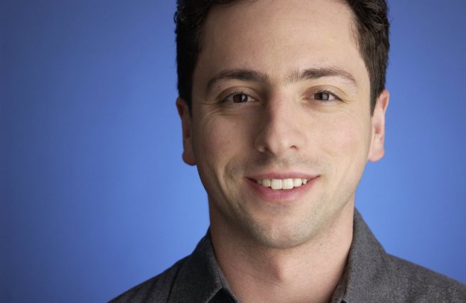
This is Sergey Brin.

sergey_brin

He’s a dude.

One of the founders of the search engine company “Google”. Having changed the world, he is now turning his attention to other projects.

One of those other projects is close to our hearts: Parkinson’s disease.

In 1996, Sergey’s mother started experiencing numbness in her hands. Initially it was believed to be RSI (Repetitive strain injury). But then her left leg started to drag. In 1999, following a series of tests, Sergey’s mother was diagnosed with Parkinson’s disease. It was not the first time the family had been affected by the condition: Sergey’s late aunt had also had Parkinson’s disease.

Both Sergey and his mother have had their DNA scanned for mutations that increase the risk of Parkinson’s disease. And they discovered that they were both carrying a mutation on the 12th chromosome, in a gene called PARK8 – one of the Parkinson’s disease associated genes. Autosomal dominant mutations (meaning if you have just one copy of the mutated gene) in the PARK8 gene dramatically increase one’s risk of developing Parkinson’s disease.
PARK8 provides the instructions for making an enzyme called Leucine-rich repeat kinase 2 (or Lrrk2).

Protein_LRRK2_PDB_2ZEJ

The structure of Lrrk2. Source: Wikipedia

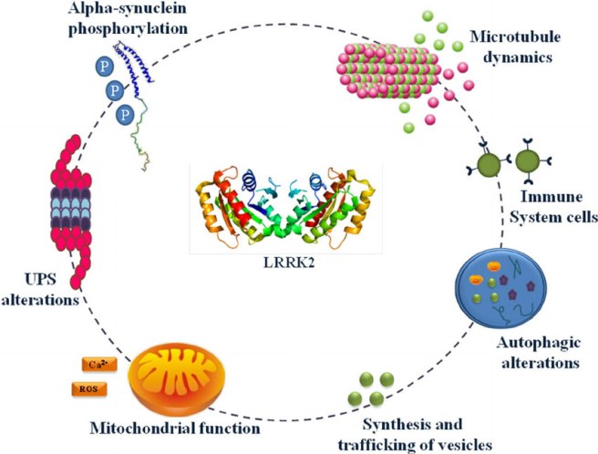
Also known as ‘Dardarin (from the Basque word “dardara” which means trembling), Lrrk2 has many functions within a cell – from helping to move things around inside the cell to helping to keep the power on (involved with mitochondrial function).

Fig-2-LRRK2-involvement-in-cellular-mechanisms-Several-data-posit-that-LRRK2-through

Source: Researchgate

NOTE: Curiously, mutations in the PARK8 gene are also associated with Crohn’s disease (Click here and here for more on this) – though the mutation is in a different location for PD.

Now, not everyone with this particular mutation will go on to develop Parkinson’s disease, and Sergey has decided that his chances are 50:50. But he does not appear to be taking any chances though. Being one of the founders of a large company like Google, has left Sergey with considerable resources at his disposal. And he has chosen to focus some of those resources on Lrrk2 research (call it an insurance  policy). He has done this via considerable donations to groups like the Michael J Fox foundation.

leadership-fox-m-img_2

Actor Michael J Fox was diagnosed at age 30. Source: MJFox foundation

So just as Pink flies derive their name from mutations in the Parkinson’s associated Pink1 gene, Lrrk2 flies have mutations in the Lrrk2 gene.

So what have the researchers at Sheffield done with the Lrrk2 flies?

In 2013, the Sheffield researchers published an interesting research report:

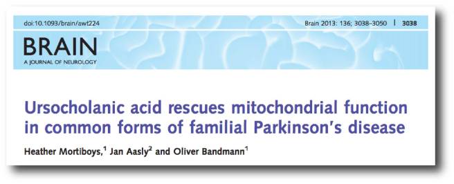
brain

Title: Ursocholanic acid rescues mitochondrial function in common forms of familial Parkinson’s disease
Authors: Mortiboys H, Aasly J, Bandmann O.
Journal: Brain. 2013 Oct;136(Pt 10):3038-50.
PMID: 24000005

In this study, the investigators took 2000 drugs (including 1040 licensed drugs and 580 naturally occurring compounds) and conducted a massive screen to identify drugs that could rescue mitochondrial dysfunction in PARK2 (Parkin) mutant cells.

Mitochondria are the power house of each cell. They keep the lights on. Without them, the lights go out and the cell dies.

Mitochondria

Mitochondria and their location in the cell. Source: NCBI

In certain genetic forms of Parkinson’s disease (such as those associated with mutations in the PARK2 gene), the mitochondria in cells becomes dysfunctional and may not be disposed of properly (Click here to read our previous post related to this).

In their huge screen of 2000 drugs, the researchers in Sheffield identified 15 drugs that could rescue the mitochondria dysfunction in the PARK2 skins cells. Of those 15 compounds, two were chosen for further functional studies. They were:

  • Ursocholanic acid
  • Dehydro(11,12)ursolic acid lactone

Neither ursocholanic acid nor dehydro(11,12)ursolic acid lactone are FDA-licensed drugs. We have little if any information regarding their use in humans. Given this situation, the researchers turned their attention to the chemically related bile acid ‘ursodeoxycholic acid’, which has been in clinical use for more than 30 years.

What is Ursodeoxycholic Acid?

Ursodeoxycholic Acid (or UDCA) is a drug that is used to to improve bile flow and reduce gallstone formation. In the USA it is also known as ‘ursodiol’.

800px-Ursodiol

Ursodiol. Source: Wikimedia

Bile is a fluid that is made and released by your liver, and it stored in the gallbladder. Its function is to help us with digestion. UDCA occurs naturally in bile – it is basically a bile acid and can therefore be useful in dissolving gallstones. UDCA has been licensed for the treatment of patients since 1980. UDCA also reduces cholesterol absorption.

So what did the Sheffield researchers find with UDCA?

The researchers tested UDCA on mitochondrial function in PARK2 skin cells, and they found that the drug rescued the cells. They then tested UDCA on skin cells from people with Parkinson’s disease who had mutations in the PARK8 (Lrrk2) gene (G2019S).

The researchers had previously found impaired mitochondrial function and morphology in skin cells taken from people with PARK8 associated Parkinson’s disease (Click here to read more about this), and other groups had reported similar findings (Click here for more on this).

And when they treated the Lrrk2 cells with UDCA, guess what happened?

UDCA was able to rescue the mitochondrial effect in those cells as well!

Obviously these results excited the Sheffield scientists and they set up a collaboration with researchers at York University and from Norway, to look at the potential of UDCA in rescuing the fate of Lrrk2 flies. The results of that study were published two years ago:

Oliver

Title: UDCA exerts beneficial effect on mitochondrial dysfunction in Lrrk2 (G2019S) carriers and in vivo.
Authors: Mortiboys H, Furmston R, Bronstad G, Aasly J, Elliott C, Bandmann O.
Journal: Neurology. 2015 Sep 8;85(10):846-52.
PMID: 26253449        (This article is OPEN ACCESS if you would like to read it).

The researchers tested UDCA on flies (or drosophila) with specific Lrrk2 mutations (G2019S) display a progressive loss of photoreceptor cell function in their eyes. The mitochondria in the photoreceptor are swollen and disorganised. When the investigators treated the flies with UDCA, they found approximately 70% rescue of the photoreceptor cells function.

The researchers in Sheffield concluded that UDCA has a marked rescue effect on cells from a Parkinson’s disease-associated gene mutation model, and they proposed that “mitochondrial rescue agents may be a promising novel strategy for disease-modifying therapy in Lrrk2-related PD, either given alone or in combination with Lrrk2 kinase inhibitors” (for more information about the Lrrk2 inhibitors they refer, click here).

And the good news regarding this line of research: other research groups have also observed similar beneficial effects with UDCA in models of Parkinson’s disease:

Low1

Title: Ursodeoxycholic acid suppresses mitochondria-dependent programmed cell death induced by sodium nitroprusside in SH-SY5Y cells.
Authors: Chun HS, Low WC.
Journal: Toxicology. 2012 Feb 26;292(2-3):105-12.
PMID: 22178905

This research group also demonstrated that UDCA could reduce cell death in a cellular model of Parkinson’s disease.

And this study was followed by another one from a different research group, which involved testing UDCA in animals:

Salem1

Title: Ursodeoxycholic Acid Ameliorates Apoptotic Cascade in the Rotenone Model of Parkinson’s Disease: Modulation of Mitochondrial Perturbations.
Authors: Abdelkader NF, Safar MM, Salem HA.
Title: Mol Neurobiol. 2016 Mar;53(2):810-7.
PMID: 25502462

These researchers found UDCA rescued a rodent model of Parkinson’s disease (involving the neurotoxin rotenone). UDCA not only improved mitochondrial performance in the rats, but also demonstrated anti-inflammatory and anti-cell death properties.

Given all this research, the Sheffield researchers are now keen to test UDCA in clinical trials for Parkinson’s disease.

Has anyone tested UDCA in the clinic for Parkinson’s disease?

Not that we are aware of, but two groups are interested in attempting it.

imgres

Firstly, the University of Minnesota – Clinical and Translational Science Institute has registered a trial (Click here to read more about this). This trial will not, however, be testing efficacy of the drug on Parkinson’s symptoms. It will focus on measuring UDCA levels in individuals after four weeks of repeated high doses of oral UDCA (50mg/kg/day), and determining the bioenergetic profile and ATPase activity in those participants. Basically, they want to see if UDCA is safe and active in people with Parkinson’s disease.

The CurePD trust (in the UK) is also currently seeking to run a clinical trial for UDCA (Click here for more on this). The group are currently organising the funding for that trial.

url-3


EDITOR’S NOTE HERE: Before we move on, the team at the SoPD would like to say that while UDCA is a clinically available drug, it is still experimental for Parkinson’s disease. There is no indication yet that it has beneficial effects in people with Parkinson’s disease. In addition, UDCA is also is known to have side effects, which include flu symptoms, nausea, diarrhea, and back pain. And individuals have been known to have allergic reactions to UDCA treatment (Click here and here for more on the side effects of UDCA). Thus we must impress caution on anyone planning to experiment with this drug. Before attempting any kind of change in a current treatment regime, PLEASE discuss your plans with a medically qualified physician who is familiar with your case history.


Ok, so that was the flies research, what about the fish? And the… uh, tigar?

Yes. The fish are called Zebrafish (or Danio rerio).

They are a tropical freshwater fish that is widely used in biological research.

Zebrafisch

Biology researchers love these little guys because their genome has been fully sequenced and they has well characterised and testable behaviours. In addition, their development is very rapid (3 months), and its embryos are large and transparent.

And the researchers at Sheffield are using these fish to study Parkinson’s disease.

How did they do that?

tiger

Title: TigarB causes mitochondrial dysfunction and neuronal loss in Pink1 deficiency
Authors: Flinn LJ, Keatinge M, Bretaud S, Mortiboys H, Matsui H, De Felice E, Woodroof HI, Brown L, McTighe A, Soellner R, Allen CE, Heath PR, Milo M, Muqit MM, Reichert AS, Köster RW, Ingham PW, Bandmann O.
Journal: Ann Neurol. 2013 Dec;74(6):837-47.
PMID:
24027110        (This article is OPEN ACCESS if you would like to read it)

Firstly, the group at Sheffield generated zebrafish that had a mutation in the Parkinson’s associated gene ‘PARK6’. This gene provides the plans for the production of a protein called Pink1 (we have previously discussed Pink1 – click here to read more on this).

In normal healthy cells, the Pink1 protein is absorbed by mitochondria and eventually degraded as it is not used. In unhealthy cells, however, this process becomes inhibited and Pink1 starts to accumulate on the outer surface of the mitochondria. Sitting on the surface, it starts grabbing another Parkinson’s associated protein called Parkin. This pairing is a signal to the cell that this particular mitochondria is not healthy and needs to be removed.

601587-fig-003

Pink1 and Parkin in normal (right) and unhealthy (left) situations. Source: Hindawi

The process by which mitochondria are removed is called mitophagy. Mitophagy is part of the autophagy process, which is an absolutely essential function in a cell. Without autophagy, old proteins and mitochondria will pile up making the cell sick and eventually it dies. Through the process of autophagy, the cell can break down the old protein, clearing the way for fresh new proteins to do their job.

Think of autophagy as the waste disposal/recycling process of the cell.

Print

The process of autophagy. Source: Wormbook

Waste material inside a cell is collected in membranes that form sacs (called vesicles). These vesicles then bind to another sac (called a lysosome) which contains enzymes that will breakdown and degrade the waste material. The degraded waste material can then be recycled or disposed of by spitting it out of the cell.

In the case of a PARK6 mutations, Pink1 protein can not function properly with Parkin and the autophagy process breaks down. As a result, the old or unhealthy mitochondria start to pile up in the cell, resulting in the cell getting sick and dying.

Now back to the Zebrafish.

When the Sheffield researchers mutated PARK6 in the zebrafish, they noticed that the fish had a very early and persistent loss of dopamine neurons in their brains. These fish also had enlarged, unhealthy mitochondria and reduced mitochondrial activity.

Given this result, the investigators next wanted to identify which genes have increased or decreased levels of activity as a result of this genetic manipulation. They identified 108 genes that were higher in the PARK6 mutant, and 146 genes had lower activity.

One gene in particular had activity levels 12 times higher in the PARK6 mutant fish than the normal zebrafish.

The name of that gene? TP53-Induced Glycolysis And Apoptosis Regulator (or Tigar).

What is Tigar?

Tigar is a gene that provides the instructions for making a protein that is activated by p53 (also known as TP53).

What does that mean?

p53 is a protein that has three major functions: controlling cell division, DNA repair, and apoptosis (or cell death). p53 performs these functions as a transcriptional activator (that is a protein that binds to DNA and helps produce RNA (the process of transcription) – see our previous post explaining this).

800px-P53

p53 protein structure, bound to DNA (in gold). Source: Wikipedia

In regulating the cell division, p53 prevents cells from dividing too much and in this role it is known as a tumour suppression – it suppresses the emergence of cancerous tumours. Genetic mutations in the p53 gene result in run away cell division, and (surprise!) as many as 50% of all human tumours contain mutations in the p53 gene.

Apop

Cancer vs no cancer. Source: Khan Academy

In DNA repair, p53 is sometimes called “the guardian of the genome” as it prevents mutations and helps to conserve stability in the genome. This function also serves to prevent the development of cancer, by helping to repair potentially cancer causing mutations….and in this role it is known as a tumour suppression. Obviously, if there is a mutation in the p53 gene, less DNA repair will occur – increasing the risk of cancer occurring.

And finally, in cell death, p53 plays a critical role in telling a cell when to die. And (continuing with the cancer theme), if there is a mutation in the p53 gene, fewer cells will be told to die – increasing the risk of cancer occurring. And in this role p53 is known as a tumour suppression.

In normal cells, the levels of p53 protein are usually low. When a cell suffers DNA damage and stress, there is often an increase in the amount of p53 protein. If this increases past a particular threshold, then the cell will be instructed to die.

If you haven’t guessed yet, p53 is a major player inside most cell, and it controls the activity of a lot of genes.

And one of those genes is Tigar.

But what does Tigar actually do?

So we have explained the “TP53-Induced” part of the “TP53-Induced Glycolysis And Apoptosis Regulator” name, let’s now focus on the “Glycolysis And Apoptosis Regulator”

Tigar is an interesting protein because it is an enzyme that primarily functions as a regulator of the breaking down of glucose (“Glycolysis” involves the conversion of glucose into a chemical called pyruvate). In addition to this role, however, Tigar acts in preventing cell death (or apoptosis).

Increased levels of Tigar protects cells from oxidative-stress induced apoptosis, by decreasing the levels of free radicals. In this way, it promotes anti-oxidant activities.

But hang on a second, anti-oxidant activity should be good for the cell right? Why are the dopamine cells are dying if Tigar levels are increasing in the PARK6 mutants?

Fantastic question!

The answer: TIGAR is also a negative regulator of a process called mitophagy. As we discussed above, mitophagy is the process of removing mitochondria by autophagy. Increases in the levels of TIGAR blocks mitophagy in a cell, and results in an increased number of swollen and unhealthy mitochondria in those cells (Click here to read more about this). These swollen mitochondria are comparable to the enlarged mitochondria identified the PARK6 zebrafish by the Sheffield researchers.

And the researchers believe that this may be the cause of the cell death in the PARK6 zebrafish – the double impact of PARK6 and Tigar induced problems with mitophagy.

NOTE: Problems with mitophagy is believed to be an important mechanism in the development of early-onset Parkinson’s disease (Click here for a recent review on this)

Ok, and what did the Sheffield researchers do next?

Given that there was such a huge increase in Tigar levels in the PARK6 zebrafish, the investigators decided to reduce Tigar levels in the PARK6 zebrafish to see what impact this would have on the fish (and their mitochondria).

Remarkably, reductions of Tigar levels resulted in complete rescue of the dopamine neurons in the PARK6 fish. It also increased mitochondrial activity in those cells, and reduced the activation of the microglia cells, which can also play a role in the removal of sick cells in the brain.

The researchers concluded that the results demonstrate that TIGAR is “a promising novel target for disease‐modifying therapy in Pink1‐related Parkinson’s disease”.

And what are the researchers planning to do next with Tigar?

Prof Oliver Bandmann, the senior scientist who ran the study, has said that they “need to finish studying TIGAR levels in the brains of people with Parkinson’s and want to better understand how this protein is involved in maintaining the cell batteries – called ‘mitochondria'” (Source).

Our guess is that the group will also be conducting studies looking at Tigar reduction in rodent models of Parkinson’s disease to determine if this is a viable target in mammals. If Tigar reduction in rodents is found to be effective, the researchers will probably turn their attention to drug screening studies to identify currently available drugs that can reduce the activity of Tigar. Such a drug would provide us with yet another potential treatment for Parkinson’s disease.

We’ll be keeping an eye out for these pieces of research.

This is all very interesting. What does the future hold for Parkinson’s research in Sheffield?

Well, in a word: Keapstone.

Que?

In March, the University of Sheffield and Parkinson’s UK have launched a new £1 million virtual biotech company called “Keapstone Therapeutics” (see the press release by clicking here).

parkinsons_virtual_biotech_graphic

Source: Parkinson’s UK

The goal of the company – the first of its kind – is to combine world-leading research from the University with funding and expertise from the charity to help develop revolutionary drugs for Parkinson’s disease.

What is virtual about it? The biotech won’t be building its own labs, employing a team of specialist laboratory scientists, or buying any high-tech equipment (which would all be incredibly expensive). Rather they will form partnerships with groups that do specific tasks the best.

Here is a video of Dr Author Roach (director of Research at Parkinson’s UK) explaining the idea behind this endeavour:

By seeking a collaboration with Sheffield in the creation of a spin-out biotech company, Parkinson’s UK is not only acknowledging Sheffield’s track record, but also making an investment in their future research. While we cannot be entirely sure of what the long-term future holds for Parkinson’s research in Sheffield, we do know that Keapstone will be an important aspect of it in the immediate future.

Could this be a model for the future of Parkinson’s disease research? Only time will tell. We will have a closer look at Keapstone Therapeutics in an upcoming post.

Click here to learn more about the virtual biotech project.

So what does it all mean?

In 2017, we here at the SoPD have decided to begin highlighting some of the Parkinson’s disease research centres as an addition feature on the blog. We have not been approached by the research group in Sheffield or the University itself, and our selection of this city as our first case study was based purely on the fact that we really like what is happening there with regards to Parkinson’s research!

The research group in Sheffield has undertaken multiple lines of research which could potentially providing us with several novel treatment options for Parkinson’s disease. These lines of research have focused not only on clinically available drugs, but also identifying novel targets. We like what they are doing and will keep a close eye on progress there.

And over the next year we will select additional centres of Parkinson’s research based on the same criteria (us liking what they are doing). Our next case study will be the Van Andel Research Institute in Grand Rapids, Michigan (we would hate to be accused of having a UK bias).


EDITORIAL NOTE:  Under absolutely no circumstances should anyone reading the material on this website consider it medical advice. The information provided here is for educational purposes only. Before considering or attempting any change in your treatment regime, PLEASE consult with your doctor or neurologist. While some of the drugs discussed on this website are clinically available, they may have serious side effects. We urge caution and professional consultation before altering any treatment regime. SoPD can not be held responsible for any actions taken based on the information provided here. 


The banner for today’s post was sourced from TotalProduceLocal

A connection between ALS & Parkinson’s disease? Oh’ll, SOD it!

604ee0d6431dbd15f686133f6fa7205c

Please excuse our use of UK slang in the title of this post, but a group of Australian researchers have recently discovered something really interesting about Parkinson’s disease.

And being a patriotic kiwi, it takes something REALLY interesting for me to even acknowledge that other South Pacific nation. This new finding, however, could be big.

In today’s post, we will review new research dealing with a protein called SOD1, and discuss what it could mean for the Parkinson’s community.


d1ea3d21c36935b85043b3b53f2edb1f87ab7fa6

The number of dark pigmented dopamine cells in the substantia nigra are reduced in the Parkinson’s disease brain (right). Source: Adaptd from Memorangapp

Every Parkinson’s-associated website and every Parkinson’s disease researchers will tell you exactly the same thing when describing the two cardinal features in the brain of a person who died with Parkinson’s disease:

  1. The loss of certain types of cells (such as the dopamine producing cells of the substantia nigra region of the brain – see the image above)
  2. The clustering (or aggregation) of a protein called Alpha synuclein in tightly packed, circular deposits, called Lewy bodies (see image below).

9-lb2

A Lewy body inside a cell. Source: Adapted from Neuropathology-web

The clustered alpha synuclein protein, however, is not limited to just the Lewy bodies. In the affected areas of the brain, aggregated alpha synuclein can be seen in the branches of cells – see the image below where alpha synuclein has been stained brown on a section of brain from a person with Parkinson’s disease.

Lewy_neurites_alpha_synuclein

Examples of Lewy neurites (indicated by arrows). Source: Wikimedia

Now, one of the problems with our understanding of Parkinson’s disease is disparity between the widespread presence of clustered alpha synuclein and very selective pattern of cell loss. Alpha synuclein aggregation can be seen distributed widely around the affected areas of the brain, but the cell loss will be limited to specific populations of cells.

If the disease is killing a particular population of cells, why is alpha synuclein clustering so wide spread?

So why is there a difference?

We don’t know.

It could be that the cells that die have a lower threshold for alpha synuclein toxicity (we discussed this is a previous post – click here?).

But this question regarding the difference between these two features has left many researchers wondering if there may be some other protein or agent that is actually killing off the cells and then disappearing quickly, leaving poor old alpha synuclein looking rather guilty.

maxresdefault

Poor little Mr “A Synuclein” got the blame, but his older brother actually did it! Source: Youtube

And this is a very serious discussion point.

This year of 2017 represents the 200th anniversary of James Parkinson’s first description of Parkinson’s disease, but it also represents the 20th anniversary since the association between alpha synuclein and PD was first established. We have produced almost 7,000 research reports on the topic of alpha synuclein and PD during that time, and we currently have ongoing clinical trials targetting alpha synuclein.

But what if our basic premise – that alpha synuclein is the bad guy – is actually wrong?

Is there any evidence to suggest this?

We are just speculating here, but yes there is.

For example, in a study of 904 brains, alpha synuclein deposits were observed in 11.3% of the brains (or 106 cases), but of those cases only 32 had been diagnosed with a neurodegenerative disorder (Click here to read more on this). The remaining 74 cases had demonstrated none of the clinical features of Parkinson’s disease.

So what else could be causing the cell death?

Well, this week some scientists from sunny Sydney (Australia) reported a protein that could fit the bill.

sydney_cruises

Sydney. Source: Vagabond

The interesting part of their finding is that the protein is also associated with another neurodegenerative condition: Amyotrophic lateral sclerosis.

Remind me again, what is Amyotrophic lateral sclerosis?

Parkinson’s disease and Amyotrophic lateral sclerosis (ALS) are the second and third most common adult-onset neurodegenerative conditions (respectively) after Alzheimer’s disease. We recently discussed ALS in a previous post (Click here to read that post).

ALS, also known as Lou Gehrig’s disease and motor neuron disease, is a neurodegenerative condition in which the neurons that control voluntary muscle movement die. The condition affects 2 people in every 100,000 each year, and those individuals have an average survival time of two to four years.

You may have heard of ALS due to it’s association with the internet ‘Ice bucket challenge‘ craze that went viral in 2014-15.

ice-bucket-challenge

The Ice bucket challenge. Source: Forbes

What is the protein associated with ALS?

In 1993, scientists discovered that mutations in the gene called SOD1 were associated with familial forms of ALS (Click here to read more about this). We now know that mutations in the SOD1 gene are associated with around 20% of familial cases of ALS and 5% of sporadic ALS.

The SOD1 gene produces an enzyme called Cu-Zn superoxide dismutase.

This enzyme is a very powerful antioxidant that protects the body from damage caused by toxic free radical generated in the mitochondria.

Protein_SOD1_PDB_1azv

SOD1 protein structure. Source: Wikipedia

One important note here regarding ALS: the genetic mutations in the SOD1 gene do not cause ALS by affecting SOD1’s antioxidant properties (Click here to read more about this). Rather, researchers believe that the cell death seen in SOD1-associated forms of ALS is the consequences of some kind of toxic effect caused by the mutant protein.

So what did the Aussie researchers find about SOD1 in Parkinson’s disease?

This week, the Aussie researchers published this research report:

SOD
Title: Amyotrophic lateral sclerosis-like superoxide dismutase 1 proteinopathy is associated withneuronal loss in Parkinson’s disease brain.
Authors: Trist BG, Davies KM, Cottam V, Genoud S, Ortega R, Roudeau S, Carmona A, De Silva K, Wasinger V, Lewis SJG, Sachdev P, Smith B, Troakes C, Vance C, Shaw C, Al-Sarraj S, Ball HJ, Halliday GM, Hare DJ, Double KL.
Journal: Acta Neuropathol. 2017 May 19. doi: 10.1007/s00401-017-1726-6.
PMID: 28527045

Given that oxidative stress is a major feature of Parkinson’s disease, the Aussie researchers wanted to investigate the role of the anti-oxidant enzyme, SOD1 in this condition. And what they found surprised them.

Heck, it surprised us!

Two areas affected by Parkinson’s disease – the substantia nigra (where the dopamine neurons reside; SNc in the image below) and the locus coeruleus (an area in the brain stem that is involved with physiological responses to stress; LC in the image below) – exhibited little or no SOD1 protein in the control brains.

But in the Parkinsonian brains, there was a great deal of SOD1 protein (see image below).

401_2017_1726_Fig1_HTML

SO1 staining in PD brain and Control brains. Source: Springer

In the image above, you can see yellowish-brown stained patches in both the PD and control images. This a chemical called neuromelanin and it can be used to identify the dopamine-producing cells in the SNc and LC. The grey staining in the PD images (top) are cells that contain SOD1. Note the lack of SOD1 (grey staining) in the control images (bottom).

Approximately 90% of Lewy bodies in the Parkinson’s affected brains contained SOD1 protein. The investigators did report that the levels of SOD1 protein varied between Lewy bodies. But the clustered (or ‘aggregated’) SOD1 protein was not just present with alpha synuclein, often it was found by itself in the degenerating regions.

The researchers occasional saw SOD1 aggregation in regions of age-matched control brains, and they concluded that a very low level of SOD1 must be inherent to the normal ageing process.

But the density of SOD1 clustering was (on average) 8x higher in the SNc and 4x higher in the LC in the Parkinsonian brain compared to age-matched controls. In addition, the SOD1 clustering was significantly greater in these regions than all of the non-degenerating regions of the same Parkinson’s disease brains.

The investigators concluded that these data suggest an association between SOD1 aggregation and neuronal loss in Parkinson’s disease. Importantly, the presence of SOD1 aggregations “closely reflected the regional pattern of neuronal loss”.

They also demonstrated that the SOD1 protein in the Parkinsonian brain was not folded correctly, a similar characteristic to alpha synuclein. A protein must fold properly to be able to do it’s assigned jobs. By not folding into the correct configuration, the SOD1 protein could not do it’s various functions – and the investigators observed a 66% reduction in SOD1 specific activity in the SNc of the Parkinson’s disease brains.

Interestingly, when the researchers looked at the SNc and LC of brains from people with ALS, they identified SOD1 aggregates matching the SOD1 clusters they had seen in these regions of the Parkinson’s disease brain.

Is this the first time SOD1 has been associated with Parkinson’s disease?

No, but it is the first major analysis of postmortem Parkinsonian brains. SOD1 protein in Lewy bodies has been reported before:

1995

Title: Cu/Zn superoxide dismutase-like immunoreactivity is present in Lewy bodies from Parkinson disease: a light and electron microscopic immunocytochemical study
Authors: Nishiyama K, Murayama S, Shimizu J, Ohya Y, Kwak S, Asayama K, Kanazawa I.
Journal: Acta Neuropathol. 1995;89(6):471-4.
PMID: 7676802

The investigators behind this study reported SOD1 protein was present in Lewy bodies, in the substantia nigra and locus coeruleus of brains from five people with Parkinson’s disease. Interestingly, they showed that SOD1 is present in the periphery of the Lewy body, similar to alpha synuclein. Both of these protein are present on the outside of the Lewy body, as opposed to another Parkinson’s associated protein, Ubiquitin, which is mainly present in the centre (or the core) of Lewy bodies (see image below).

Lewy-bodies

A more recent study also demonstrated SOD1 protein in the Parkinsonian brain, including direct interaction between SOD1 and alpha synuclein:

Alspha

Title: α-synuclein interacts with SOD1 and promotes its oligomerization
Authors: Helferich AM, Ruf WP, Grozdanov V, Freischmidt A, Feiler MS, Zondler L, Ludolph AC, McLean PJ, Weishaupt JH, Danzer KM.
Journal: Mol Neurodegener. 2015 Dec 8;10:66.
PMID: 26643113              (This article is OPEN ACCESS if you would like to read it)

These researchers found that alpha synuclein and SOD1 interact directly, and they noted that Parkinson’s disease related mutations in alpha synuclein (A30P, A53T) and ALS associated mutation in SOD1 (G85R, G93A) modify the binding of the two proteins to each other. They also reported that alpha synuclein accelerates SOD1 aggregation in cell culture. This same group of researchers published another research report last year in which they noted that aggregated alpha synuclein increases SOD1 clustering in a mouse model of ALS (Click here for more on this).

We should add that alpha synuclein aggregations in ALS are actually quite common (click here and here to read more on this).

Are there any genetic mutations in the SOD1 gene that are associated with Parkinson’s disease?

Two studies have addressed this question:

genes

Title: Sequence of the superoxide dismutase 1 (SOD 1) gene in familial Parkinson’s disease.
Authors: Bandmann O, Davis MB, Marsden CD, Harding AE.
Journal: J Neurol Neurosurg Psychiatry. 1995 Jul;59(1):90-1.
PMID: 7608718                   (This article is OPEN ACCESS if you would like to read it)

And then in 2001, a second analysis:

Genes2

Title: Genetic polymorphisms of superoxide dismutase in Parkinson’s disease.
Authors: Farin FM, Hitosis Y, Hallagan SE, Kushleika J, Woods JS, Janssen PS, Smith-Weller T, Franklin GM, Swanson PD, Checkoway H.
Journal: Mov Disord. 2001 Jul;16(4):705-7.
PMID: 11481695

Both studies found no genetic variations in the SOD1 gene that were more frequent in the Parkinson’s affected community than the general population. So, no, there are no SOD1 genetic mutations that are associated with Parkinson’s disease.

Are there any treatments targeting SOD1 that could be tested in Parkinson’s disease?

Great question. Yes there are. And they have already been tested in models of PD:

als

Title: The hypoxia imaging agent CuII(atsm) is neuroprotective and improves motor and cognitive functions in multiple animal models of Parkinson’s disease.
Authors: Hung LW, Villemagne VL, Cheng L, Sherratt NA, Ayton S, White AR, Crouch PJ, Lim S, Leong SL, Wilkins S, George J, Roberts BR, Pham CL, Liu X, Chiu FC, Shackleford DM, Powell AK, Masters CL, Bush AI, O’Keefe G, Culvenor JG, Cappai R, Cherny RA, Donnelly PS, Hill AF, Finkelstein DI, Barnham KJ.
Title: J Exp Med. 2012 Apr 9;209(4):837-54.
PMID: 22473957               (This article is OPEN ACCESS if you would like to read it)

CuII(atsm) is a drug that is currently under clinical investigation as a brain imaging agent for detecting hypoxia (damage caused by lack of oxygen – Click here to read more about this).

The researchers conducting this study, however, were interested in this compound for other reasons: CuII(atsm) is also a highly effective scavenger of a chemical called ONOO, which can be very toxic. CuII(atsm) not only inhibits this toxicity, but it also blocks the clustering of alpha synuclein. And given that CuII(atsm) is capable of crossing the blood–brain barrier, these investigators wanted to assess the drug for its ability to rescue model of Parkinson’s disease.

And guess what? It did!

And not just in one model of Parkinson’s disease, but FOUR!

The investigators even waited three days after giving the neurotoxins to the mice before giving the CuII(atsm) drug, and it still demonstrated neuroprotection. It also improved the behavioural features of these models of Parkinson’s disease.

Is CuII(atsm) being tested for anything else in Clinical trials?

Yes, there is a clinical trial ongoing for ALS in Australia.

The Phase I study, being run by Collaborative Medicinal Development Pty Limited, is a dose escalating study of Cu(II)ATSM to determine if this drug is safe for use in ALS (Click here for more on this study).

static1.squarespace

Cu(II)ATSM is an orally administered drug that inhibits the activity of misfolded SOD1 protein. It has been shown to paradoxically increase mutant SOD1 protein in a mouse model of ALS, but it also provides neuroprotection and improves the outcome for these mice (Click here to read more on this).

If this trial is successful, it would be interesting to test this drug on a cohort of people with Parkinson’s disease. Determining which subgroup of the Parkinson’s affected community would most benefit from this treatment is still to be determined. There is some evidence published last year that suggests people with genetic mutations in the Parkinson’s associated gene PARK2 could benefit from the approach (Click here to read more on this). More research, however, is needed in this area.

So what does it all mean?

Right, so summing up, a group of Australian researchers have reported that the ALS associated protein SOD1 is closely associated with the cell death that we observe in the brains of people with Parkinson’s disease.

They suggest that this could highlight a common mechanisms of toxic SOD1 aggregation in both Parkinson’s disease and ALS. Individuals within the Parkinson’s affected community do not appear to have any genetic mutations in the SOD1 gene, which makes this finding is very interesting.

What remains to be determined is whether SOD1 aggregation is a “primary pathological event”, or if it is secondary to some other disease causing agent. We are also waiting to see if a clinical trial targeting SOD1 in ALS is successful. If it is, there may be good reasons for targeting SOD1 as a novel treatment for Parkinson’s disease.


The banner for today’s post was sourced from Pinterest

Shining a light on movement

10-years-of-optogenetics

Researchers are using a powerful new tool to determine which parts of the brain are involved in movement.

The technology involves shining light on brain cells…and well, a bit of biological magic.

Today we will review some newly published research highlighting how this approach and discuss what it means for Parkinson’s disease.


crop

The Vienna city hall. Source: EUtourists

Personal story: I was at the Dopamine 2016 conference in September last year in lovely Vienna (Austria). Wonderful city, beautiful weather, and an amazing collection of brilliant researchers focused on all things dopamine-related. The conference really highlighted all the new research being done on this chemical.

There was – of course – a lots of research being presented on Parkinson’s disease, given that dopamine plays such an important role in the condition.

And it was all really interesting.

Anyways, I was sitting in one of the lecture presentation session, listening to all these new results being discussed.

And then, a lady from Carnegie Mellon University stood up and (without exaggeration) completely – blew – my – mind!

Her name is Aryn H. Gittis:

gittis_hd

She is an Assistant Professor in the Department of Biological Sciences at Carnegie Mellon University, where her group investigates the neural circuits underlying the regulation of movement, learning,  motivation, and reward.

And the ‘mind blowing‘ research that she presented in Vienna has recently been published in the journal Nature Neuroscience:

Motor.jpg
Title: Cell-specific pallidal intervention induces long-lasting motor recovery in dopamine-depleted mice
Authors: Mastro KJ, Zitelli KT, Willard AM, Leblanc KH, Kravitz AV & Gittis AH
Journal: Nature Neuroscience (2017) doi:10.1038/nn.4559
PMID: 28481350

In this report, Dr Gittis and her colleagues demonstrated that elevating the activity of one type of cell in an area of the brain called the globus pallidus, could provide long lasting relief from Parkinson’s-like motor features.

Hang on a second. What is the globus pallidus?

The globus pallidus is a structure deep in the brain and before Dr Gittis and her colleagues published their research, we already knew it played an important role in our ability to move.

Movement is largely controlled by the activity in a specific group of brain regions, collectively known as the ‘Basal ganglia‘.

B9780702040627000115_f11-01-9780702040627

The basal ganglia structures (blue) in the human brain. Source: iKnowledge

But while the basal ganglia controls movement, it is not the starting point for the movement process.

The prefrontal cortex is where we do most of our ‘thinking’. It is the part of the brain that makes decisions with regards to many of our actions, particularly voluntary movement. It is involved in what we call ‘executive functions’. It is the green area in the image below.

motor areas

Areas of the cortex. Source: Rasmussenanders

Now the prefrontal cortex might come up with an idea: ‘the left hand should start to play the piano’. The prefrontal cortex will communicate this idea with the premotor cortex and together they will send a very excited signal down into the basal ganglia for it to be considered. Now in this scenario it might help to think of the cortex as hyperactive, completely out of control toddlers, and the basal ganglia as the parental figure. All of the toddlers are making demands/proposals and sending mixed messages, and it is for the inhibiting basal ganglia to gain control and decide which is the best.

So the basal ganglia receives signals from the cortex, processes that information before sending a signal on to another important participant in the regulation of movement: the thalamus.

Brain_chrischan_thalamus

A brain scan illustrating the location of the thalamus in the human brain. Source: Wikipedia

The thalamus is a structure deep inside the brain that acts like the central control unit of the brain. Everything coming into the brain from the spinal cord, passes through the thalamus. And everything leaving the brain, passes through the thalamus. It is aware of most everything that is going on and it plays an important role in the regulation of movement. If the cortex is the toddler and the basal ganglia is the parent, then the thalamus is the ultimate policeman.

Now to complicate things for you, the processing of movement in the basal ganglia involves a direct pathway and an indirect pathway. In the simplest terms, the direct pathway encourages movement, while the indirect pathway does the opposite: inhibits it.

screen_shot_2013-02-13_at_101403_pm1360822462546

Source: Studyblue

The thalamus will receive signals from the two pathways and then decide – based on those signals – whether to send an excitatory or inhibitory message to the primary motor cortex, telling it what to do (‘tell the muscles to play the piano’ or ‘don’t start playing the piano’, respectively). The primary motor cortex is the red stripe in the image below.

motor areas

The primary motor cortex then sends this structured order down the spinal cord (via the corticospinal pathway) and all going well the muscles will do as instructed.


4c3c1107f003a36da4ace0eec928dc5c

Source: adapted from Pinterest 

Now, in Parkinson’s disease, the motor features (slowness of movement and resting tremor) are associated with a breakdown in the processing of those direct and an indirect pathways. This breakdown results in a stronger signal coming from the indirect pathway – thus inhibiting/slowing movement. This situation results from the loss of dopamine in the brain.

Pathways

Excitatory signals (green) and inhibitory signals (red) in the basal ganglia, in both a normal brain and one with Parkinson’s disease. Source: Animal Physiology 3rd Edition

Under normal circumstances, dopamine neurons release dopamine in the basal ganglia that helps to mediate the local environment. It acts as a kind of lubricant for movement, the oil in the machine if you like. It helps to reduce the inhibitory bias of the basal ganglia.

Thus, with the loss of dopamine neurons in Parkinson’s disease, there is an increased amount of activity coming out of the indirect pathway.

And as a result, the thalamus is kept in an overly inhibited state. With the thalamus subdued, the signal to the motor cortex is unable to work properly. And this is the reason why people with Parkinson’s disease have trouble initiating movement.

F1.large

Source: BJP

Now, as you can see from the basic schematic above, the globus pallidus is one of the main conduits of information into the thalamus. Given this pivotal position in the regulation of movement, the globus pallidus has been a region of major research focus for a long time.

It is also one of the sites targeted in ‘deep brain stimulation’ therapy for Parkinson’s disease (the thalamus being another target). Deep brain stimulation (or DBS) involves placing electrodes deep into the brain to help regulate activity.

F1.large-1

DBS in the globus pallidus. Source: APS

By regulating the level of activity in the globus pallidus, DBS can control the signal being sent to the thalamus, reducing the level of inhibition, and thus alleviating the motor related features of the Parkinson’s disease.

The dramatic effects (and benefits) of deep brain stimulation can be seen in this video (kindly provided by fellow kiwi Andrew Johnson):


Deep brain stimulation is not perfect, however.

The placing of the electrodes can sometimes be off target, resulting in limited beneficial effects. Plus the tuning of the device can be a bit fiddly in some cases.

A more precise method of controlling the globus pallidus would be ideal.

Ok, so the globus Pallidus region of the brain is important for movement. What did Dr Gittis and her colleagues find in their research?

They used an amazing piece of technology called ‘optogenetics‘ to specifically determine which group of cells in the globus pallidus are involved in the inhibitory signals going to the thalamus.

And their results are VERY interesting.

But what is optogenetics?

Good question.

The short answer: ‘Magic’

The long answer:  In 1979, Nobel laureate Francis Crick suggested that one of the major challenge facing the study of the brain was the need to control one type of cell in the brain while leaving others unaltered.

130628164406-watson-crick-cambridge-story-top

The DNA duo: Francis Crick (left) and James Watson. Source: CNN

Electrical stimulation cannot address this challenge because electrodes stimulate everything in the immediate vicinity without distinction. In addition the signals from electrodes lack precision; they cannot turn on/off neurons as dynamically as we require. The same problems (and more) apply to the use of drugs.

Crick later speculated that the answer might be light.

How on earth would you do that?

Well, in 1971 – eight years before Crick considered the problem – two researchers, Walther Stoeckenius and Dieter Oesterhelt, discovered a protein, bacteriorhodopsin, which acts as an ion pump on the surface of a cell membrane. Amazingly, this protein can briefly become activated by green light.

A rather remarkable property.

Later, other groups found similar proteins. One such protein, called ‘Channelrhodopsin’, was discovered in green algae (click here to read more on this). When stimulated by particular frequencies of light, these channels open up on the cell surface and allow ions to pass through. If enough channels open, this process can stimulate particular activity in the cell.

500px-ChR2_cartoon_Wong_et_al

Channelrhodopsin. Source: Openoptogenetics

Interesting, but how do you get this into the brain?

This is Karl Diesseroff:

7663_h_10630611

Source: Ozy

Looks like the mad scientist type, right? Well, remember his name, because this guy is fast heading for a Nobel prize.

He’s awesome!

He is the D. H. Chen Professor of Bioengineering and of Psychiatry and Behavioral Sciences at Stanford University. And he is one of the leading researchers in a field that he basically started.

Back in 2005, he and his collaborators published this research report:

opto
Title: Millisecond-timescale, genetically targeted optical control of neural activity
Authors: Boyden ES, Zhang F, Bamberg E, Nagel G, Deisseroth K.
Journal: Nat Neurosci. 2005 Sep;8(9):1263-8. Epub 2005 Aug 14.
PMID: 16116447

In this research report, Deisseroth and his colleagues (particularly Ed Boyden, lead author and now a professor of Biological Engineering at the McGovern Institute for Brain Research at MIT) took the short section of DNA that provides the instructions for making Channelrhodopsin from green algae and they put that piece of DNA into neurons.

And when they then shined blue light on the neurons, guess what happened? Yes, the neurons became activated – that is to say, they produced an ‘action potential’, which is one of the way information is passed from one neuron to another.

Like I said ‘Magic’!

Optogenetic-infographic

Source: Sqonline

And the best part of this biological manipulation was that Deisseroth and his colleagues could activate the neurons with absolutely amazing precision! By pulsing light on the cells for just millisecond periods, they could elicit instant action potentials:

fncir-03-021-g005

Precise control of the firing of a neuron. Source: Frontiers

And of course any surrounding cells that do not have the Channelrhodopsin DNA were not affected by the light, but were activated by the signal coming from the Channelrhodopsin+ cells.

This original research report lead to a gold rush-like search for other proteins that are light activated, and we now have an ever increasing toolbox of new proteins with curious properties. For example, we can now not only turn on neurons, but we also have proteins that can shut their activity down, blocking any action potentials (with proteins called ‘Halorhodopsin’ – click here for more on this). And many of these proteins are activated by different frequencies of light. It is really remarkable biology.

Opto2

Source: Harvard

For an excellent first-hand history of the early development of optogenetics (written by Ed Boydon who worked with Diesseroff on the first optogenetics study) – click here.

Two years after the first report of optogenetics, the first research demonstrating the use of this technology in the brain of a live animal was published (Click here and here to read more on this). And these fantastic tools are not just being used in the brain, they are being applied to tissues all over the body (for example, optogenetics can be used to make heart cells beat – click here to read more on this).

This TED talk video of Ed Boyden’s description of optogenetics is worth watching if you want to better understand the technique and to learn more about it:

Ok, so Dr Gittis and her colleagues used optogenetics in their research. What did they find?

Well, from previous research they knew that there were two types of neurons in the globus pallidus that regulate a lot of the activity in this region. The two types were identifiable by two different proteins: Lim homeobox 6 (Lhx6) and Parvalbumin (PV).

The Lhx6 neurons, which do not have any PV protein, are generally concentrated in the medial portion of the globus pallidus (closer to the centre of the brain). These Lhx6 neurons also have strong connections with the striatum and substantia nigra parts of the brain. The PV neurons, on the other hand, are more concentrated in the lateral portions of the globus pallidus (closer to the side of the brain), and they have strong connections with the thalamus (Click here to read this previous research).

In their new research report, Dr Gittis and her colleagues have used optogenetics to determine the functions of these two types of cells in the globus pallidus.

Initially, they stimulated both Lhx6 and PV neurons at the same time to see if they could restore movement in mice that had been treated with a neurotoxin (6-OHDA) that killed all the dopamine neurons. Unfortunately, they saw no rescue of the motor abilities of the mice.

They next shifted their attention to activating the two groups of cells separately to see if one of them was inhibiting the other. And when they stimulated the PV neurons alone, something amazing happened: the mice basically got up and started moving.

But the really mind blowing part: even after they turned off the stimulating light – after the pulse of light stopped – the mice were still able to keep moving around.

And this effect lasted for several hours! (note that the red line – indicating a decrease in immobility – in the image below remains stable after the stimulation of light pulses – blue lines – has stopped. Even between light pulses the mouse doesn’t return to immobility).

nn.4559-F2

Stimulation of the PV neurons. Source: Nature

The investigators then tested the reverse experiment: inhibiting the Lhx6 neurons.

And guess what?

They found that by inhibiting the Lhx6 neurons with pulses of light, they could restore movement in the dopamine-depleted mice (and again for hours beyond stimulation – note the blue line in the image below remains even after the light pulses – green lines – have stopped).

nn.4559-F4

Inhibiting of the Lhx6 neurons. Source: Nature

This result blew my mind at the conference in Vienna. And even now as I write this, I am still….well, flabbergasted! (there’s a good word).

In addition to being a very elegant experiment and use of this new optogenetic technology, this study opens new doors for us in the Parkinson’s disease research field regarding our understanding of how movement works and how we can now potentially treat PD.

Is optogentics being tested in the clinic?

The incredible answer to this question is: Yes.

Retrosense-logo

Source: Retrosense

A company in Ann Arbor (Michigan) called RetroSense Therapeutics announced in March of 2016 that they had treated their first subject in a Phase I/IIa, open-label, dose-escalation clinical study of the safety and tolerability of their lead product, RST-001 in patients with retinitis pigmentosa (Click here for the press release).

Eyeball

Source: Michiganvca

Retinitis pigmentosa is an inherited eye disease that causes severe vision impairment due to the progressive degeneration of the rod photoreceptor cells in the retina. The condition starts with patients experiencing progressive “tunnel vision” and eventual leads to blindness.

RetroSense’s lead product, RST-001 is basically a virus that infects cells with the photosensitivity gene, channelrhodopsin-2, that we discussed above. Several studies have demonstrated the ability of this approach to restore the perception of light and even vision in experimental models of blindness (Click here to read more about this).

The therapy involves injecting RST-001 into the retinas of patients who are blind. The infected cells will then fire when stimulated with blue light coming into the eye, and this information will hopefully be passed on to the brain. All going well, RetroSense plans to enroll 15 blind subjects in its trial, and they will follow them for two years. They hope to release some preliminary data, however, later this year. And a lot of people will be watching this trial and waiting for the results.

So, yes, optogenetics is being tested in humans.

Obviously, however, these are the first tentative steps in this new field. And it may be sometime before the medical regulatory bodies allow researchers to start conducting optogentic trials in the brain, let alone on people with Parkinson’s disease.

What does it all mean?

It is always rather wondrous where new discoveries take us.

A little over 10 years ago, some scientists discovered that by inserting a photosensitivity gene into brain cells they could control the firing of those cells with rapid pulses of light. And now other researchers are using that technology not only to better understand the works of our brains and how we move, but also to help make blind people see again.

Whether this technology will be able to replace therapies like deep brain stimulation with a more precise method of controlling the firing of the globus pallidus, is yet yo be seen. But this amazing new technique in our research toolbox will most certainly help to enhance our understanding of Parkinson’s disease. Taking us one step closer to ridding ourselves of it entirely.


The banner for today’s post was sourced from Scientifica

PARK2 and the big C

cancer

Recently it has been announced that the Parkinson’s disease-associated gene PARK2 was found to be mutated in 1/3 of all types of tumours analysed in a particular study.

For people with PARK2 associated Parkinson’s disease this news has come as a disturbing shock and we have been contacted by several frightened readers asking for clarification.

In today’s post, we put the new research finding into context and discuss what it means for the people with PARK2-associated Parkinson’s disease.


4bd6f881-a898-4829-8318-65e1b827d1ac

The As, the Gs, the Ts, and the Cs. Source: Cavitt


The DNA in almost every cell of your body provides the template for making a human being.

All the necessary information is encoded in that amazing molecule. The basic foundations of that blueprint are the ‘nucleotides’ – which include the familiar A, C, T & Gs – that form pairs (called ‘base pairs’) and which then join together in long strings of DNA that we call ‘chromosomes’.

Chemical-structure-of-DNA

The basics of genetics. Source: CompoundChem

If DNA provides the template for making a human being, however, it is the small variations (or ‘mutations’) in our individual DNA that ultimately makes each of us unique. And these variations come in different flavours: some can simply be a single mismatched base pair (also called a point-mutation or single nucleotide variant), while others are more complicated such as repeating copies of multiple base pairs.

nrg2554-f1

Lots of different types of genetic variations. Source: Nature

Most of the genetic variants that define who we are, we have had since conception, passed down to us from our parents. These are called ‘germ line’ mutations. Other mutations, which we pick up during life and are usually specific to a particular tissue or organ in the body (such as the liver or blood), are called ‘somatic’ mutations.

germlinesomatic1

Somatic vs germ line mutations. Source: AutismScienceFoundation

In the case of germ line mutations, there are several sorts. A variant that has to be provided by both the parents for a condition to develop, is called an ‘autosomal recessive‘ variant; while in other cases only one copy of the variant – provided by just one of the parents – is needed for a condition to appear. This is called an ‘autosomal dominant’ condition.

Auto

Autosomal dominant vs recessive. Source: Wikipedia

Many of these tiny genetic changes infer benefits, while other variants can result in changes that are of a more serious nature.

What does genetics have to do with Parkinson’s disease?

Approximately 15% of people with Parkinson disease have a family history of the condition – a grandfather, an aunt or cousin. For a long time researchers have noted this familial trend and suspected that genetics may play a role in the condition.

About 10-20% of Parkinson’s disease cases can be accounted for by genetic variations that infer a higher risk of developing the condition. In people with ‘juvenile-onset’ (diagnosed under the age 20) or ‘early-onset’ Parkinson’s disease (diagnosed under the age 40), genetic variations can account for the majority of cases, while in later onset cases (>40 years of age) the frequency of genetic variations is more variable.

For a very good review of the genetics of Parkinson’s disease – click here.

There are definitely regions of DNA in which genetic variations can increase one’s risk of developing Parkinson’s disease. These regions are referred to as ‘PARK genes’.

What are PARK genes?

We currently know of 23 regions of DNA that contain mutations associated with increased risk of developing Parkinson’s disease. As a result, these areas of the DNA have been given the name of ‘PARK genes’.

The region does not always refer to a particular gene, for example in the case of our old friend alpha synuclein, there are two PARK gene regions within the stretch of DNA that encodes alpha synuclein – that is to say, two PARK genes within the alpha synuclein gene. So please don’t think of each PARK genes as one particular gene.

There can also be multiple genetic variations within a PARK gene that can increase the risk of developing Parkinson’s disease. The increased risk is not always the result of one particular mutation within a PARK gene region (Note: this is important to remember when considering the research report we will review below).

In addition, some of the mutations within a PARK gene can be associated with increased risk of other conditions in addition to Parkinson’s disease.

And this brings us to the research report that today’s post is focused on.

One of the PARK genes (PARK2) has recently been in the news because it was reported that mutations within PARK2 were found in 2/3 of the cancer tumours analysed in the study.

Here is the research report:

MolCell2

Title: PARK2 Depletion Connects Energy and Oxidative Stress to PI3K/Akt Activation via PTEN S-Nitrosylation
Authors: Gupta A, Anjomani-Virmouni S, Koundouros N, Dimitriadi M, Choo-Wing R, Valle A, Zheng Y, Chiu YH, Agnihotri S, Zadeh G, Asara JM, Anastasiou D, Arends MJ, Cantley LC, Poulogiannis G
Journal: Molecular Cell, (2017) 65, 6, 999–1013
PMID: 28306514               (This article is OPEN ACCESS if you would like to read it)

The investigators who conducted this study had previously found that mutations in the PARK2 gene could cause cancer in mice (Click here to read that report). To follow up this research, they decided to screen the DNA from a large number of tumours (more than 20,000 individual samples from at least 28 different types of tumours) for mutations within the PARK2 region.

Remarkably, they found that approximately 30% of the samples had PARK2 mutations!

In the case of lung adenocarcinomas, melanomas, bladder, ovarian, and pancreatic, more than 40% of the samples exhibited genetic variations related to PARK2. And other tumour samples had significantly reduced levels of PARK2 RNA. For example, two-thirds of glioma tumours had significantly reduced levels of PARK2 RNA.

Hang on a second, what is PARK2?

PARK2 is a region of DNA that has been associated with Parkinson’s disease. It lies on chromosome 6. You may recall from high school science class that a chromosomes is a section of our DNA, tightly wound up to make storage in cells a lot easier. Humans have 23 pairs of chromosomes.

Several genes fall within the PARK2 region, but most of them are none-protein-coding genes (meaning that they do not give rise to proteins). The PARK2 region does produce a protein, which is called Parkin.

PARK2Fig1

The location of PARK2. Source: Atlasgeneticsoncology

Particular genetic variants within the PARK2 regions result in an autosomal recessive early-onset form of Parkinson disease (diagnosed before 40 years of age). One recent study suggested that as many as half of the people with early-onset Parkinson’s disease have a PARK2 variation.

Click here for a good review of PARK2-related Parkinson’s disease.

Ok, so if PARK2 was about Parkinson’s disease, what is it doing in cancer?

In Parkinson’s disease, Parkin – the protein of PARK2 – is involved with the removal/recycling of rubbish from the cell. But Parkin has also been found to have other functions. Of particular interest is the ability of Parkin to encourage dividing cells to…well, stop dividing. We do not see this function in neurons, because neurons do not divide. In rapidly dividing cells, however, Parkin can apparently stop the cells from dividing:

divide

Title: Parkin induces G2/M cell cycle arrest in TNF-α-treated HeLa cells
Authors: Lee MH, Cho Y, Jung BC, Kim SH, Kang YW, Pan CH, Rhee KJ, Kim YS.
Journal: Biochem Biophys Res Commun. 2015 Aug 14;464(1):63-9.
PMID: 26036576

This discovery made researchers re-designate PARK2 as a ‘tumour suppressor‘ – a gene that encodes a protein which can block the development of tumours. Now, if there is a genetic variant within a tumour suppressor – such as PARK2 – that blocks it from stopping dividing cells, there is the possibility of the cells continuing to divide and developing into a tumour.

Without a properly functioning Parkin protein, rapidly dividing cells may just keep on dividing, encouraging the growth of a tumour.

Interestingly, the reintroduction of Parkin into cancer cells results in the death of those cells – click here to read more on this.

Oh no, I have a PARK2 mutation! Does this mean I am going to get cancer?

No.

Let us be very clear: It does not mean you are ‘going to get cancer’.

And there are two good reasons why not:

Firstly, location, location, location – everything depends on where in the Parkin gene a mutation actually lies. There are 10 common mutations in the Parkin gene that can give rise to early-onset Parkinson’s disease, but only two of these are associated with an increased risk of cancer (they are R24P and R275W – red+black arrow heads in the image below – click here to read more about this).

ng.491-F2

Comparing PARK2 Cancer and PD associated mutations. Source: Nature

Parkin (PARK2) is one of the largest genes in humans (of the 24,000 protein encoding genes we have, only 18 are larger than Parkin). And while size does not really matter with regards to genetic mutations and cancer (the actual associated functions of a gene are more critical), given the size of Parkin it isn’t really surprising that it has a high number of trouble making mutations. But only two of the 13 cancer causing mutations are related to Parkinson’s.

Thus it is important to beware of exactly where your mutation is on the gene.

Second, in general, people with Parkinson’s disease actually have a 20-30% decreased risk of cancer (after you exclude melanoma, for which there is an significant increased risk and everyone in the community should be on the lookout for). There are approximately 140 genes that can promote or ‘drive’ tumour formation. But a typical tumour requires mutations in two to more of these “driver gene” for a tumour to actually develop. Thus a Parkin cancer-related mutation alone is very unlikely to cause cancer by itself.

So please relax.

The new research published this week is interesting, but it does not automatically mean people with a PARK2 mutation will get cancer.

What does it all mean?

So, summing up: Small variations in our DNA can play an important role in our risk of developing Parkinson’s disease. Some of those Parkinson’s associated variations can also infer risk of developing other diseases, such as cancer.

Recently new research suggested that genetic variations in a Parkinson’s associated genetic region called PARK2 (or Parkin) are found in many forms of cancer. While the results of this research are very interesting, in isolation this information is not useful except in frightening people with PARK2 associated Parkinson’s disease. Cancers are very complex. The location of a mutation within a gene is important and generally more than cancer-related gene needs to be mutated before a tumour will develop.

The media needs to be more careful with how they disseminate this information from new research reports. People who are aware that they have a particular genetic variation will be sensitive to any new information related to that genetic region. They will only naturally take the news badly if it is not put into proper context.

So to the frightened PARK2 readers who contacted us requesting clarification, firstly: keep calm and carry on. Second, ask your physician about where exactly your PARK2 variation is exactly within the gene. If you require more information from that point on, we’ll be happy to help.


The banner for today’s post was sourced from Ilovegrowingmarijuana

The Antibiotic and Parkinson’s: Oppsy, they got doxy!

maxresdefault

The general population are wrong to look up to scientists as the holders of the keys to some kind of secret knowledge that allows them to render magic on a semi-irregular basis.

All too often, the great discoveries are made by accident.

A while back, some researchers from Germany and Brazil made an interesting discovery that could have important implications for Parkinson’s disease. But they only made this discovery because their mice were feed the wrong food.

Today we’ll review their research and discuss what it could mean for Parkinson’s disease.


image-title1

Sir Alexander Fleming. Source: Biography

Sir Alexander Fleming is credited with discovering the antibiotic properties of penicillin.

But, as it is often pointed out, that the discovery was a purely chance event – an accident, if you like.

After returning from a two week holiday, Sir Fleming noticed that many of his culture dishes were contaminated with fungus, because he had not stored them properly before leaving. One mould in particular caught his attention, however, as it was growing on a culture plate with the bacteria staphylococcus. Upon closer examination, Fleming noticed that the contaminating fungus prevented the growth of staphylococci.

In an article that Fleming subsequently published in the British Journal of Experimental Pathology in 1929, he wrote, “The staphylococcus colonies became transparent and were obviously undergoing lysis … the broth in which the mould had been grown at room temperature for one to two weeks had acquired marked inhibitory, bactericidal and bacteriolytic properties to many of the more common pathogenic bacteria.”

photograph_from_1929_paper_by_fleming

Penicillin in a culture dish of staphylococci. Source: NCBI

Fleming isolated the organism responsible for prohibiting the growth of the staphylococcus, and identified it as being from the penicillium genus.

He named it penicillin and the rest is history.

Fleming himself appreciated the serendipity of the finding:

“When I woke up just after dawn on Sept. 28, 1928, I certainly didn’t plan to revolutionise all medicine by discovering the world’s first antibiotic, or bacteria killer. But I guess that was exactly what I did.” (Source)

And this gave rise to his famous quote:

“One sometimes finds what one is not looking for” (Source)

While Fleming’s discovery of the antibiotic properties of penicillin was made as he was working on a completely different research problem, the important thing to note is that the discovery was made because the evidence came to prepared mind.

Louis-Pasteur-Quotes-1

Pasteur knew the importance of a prepared mind. Source: Thequotes

And this is the purpose of all the training in scientific research – not acquiring ‘the keys to some secret knowledge’, but preparing the investigator to notice the curious deviation.

That’s all really interesting. But what does any of this have to do with Parkinson’s disease?

Three things:

  1. Serendipity
  2. Prepared minds
  3. Antibiotics.

Huh?

Five years ago, a group of Brazilian and German Parkinson’s disease researchers made a serendipitous discovery:

While modelling Parkinson’s disease in some mice, they noticed that only two of the 40 mice that were given a neurotoxic chemical (6-OHDA) developed the motor features of Parkinson’s disease, while the rest remained healthy. This result left them scratching their heads and trying to determine what had gone wrong.

Then it clicked:

“A lab technician realised the mice had mistakenly been fed chow containing doxycycline, so we decided to investigate the hypothesis that it might have protected the neurons.” (from the press release).

The researchers had noted the ‘curious deviation’ and decided to investigate it further.

They repeated the experiment, but this time they added another group of animals which were given doxycycline in low doses (via injection) and fed on normal food (not containing the doxycycline).

And guess what: both group demonstrated neuroprotection!

Hang on a second. Two questions: 1. What exactly is 6-OHDA?
6-hydroxydopamine (or 6-OHDA) is one of several chemicals that researchers use to cause dopamine cells to die in an effort to model the cell death seen in Parkinson’s disease. It shares many structural similarities with the chemical dopamine (which is so severely affected in the Parkinson’s disease brain), and as such it is readily absorbed by dopamine cells who unwittingly assume that they are re-absorbing excess dopamine.

Once inside the cell, 6-OHDA rapidly transforms (via oxidisation) into hydrogen peroxide (H2O2 – the stuff folk bleach their hair with) and para-quinone (AKA 1,4-Benzoquinone). Neither of which the dopamine neurons like very much. Hydrogen peroxide in particular quickly causes massive levels of ‘oxidative stress’, resulting in the cell dying.
6OHDA

Transformation of the neurotoxin 6-OHDA. Source: NCBI

Think of 6-OHDA as a trojan horse, being absorbed by the cell because it looks like dopamine, only for the cell to work out (too late) that it’s not.

Ok, and question 2. What is doxycycline?

Doxycycline is an antibiotic that is used in the treatment of a number of types of infections caused by bacteria.

doxycycline100-1-1k_1

Remind me again, what is an antibiotic?

Antibiotics are a class of drugs that either kill or inhibit the growth of bacteria. They function in one of several ways, either blocking the production of bacterial proteins, inhibiting the replication of bacterial DNA (nuclei acid in the image below), or by rupturing/inhibiting the repair of the bacteria’s outer membrane/wall.

2018_1

The ways antibiotics function. Source: FastBleep

So the researchers accidentally discovered that the a bacteria-killing drug called doxycycline prevented a trojan horse called 6-OHDA from killing dopamine cells?

Basically, yeah.

And then these prepared minds followed up this serendipitous discovery with a series of experiments to investigate the phenomenon further, and they published the results recently in the journal ‘Glial’:

Glial

Title: Doxycycline restrains glia and confers neuroprotection in a 6-OHDA Parkinson model.
Authors: Lazzarini M, Martin S, Mitkovski M, Vozari RR, Stühmer W, Bel ED.
Journal: Glia. 2013 Jul;61(7):1084-100. doi: 10.1002/glia.22496. Epub 2013 Apr 17.
PMID: 23595698

In the report of their research, the investigators noted that doxycycline significantly protected the dopamine neurons and their nerve branches (called axons) in the striatum – an area of the brain where dopamine is released – when 6-OHDA was given to mice. Both oral administration and peripheral injections of doxycycline were able to have this effect.

They also reported that doxycycline inhibited the activation of astrocytes and microglial cells in the brains of the 6-OHDA treated mice. Astrocytes and microglial cells are usually the helper cells in the brain, but in the context of disease or injury these cells can quickly take on the role of judge and executioner – no longer supporting the neurons, but encouraging them to die. The researchers found that doxycycline reduced the activity of the astrocytes and microglial cells in this alternative role, allowing the dopamine cells to recuperate and survive.

The researchers concluded that the “neuroprotective effect of doxycycline may be useful in preventing or slowing the progression of Parkinson’s disease”.

Wow, was this the first time this neuroprotective effect of doxycycline has been observed?

Curiously, No.

We have known of doxycycline’s neuroprotective effects in different models of brain injury since the 1990s (Click here, here and here for more on this). In fact, in their research report, the German and Brazilian researchers kindly presented a table of all the previous neuroprotective research involving doxycycline:

table1

And there was so much of it that the table carried on to a second page:

Table2

Source: Glia

And as you can see from the table, the majority of these reports found that doxycycline treatment had positive neuroprotective effects.

Is doxycycline the only antibiotic that exhibits neuroprotective properties?

No.

Doxycycline belongs to a family of antibiotics called ‘tetracyclines‘ (named for their four (“tetra-“) hydrocarbon rings (“-cycl-“) derivation (“-ine”)), and other members of this family have also been shown to display neuroprotection in models of Parkinson’s disease:

MPTP

Title: Minocycline prevents nigrostriatal dopaminergic neurodegeneration in the MPTP model ofParkinson’s disease.
Authors: Du Y, Ma Z, Lin S, Dodel RC, Gao F, Bales KR, Triarhou LC, Chernet E, Perry KW, Nelson DL, Luecke S, Phebus LA, Bymaster FP, Paul SM.
Journal: Proc Natl Acad Sci U S A. 2001 Dec 4;98(25):14669-74.
PMID: 11724929                    (This article is OPEN ACCESS if you would like to read it)

In this study, the researchers treated mice with an antibiotic called minocycline and it protected dopamine cells from the damaging effects of a toxic chemical called MPTP (or 1-methyl-4-phenyl-1,2,3,6-tetrahydropyridine). MPTP is also used in models of Parkinson’s disease, as it specifically affects the dopamine cells, while leaving other cells unaffected.

The researchers found that the neuroprotective effect of minocycline is associated a reduction in the activity of proteins that initiate cell death (for example, Caspace 1). This left the investigators concluding that ‘tetracyclines may be effective in preventing or slowing the progression of Parkinson’s disease’.

Importantly, this result was quickly followed by two other research papers with very similar results (Click here and here to read more about this). Thus, it would appear that some members of the tetracycline class of antibiotics share some neuroprotective properties.

So what did the Brazilian and German researchers do next with doxycycline?

They continued to investigate the neuroprotective effect of doxycycline in different models of Parkinson’s disease. They also got some Argentinians and Frenchies involved in the studies. And these lines of research led to their recent research report in the journal Scientific Reports:

Doxy1
Title: Repurposing doxycycline for synucleinopathies: remodelling of α-synuclein oligomers towards non-toxic parallel beta-sheet structured species.
Authors: González-Lizárraga F, Socías SB, Ávila CL, Torres-Bugeau CM, Barbosa LR, Binolfi A, Sepúlveda-Díaz JE, Del-Bel E, Fernandez CO, Papy-Garcia D, Itri R, Raisman-Vozari R, Chehín RN.
Journal: Sci Rep. 2017 Feb 3;7:41755.
PMID: 28155912                (This article is OPEN ACCESS if you would like to read it)

In this study, the researchers wanted to test doxycycline in a more disease-relevant model of Parkinson’s disease. 6-OHDA is great for screening and testing neuroprotective drugs. But given that 6-OHDA is not involved with the underlying pathology of Parkinson’s disease, it does not provide a great measure of how well a drug will do against the disease itself. So, the researchers turned their attention to our old friend, alpha synuclein – the protein which forms the clusters of protein (called Lewy bodies) in the Parkinsonian brain.

What the researchers found was fascinating: Doxycycline was able to inhibit the disease related clustering of alpha synuclein. In fact, by reshaping alpha synuclein into a less toxic version of the protein, doxycycline was able to enhance cell survival. The investigators also conducted a ‘dosing’ experiment to determine the most effect dose and they found that taking doxycycline in sub-antibiotic doses (20–40 mg/day) would be enough to exert neuroprotection. They concluded their study by suggesting that these novel effects of doxycycline could be exploited in Parkinson’s disease by “repurposing an old safe drug”.

Wow, has doxycycline ever been used in clinical trials for brain-related conditions before?

Yes.

From 2005-12,there was a clinical study to determine the safety and efficacy of doxycycline (in combination with Interferon-B-1a) in treating Multiple Sclerosis (Click here for more on this trial). The results of that study were positive and can be found here.

More importantly, the other antibiotic to demonstrate neuroprotection in models of Parkinson’s disease, minocycline (which we mentioned above), has been clinically tested in Parkinson’s disease:

title1

Title: A pilot clinical trial of creatine and minocycline in early Parkinson disease: 18-month results.
Authors: NINDS NET-PD Investigators..
Journal: Clin Neuropharmacol. 2008 May-Jun;31(3):141-50.
PMID: 18520981                (This article is OPEN ACCESS if you would like to read it)

This research report was the follow up of a 12 month clinical study that can be found by clicking here. The researchers had taken two hundred subjects with Parkinson’s disease and randomly sorted them into the three groups: creatine (an over-the-counter nutritional supplement), minocycline, and placebo (control). All of the participants were diagnosed less than 5 years before the start of the study. At 12 months, both creatine and minocycline were noted as not interfering with the beneficial effects of symptomatic therapy (such as L-dopa), but a worrying trend began with subjects dropping out of the minocycline arm of the study.

At the 18 month time point, approximately 61% creatine-treated subjects had begun to take additional treatments (such as L-dopa) for their symptoms, compared with 62% of the minocycline-treated subjects and 60% placebo-treated subjects. This result suggested that there was no beneficial effect from using either creatine or minocycline in the treatment of Parkinson’s disease, as neither exhibited any greater effect than the placebo. In addition, the investigators suggested that the decreased tolerability of minocycline was a concern.

Ok, so where do I sign up for the next doxy clinical trial?

Well, the researchers behind the Scientific reports research (discussed above) are hoping to begin planning clinical trials soon.

But theoretically speaking, there shouldn’t be a trial.

Huh?!?

There’s a good reason why not.

In fact, if you look at the comments section under the research article, a cautionary message has been left by Prof Paul M. Tulkens of the Louvain Drug Research Institute in Belgium. He points out that:

“…using antibiotics at sub-therapeutic doses is the best way to trigger the emergence of resistance (supported by many in vitro and in vivo studies). Using an antibiotic for other indications than an infection caused by a susceptible bacteria is something that should be discouraged”

And he is correct.

We recklessly over use antibiotics all over the world at the moment and they are one of the few lines of defence that we have against the bacterial world. Long term use (which Parkinson’s disease would probably require) of an antibiotic at sub-therapeutic levels will only encourage the rise of antibiotic resistant bacteria (possibly within individuals).

The resistance of bacteria to antibiotics can occur spontaneously via several means (for example, through random genetic mutations during cell division). With the right mutation (inferring antibiotic resistance), an individual bacteria would then have a natural advantage over their friends and it would survive our attempts to kill it with antibiotics. Being resistant to antibiotic would leave that bacteria to wreak havoc upon us.

Its the purest form of natural selection.

natural-selection_140211

How bacteria become resistant to antibiotics. Source: Reactgroup

And antibiotic resistant bacteria are fast becoming a major health issue for us, with the number of species of bacteria developing resistance increasing every year (Click here for a good review on factors contributing to the emergence of resistance, and click here for a review of the antibiotic resistant bacteria ‘crisis’).

But don’t be upset on the Parkinson’s disease side of things. Prof Tulken adds that:

“If doxycycline really acts as the authors propose, the molecular targets are probably very different from those causing antibacterial activity. it should therefore be possible to dissociate these effect from the antibacterial effects and to get active compounds devoid of antibacterial activity This is where research must go to rather than in trying to use doxycycline itself.”

And he is correct again.

Rather than tempting disaster, we need to take the more prudent approach.

Independent researchers must now attempt to replicate the neuroprotective results in carefully controlled conditions. At the same time, chemists should conduct an analysis of the structure of doxycycline to determine which parts of it are having this neuroprotective effect.

Doxycycline_structure.svg

The structure of doxycycline. Source: Wikipedia

If researchers can isolate those neuroprotective elements and those same parts are separate from the antibiotic properties, then we may well have another experimental drug for treating Parkinson’s disease.

And the good news is that researchers are already reasonably sure that the mechanisms of the neuroprotective effect of doxycycline are distinct from its antimicrobial action.

So what does it all mean?

Researchers have once again identified an old drug that can perform a new trick.

The bacteria killing antibiotic, doxycycline, has a long history of providing neuroprotection in models of brain disease, but recently researchers have demonstrated that doxycycline may have beneficial effects on particular aspects of Parkinson’s disease.

Given that doxycycline is an antibiotic, we must be cautious in our use of it. It will be interesting to determine which components of doxycycline are neuroprotective, and whether other antibiotics share these components. Given the number of researchers now working in this area, it should not take too long.

We’ll let you know when we hear something.


EDITOR’S NOTE: Under absolutely no circumstances should anyone reading this material consider it medical advice. The material provided here is for educational purposes only. Before considering or attempting any change in your treatment regime, PLEASE consult with your doctor or neurologist. While some of the drugs discussed on this website are clinically available, they may have serious side effects. We therefore urge caution and professional consultation before any attempt to alter a treatment regime. SoPD can not be held responsible for any actions taken based on the information provided here. 


The banner for today’s post was sourced from Youtube

Trying to digest gut research

2015-02-16-BrainvsStomachImage.png

Our first ever posting here on the SoPD dealt with the curious relationship between the gut and Parkinson’s disease (Click here to see that post). Since then, there have been a string of interesting research reports adding to the idea that the gastrointestinal system may be somehow influencing the course of Parkinson’s disease.

In today’s post we will review the most recent helpings and discuss how they affect our understanding of Parkinson’s disease.


Qz

Source: Qz

Interesting fact: The human digestive system is about 26 feet long – approximately 8 meters – from mouth to anus.

Recent research indicates that our brains are heavily influenced by the activities of this food consuming tract. Not just the nutrients that it takes in, but also by the bugs that live within those 26 feet.

Another interesting fact: The human gut hosts tens of trillions of microorganisms, including at least 1000 species of bacteria (which is a guess-timate as we are not really sure how many species there are). They make up as much as 2 kg of your total weight.

And those bacteria have influence!

In December of last year, we reviewed a study in which the researchers demonstrated that mice genetically engineered to display features of Parkinson’s disease performed as well as normal mice if they were raised with reduced levels of bacteria in their gut (either in a germ-free environment or using antibiotics). That study also showed that transplanting bacteria from the gut of people with Parkinson’s disease into mice raised in a germ-free environment resulted in those mice performing worse on the behavioural tasks than mice injected with gut samples from healthy human subjects (Click here to read that post).

Wow, so what new gut research has been reported?

A little bit of history first:

Two years ago, some Danish researchers published this research report:

Gut3

Title: Vagotomy and Subsequent Risk of Parkinson’s Disease.
Authors: Svensson E, Horváth-Puhó E, Thomsen RW, Djurhuus JC, Pedersen L, Borghammer P, Sørensen HT.
Journal: Annals of Neurology, 2015, May 29. doi: 10.1002/ana.24448.
PMID: 26031848

In their report, the researchers highlighted the reduced risk of Parkinson’s disease following a truncal vagotomy.

So what’s a truncal vagotomy?

A vagotomy is a surgical procedure in which the vagus nerve is cut. It is typically due to help treat stomach ulcers.

The vagus nerve runs from the lining of the stomach to the brain stem, near the base of the brain.

gut_aid_in_PD
A diagram illustrating the vagal nerve connection with the enteric nervous system which lines the stomach. Source: NCBI

A vagotomy comes in two forms: it can be ‘truncal‘ (in which the main nerve is cut) or ‘superselective’ (in which specific branches of the nerve are cut, which the main nerve is left in tact).

Vagotomy

A schematic demonstrating the vagal nerve surrounding the stomach. Image A. indicates a ‘truncal’ vagotomy, where the main vagus nerves are cut above the stomach; while image B. illustrates the ‘superselective’ vagotomy, cutting specific branches of the vagus nerve connecting with the stomach. Source: Score

And what did the Danish scientists find?

Exploring the public health records, the Danish researcher found that between 1975 and 1995, 5339 individuals had a truncal vagotomy and 5870 had superselective vagotomy. Using the Danish National registry (which which stores all of Denmark’s medical information), they then looked for how many of these individuals went on to be diagnosed with Parkinson’s disease. They compared these vagotomy subjects with more than 60,000 randomly-selected, age-matched controls.

They found that subjects who had a superselective vagotomy had the same chance of developing Parkinson’s disease as anyone else in the general public (a hazard ratio (or HR) of 1 or very close to 1).

But when they looked at the number of people in the truncal vagotomy group who were later diagnosed with Parkinson’s disease, the risk had dropped by 35%. Furthermore, when they followed up the truncal group 20 years later, checking to see who had been diagnosed with Parkinson’s in 2012, they found that their rate was half that of both the superselective group and the control group (see table below; HR=0.53). The researchers concluded that a truncal vagotomy reduces the risk of developing Parkinson’s disease.

Svensson_Table2

Source: Svensson et al (2015) Annals of Neurology – Table 2.

Then last year, at the meeting in Berlin, data was presented that failed to replicate the findings in a separate group of people (Sweds).

Vagotomy

Title: Vagotomy and Parkinson’s disease risk: A Swedish register-based matched cohort study
Authors: B. Liu, F. Fang, N.L. Pedersen, A. Tillander, J.F. Ludvigsson, A. Ekbom, P. Svenningsson, H. Chen, K. Wirdefeldt
Abstract Number: 476 (click here to see the original abstract – OPEN ACCESS)

The Swedish researchers collected information regarding 8,279 individuals born in Sweden between 1880 and 1970 who underwent vagotomy between 1964 and 2010 (3,245 truncal and 5,029 selective). For each vagotomized individual, they  collected medical information for 40 control subjects matched for sex and year of birth (at the date of surgery). They found that vagotomy was not associated with Parkinson’s disease risk.

Truncal vagotomy was associated with a lower risk more than five years after the surgery, but that result was not statistically significant. The researcher suggested that the findings needs to be verified in larger samples.

The results of that study have now been published (this week):

Swedish
Title: Vagotomy and Parkinson disease: A Swedish register-based matched-cohort study
Authors: Liu B, Fang F, Pedersen NL, Tillander A, Ludvigsson JF, Ekbom A, Svenningsson P, Chen H, Wirdefeldt K.
Journal: Neurology. 2017 Apr 26. pii: 10.1212/WNL.0000000000003961.
PMID: 28446653             (This article is OPEN ACCESS if you would like to read it)

In this report, the researchers suggest that “there was a suggestion of lower risk among patients with truncal vagotomy” and they note that the hazard ratio (or HR) is 0.78 for this group (ranging between 0.55-1.09), compared to the HR of 0.96 (ranging between 0.78-1.17) for all of the vagotomy group combined. And they not that this trend is further apparent when the truncal vagotomy was conducted at least 5 years before Parkinson’s disease diagnosis (HR = 0.59, ranging between 0.37-0.93). These numbers are not statistically significant, so the investigators could only suggest that there was a trend towards truncal vagotomy lowering the risk of Parkinson’s disease.

What are the differences between the studies?

The Danish researcher analysed medical records between 1975 and 1995 from 5339 individuals had a truncal vagotomy and 5870 had superselective vagotomy. The Sweds on the other hand, looked over a longer period (1964 – 2010) but at a smaller sample size for the truncal group (3,245 truncal and 5,029 selective). Perhaps if the truncal group in the Swedish study was higher, the trend may have become significant.

So should we all rush out and ask our doctors for a vagotomy?

No.

That would not be advised (though I’d love to be a fly on the wall for that conversation!).

It is important to understand that a vagotomy can have very negative side-effects, such as vomiting and diarrhoea (Click here to read more on this).

Plus, while the results are interesting, we really need a much larger study for definitive conclusions to be made. You see, in the Danish study (the first report above) the number of people that received a truncal vagotomy (total = 5339) who then went on develop Parkinson’s disease 20 years later was just 10 (compared with 29 in the superselective group). And while that may seem like a big difference between those two numbers, the numbers are still too low to be truly conclusive. We really need the numbers to be in the hundreds.

Plus, it is important to determine whether this result can be replicated in other countries. Or is it simply a Scandinavian trend?

Mmm, interesting. So what does it all mean?

No, stop. We’re not summing up yet. This is one of those ‘but wait there’s more!’ moments.

It has been a very busy week for Parkinson’s gut research.

A German research group published a report about their analysis of the microbes in the gut and how they differ in Parkinson’s disease (when compared to normal healthy controls).

maxresdefault

Microbes. Source: Youtube

Regular readers of this blog will realise that we have discussed this kind of study before in a previous post (Click here for that post).

This type of study – analysing the bacteria of the gut – has now been done not just once:

biota-title

Title: Gut microbiota are related to Parkinson’s disease and clinical phenotype.
Authors: Scheperjans F, Aho V, Pereira PA, Koskinen K, Paulin L, Pekkonen E, Haapaniemi E, Kaakkola S, Eerola-Rautio J, Pohja M, Kinnunen E, Murros K, Auvinen P.
Journal: Mov Disord. 2015 Mar;30(3):350-8.
PMID: 25476529

Nor twice:

Fecal3

Title: Short chain fatty acids and gut microbiota differ between patients with Parkinson’s disease andage-matched controls.
Authors: Unger MM, Spiegel J, Dillmann KU, Grundmann D, Philippeit H, Bürmann J, Faßbender K, Schwiertz A, Schäfer KH.
Journal: Parkinsonism Relat Disord. 2016 Nov;32:66-72.
PMID: 27591074

Not three times:

Kesh1

Title: Colonic bacterial composition in Parkinson’s disease
Authors: Keshavarzian A, Green SJ, Engen PA, Voigt RM, Naqib A, Forsyth CB, Mutlu E, Shannon KM.
Journal: Mov Disord (2015) 30, 1351-1360.
PMID: 26179554

Not even four times:

Plos1

Title: Intestinal Dysbiosis and Lowered Serum Lipopolysaccharide-Binding Protein in Parkinson’s Disease.
Authors: Hasegawa S, Goto S, Tsuji H, Okuno T, Asahara T, Nomoto K, Shibata A, Fujisawa Y, Minato T, Okamoto A, Ohno K, Hirayama M.
Journal: PLoS One. 2015 Nov 5;10(11):e0142164.
PMID: 26539989                    (This article is OPEN ACCESS if you would like to read it)

But FIVE times now (all the results published in the 2 years):

gut-title

Title: Parkinson’s disease and Parkinson’s disease medications have distinct signatures of the gut microbiome.
Authors: Hill-Burns EM, Debelius JW, Morton JT, Wissemann WT, Lewis MR, Wallen ZD, Peddada SD, Factor SA, Molho E, Zabetian CP, Knight R, Payami H.
Journal: Mov Disord. 2017 Feb 14. [Epub ahead of print]
PMID: 28195358

(And we apologies to any researchers not mentioned here – these are simply the studies we are aware of).

The researchers in the study published this week, however, did something different to these previous studies:

Gut1
Title: Functional implications of microbial and viral gut metagenome changes in early stage L-DOPA-naïve Parkinson’s disease patients
Authors: Bedarf JR, Hildebrand F, Coelho LP, Sunagawa S, Bahram M, Goeser F, Bork P, Wüllner U.
Journal: Genome Med. 2017 Apr 28;9(1):39.
PMID: 28449715            (This article is OPEN ACCESS if you would like to read it)

The researchers in this study focused their analysis on 31 people with early stage Parkinson’s disease. In addition, all of those subjects were not taking any L-DOPA. The fecal samples collected from these subjects was compared with samples from 28 age-matched controls.

And what did they find?

In the early-stage, L-dopa-naïve Parkinson’s disease fecal samples, the researchers found increased levels of two families of microbes (Verrucomicrobiaceae and unclassified Firmicutes) and lower levels of two other familes (Prevotellaceae and Erysipelotrichaceae). And these differences could be used to reliably differentiate between the two groups (PD and control) to an accuracy of 84%.

In addition, the investigators found that the total virus abundance was decreased in the Parkinsonian participants. The researchers concluded that their study provides evidence of differences in the microbiome of the gut in Parkinson’s disease at a very early stage in the course of the condition, and that exploration of the Parkinson’s viral populations “is a promising avenue to follow up with more specific research” (we here at SoPD are particularly intrigued with this statement!).

So is there a a lot of consensus between the studies? Any new biomarkers?

(Big sigh) Yes….. and no on the consensus question.

The good news is that all of the studies agree that there is a difference between the abundance of different groups of bacteria in the Parkinsonian gut.

BUT only three of the six studies studies demonstrate any agreement as to which groups of bacteria. And those three studies could only agree on one family of bacteria. The recent study (Bedarf et al) agreed with the Scheperjans et al and Unger et al studies in that they all observed found reduced levels of Prevotellaceae bacteria in the gut of people with Parkinson’s disease.

216-5198-1-pb

The Prevotellaceae family of bacteria. Source: MindsofMalady

Unfortunately, the reduction in abundance of this particular bacteria does not appear to be specific to Parkinson’s disease, as similar reduced levels have been observed in Japanese multiple sclerosis patients and in autistic children (Click here and here to read more about those studies).

This lack of agreement between the studies with regards to the difference in the abundance of the families of bacteria may reflect the complexity of the gut microbiome. Alternatively, it could also reflect regional differences (the Keshavarzian et al. study was conducted in Chicago, the Bedarf et al and Unger et al studies were in Germany, Scheperjans et al was in Finland, Hill-Burn et al in Alabama, and the Hasegawa et al study was in conducted in Japan).

Either way, it leaves the field lacking agreement as to which families of bacteria should be followed up in future research.


So what does it all mean?

Right, so summing up, researchers are trying to determine what role the gut may play the course of Parkinson’s disease. There is evidence that the nerves connecting the digestive organ to the brain may act as some kind of gate way for an unknown agent or simply a provocative element in the condition. Severing those nerves to the gut appears to reduce the risk of developing Parkinson’s disease.

And the bacteria populating the gut appears to be different in people with Parkinson’s disease, but there does not seem to be consistency between studies, leaving the search for biomarkers in this organ sadly lacking. Maybe it reflects regional differences, perhaps it reflects the complexity of Parkinson’s disease. Hopefully as follow up research into this particular field continues, a consensus will begin to appear. Admittedly, most of these studies are based on single fecal samples collected from individuals at just one time point. A better experimental design would be to collect multiple samples over time, allowing for variability within and between individuals to be ironed out.

Despite all of these cautionary comments, there does appear to be some smoke here. And we will be watching the gut with great interest as more research comes forward.


The banner for today’s post was sourced from the HuffingtonPost

Improving Patient Education – Introducing Eirwen Malin

1352_Patient_Education_1140x400

Today’s post is something a bit different from our usual fodder.

Here at SoPD HQ, we like to throw our support behind worthy projects. And we were recently contacted by Eirwen Malin regarding an idea that we were genuinely passionate about: Improving patient education

Eirwen is being supported by the Winston Churchill Memorial Trust and in the second half of the year she will travel to the USA and then later to Argentina to find new ideas for Patient Education.

In today’s post, we are handing over the keys to the car to Eirwen and we will let her explain the project that she is about to undertake. The goal of this post is to get feedback, ideas and thoughts about the plans for her project. We also encourage all our readers to follow Eirwen (contact details at the bottom of the post) as she undertakes this exciting endeavour.


UntitledHello, this is me – Eirwen Malin. I’m not prepared to own up to quite how many years I worked in the Third sector in Wales but take it from me (and my photo) quite a lot. I worked mostly trying to influence policy and practice, advocating on behalf of a range of different groups and issues, researching, running demonstration projects, that type of thing. Trying to get the issues heard above the clamour and competing for funding which would hopefully make a difference.

In 2014 I was diagnosed with Parkinson’s and life changed a lot.

It took me a while to realise it but apart from threatening my physical voice, (it’s suggested that 75-90% of people with Parkinson’s have some sort of voice, speech or communication difficulties, see here for more information) having Parkinson’s gave me a new and more powerful voice. I could now speak with the authority of “lived experience”, which might help me make a difference for me and my fellow Parkies.

My own experience of diagnosis was not good. I’d been referred to a neurologist to “put my mind at rest”, so it was completely unexpected. I was not diagnosed by a PD specialist and had to be referred on to a clinic, I was told what I needed was information and then sent off to wait for an appointment with no phone number, website address, fact sheet, nothing!

While I waited, for nearly 6 months, I found masses of information, some of it well expressed and clear, some incomprehensible, some coming from authoritative sources, some from people who were living with the condition, some contradictory, some pseudo-scientific, some completely off the wall yet plausible, in short a potential minefield! Now I am a reasonably competent person, who quite enjoys and is able to read and make sense of research papers, understand the statistics, weigh up the arguments and generally make sense of what’s available. However lots of people with Parkinson’s will not be like me.

So, now I thought maybe I could use my new voice to shout out for the need for Patient Education.

I signed up as a volunteer facilitator for Parkinson’s UK’s Self-Management Programme.

parkinsonsuk_logolong_4cp

It’s an excellent course, based on work long championed by Dr Kate Lorig at Stanford:

static1.squarespace

I have seen it help people to gain confidence and regain a measure of control over their lives. I would thoroughly recommend it, however, 6 half day sessions can’t provide the on-going information about medical, social and lifestyle adjustments that are required to live as well as possible with Parkinson’s. Sometimes one needs almost daily updates to manage the complex and peskily changing symptoms. After all the patient is the only one who is there 24/7/365! They must know where to find information and importantly what questions to ask. I meet far too many people desperately seeking guidance.

The opportunity arose to apply for a Winston Churchill Travel Fellowship, as the strapline says the idea is “Travel to learn – return to inspire”.

Logo + image resized_11

Established in 1965, following the death of Sir Winston Churchill, the Winston Churchill Memorial Trust hands out 150 fellowships each year providing a unique opportunity for UK citizens to travel overseas with the goal of bringing back fresh ideas and new solutions to today’s issues, for the benefit of others in the UK.

At the end of a quite lengthy application process which has whittled over 1000 application down to about 150 Fellowships I feel very honoured, “Yippee”, to have been successful in the Medical Practice and Education category alongside a CEO of an NHS Trust, Doctors, Nurses, Researchers etc, Yikes!

My quest is to find new ideas for Patient Education. I am focussing particularly in the field of degenerative neurological conditions. This area is particularly tricky because I think it’s reasonable to say that even the experts still have much to learn and the multi-faceted nature of the conditions and their symptoms result in the need for a team of medical practitioners to support the patient making it even more difficult to provide consistent information. The patient has to know whats going on.

For my fellowship, I will be travelling to USA and Argentina.

map-canada-usa-argentina

Yippee, exciting trips, but once again Yikes, the journeys are long, I’ll get even stiffer, and I don’t do well in crowds or queues. It’s a good job that my partner can come and help with the stressful bits.

I’ve tried to cover as many perspectives as I can think of but I’d welcome ideas from readers. I’ll try to fit them in.

A bit more detail on the US trip July-August 2017.

New York

A meeting with the Michael J Fox Foundation. I found lots of information on their website, it seems like a good place to start.

MJFFLogo

I’m hoping to visit New York University’s Electronic Media Patient Education Initiative but still waiting for confirmation

I’m excited to be going to visit Dance for PD in Brooklyn. They do some fantastic work getting people moving and I’m sure there is much to learn.

DFPDlogo400x129

I’ll be talking with the charismatic David Leventhal and other staff to ask how they see their educational role. Most important I want to get feedback from their participants. I have developed a one-woman storytelling performance Sorting the Sock Drawer which I will use to stimulate discussion.

San Francisco area

I’m going to see Dr Kate Lorig (mentioned above) and hoping to talk to some people who will be at Stanford on a one-week course.

University of California San Francisco Parkinson’s Disease Clinic and Research Centre. Really clearly written information on their website that I wish I had found earlier.

UCSF_sublogo_ParkinsonsDiseaseClinic_navy_RGB

By the way I’ll be packing my cheesecloth tunic and flares – it’s the 50th anniversary of the “Summer of Love” with apologies to younger readers who don’t remember!

Denver

I will meet Professor Cynthia McRae, a behavioural psychologist who focusses on the impact of non-medical symptoms such as quality of life, depression, loneliness, and other psychological factors that are often associated with Parkinson’s disease, but are not always included within medical research.

cMcRae_000

Professor Cynthia McRae

She said “If there is such a thing as a good place to have Parkinson’s then Denver is it!” and introduced me to the Parkinson’s Association of the Rockies. They have so much going on I shall spend some time with them. Once again I’ll be using my performance to stimulate a discussion.

Dallas

A flying visit to spend a day with the Parkinson’s Voice Project, their mantra about people with Parkinson’s living “with intent” really speaks to me.

logoxx

Pittsburgh and around

University of Pittsburgh Institute for Neurodegenerative Diseases to be confirmed

I will visit the Wheeling Hospital Parkinson’s Education Centre. Also at Wheeling I will be filling a gap in the schedule to speak with general medical practitioners to ask about issues for them in helping patients with neurodegenerative conditions

Finally Health Plan, in St Clairesville Ohio. Health Plan is a not-for profit health insurance provider that calls itself a health maintenance organisation. It will be interesting to get a business perspective on Patient Education.

url

Alongside the formal meetings I hope to just talk to people I meet, tell them what I am doing and get their views. In the end that might be as important as the planned programme.

Planning for Argentina in Oct-Nov still to be finalised.

So once again Yippee it’s going to be fascinating and exciting but Yikes I really do hope I can come home with the goods and help influence the provision of a better system of educating patients than I encountered. I feel the sense of responsibility to the Winston Churchill Memorial Trust who have invested faith and funds in my idea and my ability to deliver and even more so to my fellow Parkies diagnosed or not for whom I’d like to make a difference.

I’ll be posting activities, photos, videos of the formal meetings and the more touristy parts of the trips on facebook https://www.facebook.com/EirwenWCTF you can follow my activities, send me messages there os send messages via e-mail eirwenwctf@gmail.com