Stress and Parkinson’s disease

stress_general_shutterstock-ollyy_0

Stress.

We all suffer it. Whether it is work related, relationship related, or simply self-induced, we humans foolishly put a great deal of pressure on our bodies.

Many pieces of research suggest that this pressure takes a toll on our health, which could lead to long-term conditions like Parkinson’s disease.

Recently some Korean researchers have identified a stress-related hormone that could have beneficial effects for Parkinson’s disease.

In today’s post, we will review their recently published research and look at what it means for people with Parkinson’s disease.


timthumb

Source: Islamicity

Shortly before leaving the role of President of the United States of America, ex-President Barrack Obama was asked about the stress that comes with the job, and his answer was interesting. He suggested that it is important to take a ‘long view’ of events and not to get bogged down by the weight of everything going on around you:

Despite these sage words, it is difficult not to notice the impact that his previous job has had on the man:

mfxiqpy

What a stress can do to a person. Source: Reddit

Stress seems to be a major part of modern life for many people – some people even indicate that they need it and that they thrive on it. But this pressure that we put on our bodies tends to have a damaging effect on our general health. And there is evidence that that stress may even lead to long term consequences such as cancer and neurodegenerative conditions such as Parkinson’s disease.

Causality, however, is very difficult to determine in science.

The best we can do is suggest that a particular variable (such as stress) may increase one’s risk of developing a particular condition (such as Parkinson’s disease).

So what do we know about stress and Parkinson’s disease?

This is Professor Bas Bloem.

0903tzbasbloem

Prof Bloem – no stress here. Source: NRC

He’s awesome.

Professor Bloem is a consultant neurologist at the Department of Neurology, Radboud University Nijmegen Medical Centre (the Netherlands). He is also one of the researchers behind ParkinsonNet – an innovative healthcare concept that now consists of 64 professional networks for people with Parkinson’s disease covering all of the Netherlands.

In 2010, his research group noticed something interesting:

Bloem

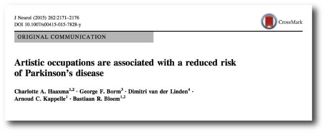
Title: Artistic occupations are associated with a reduced risk of Parkinson’s disease.
Authors: Haaxma CA, Borm GF, van der Linden D, Kappelle AC, Bloem BR.
Journal: J Neurol. 2015 Sep;262(9):2171-6.
PMID: 26138540               (This article is OPEN ACCESS if you would like to read it)

In their study, Prof Bloem and his colleagues conducted a case–controlled analysis of 750 men with Parkinson’s disease (onset ≥40 years) and 1300 healthy men, which involved the participants completing a questionnaire about their occupational history. As expected (based on previous reports), they found that farming was associated with an increased risk of developing Parkinson’s disease (click here for more on this).

Interestingly, artistic occupations late in life were associated with a reduced risk of subsequent Parkinson’s disease. Another interesting observation from the study was that no initial occupation (early in life) predicted Parkinson’s disease, which the researchers proposed indicated that the premotor phase of the disease starts later in life.

One interpretation of this finding is that creative people are less likely to develop Parkinson’s disease. An alternative theory, however, may be that artist jobs are associated with a less stressful, more relaxed lifestyle.

Could it be that the lower levels of stress associated with artistic occupations may be having an impact on the risk of developing Parkinson’s disease?

This idea is not as crazy as it sounds.

Consider different kinds of stress. Research suggests that people who undergo tremendous emotional stress have a higher risk of developing Parkinson’s disease. For example, there is the case of ex-prisoners of war:

Prisoner

Title:Neurological disease in ex-Far-East prisoners of war
Authors: Gibberd FB, Simmonds JP.
Journal: Lancet. 1980 Jul 19;2(8186):135-7.
PMID: 6105303

At the end of World war II, a neurological unit was set up at Queen Mary’s Hospital (Roehampton) to treat Ex-Far East prisoners of war. 4684 individuals were referred to the unit, of these 679 were found to have neurological disease (most of these – 593 cases – were loss of sight and peripheral nerve damage).

In follow up work in the 1970s, however, it was found that many of these individuals had gone on to develop other neurological conditions (dementia, multiple sclerosis, etc). Of interest to us, though was the finding that across the entire group of ex-prisoners investigated (4684 individuals), Parkinson’s disease was apparent in 24 of them – this is a frequency 5x that of the general population!

Even in animal models of Parkinson’s disease, emotional stress seems to exaccerbate the neurodegeneration that is being modelled:

Canada
Title: Stress accelerates neural degeneration and exaggerates motor symptoms in a rat model ofParkinson’s disease.
Authors: Smith LK, Jadavji NM, Colwell KL, Katrina Perehudoff S, Metz GA.
Journal: Eur J Neurosci. 2008 Apr;27(8):2133-46.
PMID: 18412632                  (This article is OPEN ACCESS if you would like to read it)

The investigators in this study demonstrated that chronic stress exaggerates the motor/behavioural deficits in a rat model of Parkinson’s disease. In addition, the stress resulted in a greater loss of dopamine neurons in the brains of these rats.

For an interesting review of the effect of stress in Parkinson’s disease – Click here.

Interesting. So what did the Korean researchers – you mentioned above – find this week?

Something interesting.

This is Dr Yoon-Il Lee.

Lee


Source: Dgist

He’s a dude.

He is a senior research scientists at the Daegu Gyeongbuk Institute of Science and Technology (DGIST) in Daegu Metropolitan City, South Korea.

Recently, his group has collaborated with Professor Yunjong Lee’s research team published this research report:

Lee

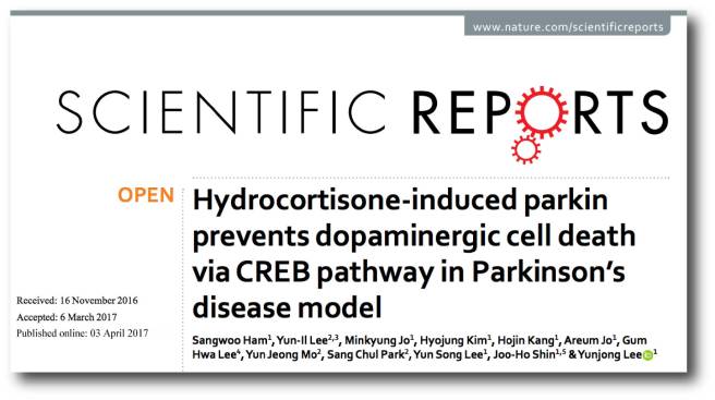
Title: Hydrocortisone-induced parkin prevents dopaminergic cell death via CREB pathway inParkinson’s disease model
Authors: Ham S, Lee YI, Jo M, Kim H, Kang H, Jo A, Lee GH, Mo YJ, Park SC, Lee YS, Shin JH, Lee Y.
Journal: Sci Rep. 2017 Apr 3;7(1):525. doi: 10.1038/s41598-017-00614-w.
PMID: 28366931         (This article is OPEN ACCESS if you would like to read it)

Dr Lee and his colleagues began this study with cells were engineered to produce a bioluminescent signal when a gene called Parkin was activated. Parkin is a Parkinson’s associated gene as genetic mutations in this gene can result in carriers developing a juvenile-onset/early-onset form of Parkinson’s disease.

The researchers then conducted an enormous screening experiment to find agents that turn on the Parkin gene. They applied a library of 1172 FDA-approved drugs (from Selleck Chemicals) to these cells – one drug per cell culture – and looked at which cell cultures began to produce a bioluminescent signal. They found 5 drugs that not only made the cells bioluminescent, but also resulted in Parkin protein being produced at levels 2-3 times higher than normal. Those drugs were:

  • Deferasirox – an iron chelator (interesting considering our previous post)
  • Vorinostat – a cancer drug (for treating lymphoma)
  • Metformin – a diabetes medication
  • Clindamycin – an antibiotic
  • Hydrocortisone

Hydrocortisone produced the highest levels of Parkin (Interestingly, hydrocortisone also did not increase the activity of PERK, an indicator of endoplasmic reticulum stress, while the other drugs did).

What is Hydrocortisone?

Hydrocortisone is the name for the hormone ‘cortisol’ when supplied as a medication.

Ok, so what is cortisol?

Cortisol is a glucocorticoid (a type of hormone) produced from cholesterol by enzymes in the cortex of the adrenal gland, which sits on top of the kidneys. It is produced in response to stress (physical or emotional)

CDR739009

The location of the adrenal glands. Source: Cancer

Cortisol helps us to deal with physical or emotional stress by reducing the activity of certain bodily functions – such as the immune system – so that the body can focus all of it’s energies toward dealing with the stress at hand.

Now generally, the functions of cortisol are supposed to be short-lived – long enough for the body to deal with the offending stressor and then levels go back to normal. But the normal levels of cortisol also fluctuate across the span of the day, with levels peaking around 8-9am:


open-uri20120801-11642-h5dnby

A graph of cortisol levels over the day. Source: HealthTap


Ok, so what did the Korean researchers do next?

Dr Lee and his colleagues gave the hydrocortisone drug to cell cultures which they then stressed (causing cell death). Hydrocortisone protected the cells from dying, and (importantly) it achieved this feat in a manner that was dependent on parkin activation. In cells that do not naturally have parkin, hydrocortisone was found to have no effect on cell survival.

Next the researchers treated mice with hydrocortisone before they then modelled Parkinson’s disease using the neurotoxin 6-OHDA. Hydrocortisone treatment resulted in approximate a two-fold increase in levels of parkin within particular areas of the brain. Without hydrocortisone treatment, the mice suffered the loss of approximately 45% of their dopamine neurons. Mice pre-treated with hydrocortisone, however, demonstrated enhanced dopamine neuron survival.

The researchers concluded that a sufficient physiological supply of hydrocortisone was required for protection of the brain, and that hydrocortisone treatment could be further tested as a means of maintaining high levels of parkin in the brain.

So what do we know about cortisol in Parkinson’s disease?

So this is where the story gets interesting;

Dobbs

Title: Cortisol is higher in parkinsonism and associated with gait deficit.
Authors: Charlett A, Dobbs RJ, Purkiss AG, Wright DJ, Peterson DW, Weller C, Dobbs SM.
Journal: Acta Neurol Scand. 1998 Feb;97(2):77-85.
PMID: 9517856

The researchers who conducted this study were interested in the role of cortisol in Parkinson’s disease. They measured cortisol levels in the blood of 96 subjects with Parkinson’s disease and 170 control subjects.  They found that cortisol levels were 20% higher in the subjects with Parkinson’s disease, and that MAO-B inhibitor treatment for Parkinson’s (Selegiline) reduced cortisol levels.
And MAO-B inhibitors are not the only Parkinson’s medication associated with reduced levels of cortisol:

Muller

Title: Acute levodopa administration reduces cortisol release in patients with Parkinson’s disease.
Authors: Müller T, Welnic J, Muhlack S.
Journal: J Neural Transm (Vienna). 2007 Mar;114(3):347-50.
PMID: 16932991

In this study the researchers found that cortisol levels started to decrease significantly just 30 minutes after L-dopa was taken.

Whether this lowering of cortisol levels may have any kind of detrimental effect on Parkinson’s disease is yet to be determined and required further investigation.

Is hydrocortisone or cortisol used in the clinic?

Yes it is.

Hydrocortisone is used to treat rheumatism, skin diseases, and allergies.

white-tablet-pills-on-reflective-surface

Hydrocortisone tablets. Source: Wisegeeks

Thus, there is the potential for another example of drug repurposing here. But the drug is not without side effects, which include:

  • Sleep problems (insomnia)
  • Mood changes
  • Acne, dry skin, thinning skin, bruising or discoloration;
  • Slow wound healing
  • Increased sweating
  • Headache, dizziness, spinning sensation;
  • nausea, stomach pain

For the full list of potential side effects – click here.

So what does it all mean?

Researchers in Korea have recently found that hydrocortisone (cortisol) can increase levels of Parkinson’s associated protein Parkin in cells, which in turn has a positive, neuroprotective effect on models of Parkinson’s disease.

We will now wait to see if the results can be independently replicated before attempting to take this drug to clinical trials for Parkinson’s disease. Any replication of the study should involve a range of treatment regimes so that we can determine if delayed administration can also be beneficial (this would involve delaying hydrocortisone treatment until after the neurotoxin has been given). Those studies could also look at the inflammatory effect in the brains as hydrocortisone has previously been demonstrated to have anti-inflammatory effects.
Interesting times. Stay tuned.


EDITOR’S NOTE: Under absolutely no circumstances should anyone reading this material consider it medical advice. The material provided here is for educational purposes only. Before considering or attempting any change in your treatment regime, PLEASE consult with your doctor or neurologist. While some of the drugs discussed on this website are clinically available, they may have serious side effects. We urge caution and professional consultation before altering any treatment regime. SoPD can not be held responsible for any actions taken based on the information provided here. 


The banner for today’s post was sourced from ZetaYarwood

Iron, life force, and Parkinson’s disease

pranaLogo

‘Prana’ is a Hindu Sanskrit word meaning “life force”.

An Australian biotech company has chosen this word for their name.

Recently Prana Biotechnology Ltd announced some exciting results from their Parkinson’s disease research programme.

In today’s post we will look at what the company is doing, the science underlying the business plan, and review the results they have so far.


adpd2017

Source: ADPD2017

At the end of March, over 3000 researchers in the field of neurodegeneration gathered in the Austrian capital of Vienna for the 13th International Conference on Alzheimer’s and Parkinson’s Diseases and Related Neurological Disorders (also known as ADPD2017).

crop

The Vienna city hall. Source: EUtourists

A lot of interesting new research in the field of Parkinson’s disease was presented at the conference (we will look at some other presentation in future posts), but one was of particular interest to us here at SoPD HQ.

The poster entitled: Abstract: 104 – PBT434 prevents neuronal loss, motor function and cognitive impairment in preclinical models of movement disorders by modulation of intracellular iron’, was presented by Associate Professor David Finkelstein, of the Florey Institute of Neuroscience and Mental Health (Melbourne, Australia).

Unfortunately the ADPD2017 conference’s scientific programme search engine does not allow for individual abstracts to be linked to on the web so if you would like to read the abstract, you will need to click here for the search engine page and search for ‘PBT434’ or ‘Finkelstein’ in the appropriate boxes.

Prof Finkelstein was presenting preclinical research that had been conducted by an Australian biotech company called Prana Biotechnology Ltd.

promo1

Source: Prana Biotechnology Ltd

What does the company do?

Prana Biotechnology Ltd has a large portfolio of over 1000 small chemical agents that they have termed ‘MPACs’ (or Metal Protein Attenuating Compounds). These compounds are designed to interrupt the interactions between particular metals and target proteins in the brain. The goal of this interruption is to prevent deterioration of brain cells in neurodegenerative conditions.

For Parkinson’s disease, the company is proposing a particular iron chelator they have called PBT434.

What is an iron chelator?

Iron chelator therapy involves the removal of excess iron from the body with special drugs. Chelate is from the Greek word ‘chela’ meaning “claw”.

chelationtherapy_edited-01

Chelator therapy. Source: Stanford

Iron overload in the body is a common medical problem, sometimes arising from disorders of increased iron absorption such as hereditary haemochromatosis. Iron chelator therapy represents one method of reducing the levels of iron in the body.

But why is iron overload a problem?

iron

Iron. Source: GlobalSpec

Good question. It involves the basic properties of iron.

Iron is a chemical element (symbol Fe). It has the atomic number 26 and by mass it is the most common element on Earth (it makes up much of Earth’s outer and inner core). It is absolutely essential for cellular life on this planet as it is involved with the interactions between proteins and enzymes, critical in the transport of oxygen, and required for the regulation of cell growth and differentiation.

So why then – as Rosalind asked in Shakespeare’s As You Like It – “can one desire too much of a good thing?”

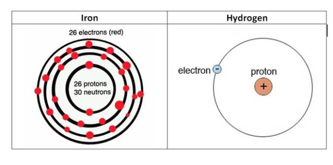
Well, if you think back to high school chemistry class you may recall that there are these things called electrons. And if you have a really good memory, you will recall that the chemical hydrogen has one electron, while iron has 26 (hence the atomic number 26).

atoms

The electrons of iron and hydrogen. Source: Hypertonicblog

Iron has a really interesting property: it has the ability to either donate or take electrons. And this ability to mediate electron transfer is one of the reasons why iron is so important in the body.

Iron’s ability to donate and accept electrons means that when there is a lot of iron present it can inadvertently cause the production of free radicals. We have previously discussed free radicals (Click here for that post), but basically a free radical is an unstable molecule – unstable because they are missing electrons.

imgres

How free radicals and antioxidants work. Source: h2miraclewater

In an unstable format, free radicals bounce all over the place, reacting quickly with other molecules, trying to capture the much needed electron to re-gain stability. Free radicals will literally attack the nearest stable molecule, to steal an electron. This leads to the “attacked” molecule becoming a free radical itself, and thus a chain reaction is started. Inside a living cell this can cause terrible damage, ultimately killing the cell.

Antioxidants can help try and restore the balance, but in the case of iron overload iron doctors will prescribe chelator treatment to deal with the situation more efficiently. By soaking up excess iron, we can limit the amount of damage caused by the surplus of iron.

So what research has been done regarding iron content and the Parkinsonian brain?

Actually, quite a lot.

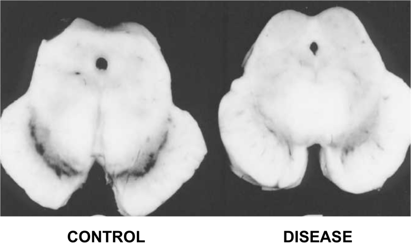
In 1968, Dr Kenneth Earle used an X-ray based technique to examine the amount of iron in the substantia nigra of people with Parkinson’s disease (Source). The substantial nigra is one of the regions in the brain most badly damaged by the condition – it is where most of the brain’s dopamine neurones resided.

d1ea3d21c36935b85043b3b53f2edb1f87ab7fa6

The dark pigmented dopamine neurons in the substantia nigra are reduced in the Parkinson’s disease brain (right). Source:Memorangapp

Earle examined 11 samples and compared them to unknown number of control samples and his results were a little startling:

The concentration of iron in Parkinsonian samples was two times higher than that of the control samples.

Since that first study, approximately 30 investigations have been made into levels of iron in the Parkinsonian brain. Eleven of those studies have replicated the Earle study by looking at postmortem tissue. They have used different techniques and the results have varied somewhat:

  • Sofic et al. (1988)                             1.8x increase in iron levels
  • Dexter et al. (1989)                         1.3x increase in iron levels
  • Uitti et al. (1989)                              1.1x increase in iron levels
  • Riederer et al 1989                         1.3x increase in iron levels
  • Griffiths and Crossman (1993)     2.0x increase in iron levels
  • Mann et al. (1994)                           1.6x increase in iron levels
  • Loeffler et al. (1995)                       0.9   (lower)
  • Galazka-Friedman et al., 1996     1.0   (no difference)
  • Wypijewska et al. (2010)               1.0   (no difference)
  • Visanji et al, 2013                            1.7x increase in iron levels

Overall, however, there does appear to be a trend in the direction of higher levels of iron in the Parkinsonian brains. A recent meta-analysis of all this data confirmed this assessment as well as noting an increase in the caudate putamen (the region of the brain where the dopamine neuron branches release their dopamine – Click here for that study).

Brain imaging of iron (using transcranial sonography and magnetic resonance imaging (MRI)) has also demonstrated a strong correlation between iron levels in the substantia nigra region and Parkinson’s disease severity/duration (Click here and here to read more on this).

Thus, there appears to be an increase of iron in the regions most affected by Parkinson’s disease and this finding has lead researchers to ask whether reducing this increase in iron may help in the treatment of Parkinson’s disease.

How could iron overload be bad in Parkinson’s disease?

Well in addition to causing the production of free radicals, there are many possible ways in which iron accumulation could be aggravating cell loss in Parkinson’s disease.

983245.fig.001

Possible causes and consequences of iron overload in Parkinson’s disease. Source: Hindawi

High levels of iron can cause the oxidation of dopamine, which results in the production of hydrogen peroxide (H2O– a reactive oxygen species – the stuff that is used to bleach hair and is also used as a propellant in rocketry!). This reaction can cause further oxidative stress that can then lead to a range of consequences including protein misfolding, lipid peroxidation (which can cause the accumulation of the Parkinson’s associated protein alpha synuclein), mitochondrial dysfunction, and activation of immune cells in the brain.

And this is just a taster of the consequences.

For further reading on this topic we recommend two very good reviews – click here and here.

Ok, so iron overload is bad, but what was the research presented in Austria?

The abstract:

Title: PBT434 prevents neuronal loss, motor function and cognitive impairment in preclinical models of movement disorders by modulation of intracellular iron
Authors: D. Finkelstein, P. Adlard, E. Gautier, J. Parsons, P. Huggins, K. Barnham, R. Cherny
Location: C01.a Posters – Theme C – Alpha-Synucleinopathies

The researchers at Prana Biotechnology Ltd assessed the potential of one of their candidate drugs, PBT434, in both cell culture and animal models of Parkinson’s disease. The PBT434 drug was selected for further investigation based on its performance in cell culture assays designed to test the inhibition of oxidative stress and iron-mediated aggregation of Parkinson’s associated proteins like alpha synuclein.

PBT434 significantly reduced the accumulation of alpha synuclein and markers of oxidative stress, and prevented neuronal loss.

The investigators also demonstrated that orally administered PBT434 readily crossed the blood brain barrier and entered the brain. In addition the drug was well-tolerated in the experimental animals and improved motor function in toxin-induced (MPTP and 6-hydroxydopamine) and transgenic mouse models of Parkinson’s disease (alpha synuclein -A53T and tau – rTg4510).

These results are in agreement with previous studies that have looked at iron chelator therapy in models of Parkinson’s disease (Click here, here and here for some examples)

Interestingly, PBT434 also demonstrated neuroprotective properties in animal models of multiple systems atrophy (or MSA). Suggesting that perhaps iron chelation could be a broad neuroprotective approach.

The researchers concluded that this preclinical data demonstrates the efficacy of PBT434 as a clinical candidate for Parkinson’s disease. PBT434 shows a strong toxicology profile and favourable therapeutic activity.  Prana is preparing its pre-clinical development package for PBT434 to initiate human clinical trials.

Does Prana have any other drugs in clinical trials?

Yes, they do.

pipeline-assets07-1024x571

Source: Prana

Prana Biotechnology has another product called PBT2.

The company currently has two clinical trial programs for PBT2 focused on two other neurodegenerative diseases: Alzheimer’s disease and Huntington’s disease.

The Alzheimer’s study was called the IMAGINE Trial, but (there is always a ‘but’) recently PBT2 failed to meet its primary endpoint (significantly reducing levels of beta-amyloid  – the perceived bad guy in Alzheimer’s disease) in a phase III trial of mild Alzheimer’s disease. PBT2 was, however, shown to be safe and very well tolerated over the 52 week trial, with no difference in the occurrence of adverse events between the placebo and treated groups.

In addition, there was less atrophy (shrinkage) in the brains of those patients treated with PBT2 when compared to control brains, 2.6% and 4.0%, respectively (based on brain imaging).  The company is tracking measures of brain volume and cognition in a 12 month extension study. It could be interesting to continue that follow up long term to evaluate the consequences of long term use of this drug on Alzheimer’s disease – even if the effect is minimal, any drug that can slow the disease down is useful and could be used in conjunction with other neuroprotective medications.

For Huntington’s disease, the company is also using the PBT2 drug and this study has had a bit more success. The study, called Reach2HD, was a six month phase II clinical trial in 109 patients with early to mid-stage Huntington’s disease, across 20 sites in the US and Australia. The company was aiming to assess the safety profile of this drug in this particular condition, as well as determining the motor and behavioural benefits.

In the ReachHD study, PBT2 showed signs of improving some aspects of cognitive function in the study, which potentially represents a major event for a disease for which there is very little in the way of medical treatments.

For a full description of the PBT2 trials, see this wikipedia page on the topic.

Is Prana the only research group working on iron chelators technology for Parkinson’s disease?

No.

There is a large EU-based consortium called FAIR PARK II, which is running a five year trial (2015 – 2020) of the iron chelator deferiprone (also known as Ferriprox). The study is a multi-centre, placebo-controlled, randomised clinical trial involving 338 people with recently diagnosed Parkinson’s disease.

LOGO_FAIR_PARK_TIME1

The population will be divided into two group (169 subjects each). They will then be assigned either deferiprone (15 mg/kg twice a day) or a placebo. Each subject will be given 9-months of treatment followed by a 1-month post-treatment monitoring period, in order to assess the disease-modifying effect of deferiprone (versus placebo).

Product-14303066240

Deferiprone. Source: SGPharma

As far as we are aware, this FAIR PARK II clinical trial is still recruiting participants – please click here to read more about this – thus it will most likely be some time before we hear the results of this study.

Are there natural sources of chelators?

Yes there are. In fact, many natural antioxidants exert some chelating activities.

Prominent among the natural sources of chelators: Green tea has components of plant extracts, such as epigallocatechin gallate (EGCG – which we have previously discussed in regards to Parkinson’s disease, click here to read that post) which possess structures which infer metal chelating properties.

As we have said before people, drink more green tea!

cup and teapot of linden tea and flowers isolated on white

Anyone fancy a cuppa? Source: Expertrain

So what does it all mean?

Summing up: We do not know what causes Parkinson’s disease. Most of our experimental treatments are focused on the biological events that occur in the brain around and after the time of diagnosis. These include an apparent accumulation of iron in affected brain regions.

Research groups are currently experimenting with drugs that reduce the levels of iron in the brain as a potential treatment for Parkinson’s disease. Preclinical data certainly look positive. We will now have to wait and see if those results translate into the human.

Previous clinical trials of metal chelators in neurodegeneration have had mixed success in demonstrating positive benefits. It may well be, however, that this treatment approach should be used in conjunction with other neuroprotective approaches – as a supplement. It will be interesting to see how Prana Biotechnology’s drug PBT434 fares in human clinical trials for Parkinson’s disease.

Stay tuned for more on this.


UPDATE – 3rd May 2017

Today the results of a double-blind, phase II clinical trial of iron chelator deferiprone in Parkinson’s disease were published. The results of the study indicate a mildly positive effect (though not statistically significant) after 6 months of daily treatment.

Iron1
Title: Brain iron chelation by deferiprone in a phase 2 randomised double-blinded placebo controlled clinical trial in Parkinson’s disease
Authors: Martin-Bastida A, Ward RJ, Newbould R, Piccini P, Sharp D, Kabba C, Patel MC, Spino M, Connelly J, Tricta F, Crichton RR & Dexter DT
Journal: Scientific Reports (2017), 7, 1398.
PMID: 28469157        (This article is OPEN ACCESS if you would like to read it)

In this Phase 2 randomised, double-blinded, placebo controlled clinical trial, the researchers recruited 22 people with early stage Parkinson’s disease (disease duration of less than 5 years; 12 males and 10 females; aged 50–75 years). They were randomly assigned to either a placebo group (8 participants), or one of two deferiprone treated groups: 20mg/kg per day (7 participants) or 30mg/kg per day (7 participants). The treatment was two daily oral doses (taken morning and evening), and administered for 6 months with neurological examinations, brain imaging and blood sample collections being conducted at 0, 3 and 6 months.

Deferiprone therapy was well tolerated and brain imaging indicated clearance of iron from various parts of the brain in the treatment group compared to the placebo group. Interestingly, the 30mg/kg deferiprone treated group demonstrated a trend for improvement in motor-UPDRS scores and quality of life (although this was not statistically significance). The researchers concluded that “more extensive clinical trials into the potential benefits of iron chelation in PD”.

Given the size of the groups (7 people) and the length of the treatment period (only 6 months) in this study it is not really a surprise that the researchers did not see a major effect. That said, it is very intriguing that they did see a trend towards motor score benefits in the  30mg/kg deferiprone group – remembering that this is a double blind study (so even the investigators were blind as to which group the subjects were in).

We will now wait to see what the FAIR PARK II clinical trial finds.


UPDATE: 28th June 2017

Today, the research that Prana biotechnology Ltd was presenting in Vienna earlier this year was published:

Prana

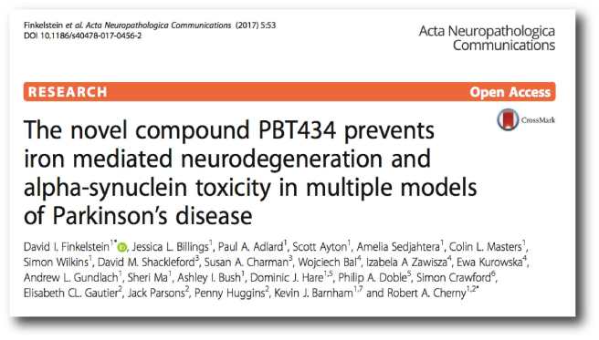
Title: The novel compound PBT434 prevents iron mediated neurodegeneration and alpha-synuclein toxicity in multiple models of Parkinson’s disease.
Authors: Finkelstein DI, Billings JL, Adlard PA, Ayton S, Sedjahtera A, Masters CL, Wilkins S, Shackleford DM, Charman SA, Bal W, Zawisza IA, Kurowska E, Gundlach AL, Ma S, Bush AI, Hare DJ, Doble PA, Crawford S, Gautier EC, Parsons J, Huggins P, Barnham KJ, Cherny RA.
Journal: Acta Neuropathol Commun. 2017 Jun 28;5(1):53.
PMID: 28659169             (This article is OPEN ACCESS if you would like to read it)

The results suggest that PBT434 is far less potent than deferiprone or deferoxamine at lowering cellular iron levels, but this weakness is compensated by the reduced levels of alpha synuclein accumulation in models of Parkinson’s disease. PBT434 certainly appears to be neuroprotective demonstrating improvements in motor function, neuropathology and biochemical markers of disease state in three different animal models of Parkinson’s disease.

The researchers provide little information as to when the company will be exploring clinical trials for this drug, but in the press release associated with the publication, Dr David Stamler (Prana’s Chief Medical Officer and Senior Vice President, Clinical Development) was quoted saying that they “are eager to begin clinical testing of PBT434”. We’ll keep an eye to the ground for any further news.


FULL DISCLOSURE: Prana Biotechnology Ltd is an Australasian biotechnology company that is publicly listed on the ASX. The information presented here is for educational purposes. Under no circumstances should investment decisions be made based on the information provided here. The SoPD website has no financial or beneficial connection to either company. We have not been approached/contacted by the company to produce this post, nor have we alerted them to its production. We are simply presenting this information here as we thought the science of what the company is doing might be of interest to other readers. 

In addition, under absolutely no circumstances should anyone reading this material consider it medical advice. The material provided here is for educational purposes only. Before considering or attempting any change in your treatment regime, PLEASE consult with your doctor or neurologist. Metal chelators are clinically available medications, but it is not without side effects (for more on this, see this website). We urge caution and professional consultation before altering a treatment regime. SoPD can not be held responsible for any actions taken based on the information provided here. 


The banner for today’s post was sourced from Prana

An Ambroxol update – active in the brain

Ambroxol-800x400

This week pre-clinical data was published demonstrating that the Ambroxol is active in the brain.

This is important data given that there is currently a clinical trial being conducted for Ambroxol in Parkinson’s disease.

Today’s post will review the new data and discuss what is happening regarding the clinical trial.


1082760

Ambroxol. Source: Skinflint

We have previously discussed the potential use of Ambroxol in the treatment of Parkinson’s disease (Click here to read that post). Today we follow up that post with new data that provides further support for an on-going clinical trial.

Firstly, what is Ambroxol?

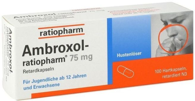
Ambroxol is a commonly used treatment for respiratory diseases (the respiratory system being the lungs and related components required for breathing). Ambroxol promotes the clearance of mucus and eases coughing. It also has anti-inflammatory properties, reducing redness in a sore throat. It is the active ingredient of products like Mucosolvan, Mucobrox, and Mucol.


What is the connection between Ambroxol and Parkinson’s disease?

So this is where a gene called GBA comes into the picture.

Genetic mutations in the GBA (full name: Glucosylceramidase Beta) gene are the most common genetic anomaly associated with Parkinson’s disease. People with a mutation in their GBA gene have a higher risk of developing Parkinson’s disease than the general population. And interestingly, people with Parkinson’s disease are approximately five times more likely to carry a GBA mutation than healthy control subjects.

What does GBA do?

The GBA gene provides the instructions for making an enzyme (called glucocerebrosidase) that helps with the digestion and recycling of waste inside cells. The enzyme is located and active inside ‘lysosomes‘.

What are Lysosomes?

Lysosomes are small bags of digestive enzymes that can be found inside cells. They help to break down proteins that have either been brought into the cell or that have served their function and need to be digested and disposed of (or recycled).

Lysosomes

How lysosomes work. Source: Prezi

Inside the lysosomes are enzymes like glucocerebrosidase which help to break material down into useful parts. The lysosome will fuse with other small bags (called vacuole) that act as storage vessels of material inside a cell. The enzymes from the lysosome will mix with the material in the vacuole and digest it (or it break down into more manageable components).

Now people with a genetic mutation in their GBA gene will often have an abnormally short, non-functioning version of the glucocerebrosidase enzyme. In those cases the breaking down of waste inside the lysosome becomes inhibited. And if waste can’t be disposed of or recycled properly, things start to go wrong in the cell.

How does Ambroxol correct this?

It was recently shown that Ambroxol triggers exocytosis of lysosomes (Source). Exocytosis is the process by which waste is exported out of the cell.

exocytosis

Exocytosis. Source: Socratic

Thus by encouraging lysosomes to undergo exocytosis and spit their contents out of the cell – digested or not – Ambroxol allows the cell to remove waste effectively and therefore function in a more normal fashion. This mechanism of treatment seemingly bi-passes the faulty glucocerebrosidase digestion enzyme entirely.

Until recently, two important questions, however, have remained unanswered:

  1. Does Ambroxol enter the brain and have this function there?
  2. What are the consequences of long term Ambroxol use?

We now have an answer for question no. 1:

Amb2

Title: Ambroxol effects in glucocerebrosidase and α-synuclein transgenic mice.
Authors: Migdalska-Richards A, Daly L, Bezard E, Schapira AH.
Journal: Ann Neurol. 2016 Nov;80(5):766-775.
PMID: 27859541            (This article is OPEN ACCESS if you would like to read it)

In this study, the researchers treated mice with Ambroxol for 12 days and then measured the level of glucocerebrosidase activity in the brain. They gave Ambroxol to three different groups of mice:

  • a group of normal mice,
  • a group of mice which had been genetically engineered with a specific mutation in their GBA gene (the heterozygous L444P mutation)
  • a group of mice that produced human alpha synuclein (the protein closely associated with Parkinson’s disease).

When they looked at the level of glucocerebrosidase enzyme activity in normal mice, they found an increase of approximately 20% (in mice treated with 4mM Ambroxol). One curious finding was that this dose was the only dose that increase glucocerebrosidase activity (1, 3, and 5mM of Ambroxol had no effect). The investigators noted, however, a reduction in water drinking of mice receiving 5mM in their drinking water (maybe they didn’t like the taste of it!), suggesting that they were not getting as much Ambroxol as the 4mM group.

The 4mM level of of Ambroxol also increased glucocerebrosidase activity in the L444P mutation mice and the alpha-synuclein mice (which interestingly also has reduced levels of glucocerebrosidase activity). One important observation in the alpha synuclein mice was the finding that Ambroxol was able to reduce the levels of alpha synuclein in the cells, indicating better clearance of un-wanted excess of proteins.

These combined results suggested to the investigators that Ambroxol is entering the brain of mice (passing through the protective blood brain barrier) and able to be effective there. In addition, they did not witness any serious adverse effects of ambroxol administration in the mice – an observation made in other studies of Ambroxol in normal mice (Click here to read more about this).

These studies have been followed up by a dosing study in primates which was just published:

Ambrox

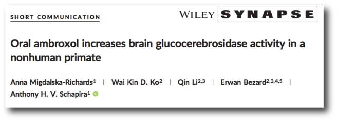
Title: Oral ambroxol increases brain glucocerebrosidase activity in a nonhuman primate.
Authors: Migdalska-Richards A, Ko WK, Li Q, Bezard E, Schapira AH.
Journal: Synapse. 2017 Mar 12. doi: 10.1002/syn.21967.
PMID: 28295625            (This article is OPEN ACCESS if you would like to read it)

In this study, the investigators analysed the effect of Ambroxol treatment on glucocerebrosidase activity in three healthy non-human primates. One subject was given an ineffective control solution vehicle, another subject received 22.5 mg/day of Ambroxol and the third subject received 100 mg/day of Ambroxol. They showed that daily administration 100 mg/day of Ambroxol results in increased levels of glucocerebrosidase activity in the brain (approximately 20% increase on average across different areas of the brain). Importantly, the 22.5 mg treatment did not result in any increase.

The investigators wanted to determine if the effect of Ambroxol was specific to glucocerebrosidase, and so they analysed the activity of another lysosome enzyme called beta-hexosaminidase (HEXB). They found that 100 mg/day of Ambroxol also increased HEXB activity (again by approximately 20%), suggesting that Ambroxol may be having an effect on other lysosome enzymes and not just glucocerebrosidase.

The researches concluded that these results provide the first data of the effect of Ambroxol treatment on glucocerebrosidase activity in the brain of non-human primates. In addition, the results indicate that Ambroxol is active and as the researchers wrote “should be further investigated in the context of clinical trials as a potential treatment for Parkinson’s disease”.

And there is a clinical trial currently underway?

Yes indeed.

Funded by the Cure Parkinson’s Trust and the Van Andel Research Institute (USA), there is currently a phase I clinical trial with 20 people with Parkinson’s disease receiving Ambroxol over 24 months. Importantly, the participants being enrolled in the study have both Parkinson’s disease and a mutation in their GBA gene. The study is being led by Professor Anthony Schapira at the Royal Free Hospital (London).

EDITORS NOTE HERE: Readers may be interested to know that Prof Schapira is also involved with another clinical trial for GBA-associated Parkinson’s disease. The work is being conducted in collaboration with the biotech company Sanofi Genzyme, and involves a phase II trial, called MOVE-PD, which is testing the efficacy, and safety of a drug called GZ/SAR402671 (Click here to read more about this clinical trial). GZ/SAR402671 is a glucosylceramide synthase inhibitor, which will hopefully reduce the production and consequent accumulation of glycosphingolipids in people with a mutation in the GBA gene. This approach is trying to reduce the amount of protein that can not be broken down by the faulty glucocerebrosidase enzyme. The MOVE-PD study will enroll more than 200 patients worldwide (Click here and here to read more on this).

The current Phase 1 trial at the Royal Free Hospital will be primarily testing the safety of Ambroxol in GBA-associated Parkinson’s disease. The researchers will, however, be looking to see if Ambroxol can increase levels of glucocerebrosidase and also assess whether this has any beneficial effects on the Parkinson’s features.

So what does it all mean?

There is a major effort from many of the Parkinson’s disease related charitable groups to clinically test available medications for their ability to slow this condition. Big drug companies are not interested in this ‘re-purposing effort’ as many of these drugs are no longer patent protected and thus providing limited profit opportunities for them. This is one of the unfortunate realities of the pharmaceutical industry business model.

One of the most interesting drugs being tested in this re-purposing effort is the respiratory disease-associated treatment, Ambroxol. Recently new research has been published that indicates Ambroxol is able to enter the brain and have an impact by increasing the level of protein disposal activity.

A clinical trial testing Ambroxol in Parkinson’s disease is underway and we will be watching for the results when they are released (most likely late 2019/early 2020, though preliminary results may be released earlier).

This trial is worth watching.

Stay tuned.


EDITOR’S NOTE: Under absolutely no circumstances should anyone reading this material consider it medical advice. The material provided here is for educational purposes only. Before considering or attempting any change in your treatment regime, PLEASE consult with your doctor or neurologist. Amboxol is a commercially available medication, but it is not without side effects (for more on this, see this website). We urge caution and professional consultation before altering a treatment regime. SoPD can not be held responsible for any actions taken based on the information provided here. 


The banner for today’s post was sourced from Pharmacybook

Stimulating research in London (Canada)

Spinal-Cord-final

Recently the SoPD has been contacted by readers asking about this video:

http://london.ctvnews.ca/video?clipId=1080895

The video presents a news article from Canada describing a clinical study of spinal cord stimulation for Parkinson’s disease.

In today’s post we review what spinal cord stimulation is and what research has been done in Parkinson’s disease.


should-say-50th-birthday-speech_67e6879f1e6fbd7

50 years celebration. Source: Reference

As many readers will be aware from 2017 represents the 200 year anniversary of the first description of Parkinson’s disease by one Mr James Parkinson.

Many readers will not be aware, however, that 2017 is also represents the 50th anniversary of the first use of a technique called spinal cord stimulation:

What is spinal cord stimulation?

Anterior_thoracic_SCS

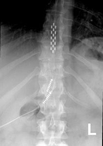
An x-ray of the spine with a stimulator implanted (towards the top of the image, and cords leading off to the bottom left). Source: Wikipedia

A spinal cord stimulator involves a small device being used to apply pulsed electrical signals to the spinal cord. It is generally used for pain relief, but it has recently been tested in a variety of other medical conditions.

The device is a column of stimulating electrodes that is surgically implanted in the epidural space of the spine. And before you ask: the epidural space is the area between the outer protective skin of the spinal cord (called the dura mater) and the surrounding vertebrae. So the device lies against the spinal cord, and is protected by the bones that make up the spine (as shown in the image below).

stimimplanttrial_1280

The stimulating electrodes within the epidural space. Source: SpineOne

An electrical pulse generator is implanted in the lower abdomen and conducting wires are connected between the electrodes to the generator. Much like deep brain stimulation, the system is entirely enclosed in the body and operated with a remote control.

How does spinal cord stimulation work?

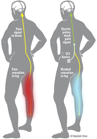
The stimulation basically interrupts the feeling of pain – blocking it from reaching the brain – substituting it with a more pleasing sensation called paresthesia (a kind of tingling or numbness).

PE-SCS Fig1

Source: MayoClinic

The stimulation does not eliminate the source of pain, it simply masks it by interfering with the signal going to the brain.  As a result the amount of relief from pain varies from person to person. In general, spinal cord stimulation resulting in a 50-70% reduction in pain.

But Parkinson’s results from inability to move, how would spinal cord stimulation work in Parkinson’s disease?

Yeah, this is a good question and the answer is not entirely clear, but the researchers (behind the research we discuss below) suggest that beneficial effects from spinal cord stimulation in Parkinson’s disease could be coming from direct activation of ascending pathways reaching thalamic nuclei and the cerebral cortex. That is to say (in plain English): activation of the spinal cord results in a signal going up into the brain where it alters the interaction between two of the regions involved in the initiation of movement (the thalamus and the cortex). And as we shall discuss below, there is evidence backing this idea.

Ok, so how much research has been done on spinal cord stimulation for Parkinson’s disease?

Actually quite a bit (in fact, for a good early review on the topic – click here).

The first real attempt at spinal cord stimulation for Parkinson’s disease was this report here:

Spinal1

Title: Spinal Cord Stimulation Restores Locomotion in Animal Models of Parkinson’s Disease
Authors: Fuentes, R., Petersson, P., Siesser, W. B., Caron, M. G., & Nicolelis, M. A. L.
Journal: Science (2009) 323(5921), 1578-1582.
PMID: 19299613                   (This article is OPEN ACCESS if you would like to read it)

It was conducted by Prof Miguel Nicolelis and his colleagues at Duke University. Duke were kind enough to make this short video about the research:

In their research report, the scientists injected mice with a drug that reduced the level of dopamine in the brain (the tyrosine hydroxylase inhibitor alpha-methyl-para-tyrosine  or AMPT). Similar to Parkinson’s disease, this resulted in a significant reduction in the movements of those mice. It also resulted in changes in the neuronal activity patterns of cells in an area of the brain called the motor cortex (we have talked about the motor cortex in a previous post). When the researchers then conducted spinal cord stimulation on these mice, they found that stimulation corrected both the loss of movement and the altered activity in the motor cortex.

The researchers then tested spinal cord stimulation in rats which had their dopamine system severely depleted (using the neurotoxin 6-OHDA), and they again found that the treatment could rescue the loss of locomotor ability. Curiously, spinal cord stimulation in the rats also caused an increase in locomotion activity after the stimulation period had stopped. On top of this, the researchers found that spinal cord stimulation aided the effect of L-dopa, allowing lower doses of L-dopa to achieve the same behavioural results as higher doses in animals not receiving spinal cord stimulation.

These initial results were then replicated in primates:

Monkey

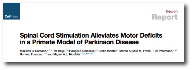
Title: Spinal cord stimulation alleviates motor deficits in a primate model of Parkinson disease.
Authors: Santana MB, Halje P, Simplício H, Richter U, Freire MA, Petersson P, Fuentes R, Nicolelis MA.
Journal: Neuron. 2014 Nov 19;84(4):716-22.
PMID: 25447740              (This article is OPEN ACCESS if you would like to read it)

In this study, the researchers modelled Parkinson’s disease in five adult marmosets using the neurotoxin 6-OHDA, which resulted in a reduction in spontaneous behaviour and a significant loss of dopamine neurons in the brain. They then implanted a spinal cord stimulator in each of the animals, which once activated resulted in a 200% improvement in some aspects of behavioural activity. Improvements observed in Parkinson’s-like features included freezing (31%), hypokinesia (23%), posture (23%), and bradykinesia (21%) as calculated by investigators blind to the treatment conditions of each subject.

In the brain, the researchers found that spinal cord stimulation resulted in similar improvements in neural activity as that seen with L-dopa treatment. Given all of these results, the investigators concluded that spinal cord stimulation “should be further tested in clinical studies aimed at measuring its long-term efficacy as a less invasive, long-term therapy for” people with Parkinson’s disease.

And it was not just Prof Nicolelis’ group that has achieved these results. Japanese researchers have also reported spinal cord stimulation having beneficial effects in models of Parkinson’s disease:

NeuoroProtect

Title: Spinal cord stimulation exerts neuroprotective effects against experimental Parkinson’s disease.
Authors: Shinko A, Agari T, Kameda M, Yasuhara T, Kondo A, Tayra JT, Sato K, Sasaki T, Sasada S, Takeuchi H, Wakamori T, Borlongan CV, Date I.
Journal: PLoS One. 2014 Jul 10;9(7):e101468.
PMID: 25009993           (This article is OPEN ACCESS if you would like to read it)

In this report, the researchers actually found that spinal cord stimulation resulted in neuroprotection in a classical model of Parkinson’s disease (rodent 6-OHDA striatal delivery). Across three different levels of stimulation, the researchers reported better rescue of motor deficits and protection of dopamine neurons (particularly for 50Hz stimulation). The researchers also provided evidence suggesting that the neuroprotective effect might have something to do with a protein called Vascular endothelial growth factor (or VEGF). Interestingly, they found that the neuroprotective protein GDNF (that we have discussed before – click here for that post) was not involved.

So has this spinal stimulation procedure ever been conducted in humans with Parkinson’s disease before?

Yes, it has. But the results were a bit disappointing.

Stim1

Title: Spinal cord stimulation failed to relieve akinesia or restore locomotion in Parkinson disease.
Authors: Thevathasan W, Mazzone P, Jha A, Djamshidian A, Dileone M, Di Lazzaro V, Brown P.
Journal: Neurology. 2010 Apr 20;74(16):1325-7.
PMID: 20404313          (This article is OPEN ACCESS if you would like to read it)

In this very small clinical study, just two people (both 75+ years of age) with Parkinson’s disease were fitted with spinal cord stimulators. Ten days after the surgery, the subjects participated in a blind analysis of the motor effects of spinal stimulation (blind analysis meaning that the assessors were not aware of their surgical treatment). The assessors, however, found no improvements as a result of the stimulation treatment.

This report lead to a letter to the journal from Prof Nicolelis and his colleagues:

Neurol

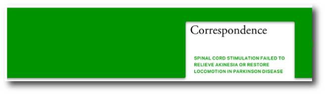
In their letter, Prof Nicolelis and co point out several issues with the clinical study that may impact the final results (such as the tiny size of the study (only two participants) and the fact that the electrodes were located at a high cervical level, while in the rodent study they were located at a high thoracic level). In addition, the commercially available electrodes used in the human clinical study did not match the relative size or orientation of the electrodes used in the rodent study.

The researchers of the clinical study suggested that the beneficial motor effect described in the rodent study may be due to an increase in arousal (as a result of higher stimulation). But Prof Nicolelis and colleagues pointed out in their letter that their rodent study included three control experiments (including air puffs, trigeminal stimulation at the highest intensity tolerated by the animals, and direct measurements of changes in heart rate following spinal stimulation) which did not find a strong connection between arousal response and recovery seen in the level of locomotion.

The letter concluded that the results of the small clinical trial were inconclusive, and that further research in nonhuman primate models of Parkinson’s are required to determine the effects of electrode design and stimulation parameters. The doctors behind the clinical study agreed that more research is required.

And what do we know about this new clinical study?

Unfortunately, not very much.

The study is being conducted by Prof. Mandar Jog of Western University. Recently the Parkinson’s Society Southwestern Ontario provided some funding towards the study (Click here for more on this), but that is about as much as we could find on the work.

So what does it all mean?

Summing up: Spinal cord stimulation is a technique that is used to alleviate severe back pain. It has recently been proposed for Parkinson’s disease, resulting in several clinical trials. Here at the SoPD we are not sure what our opinion on spinal cord stimulation is at present, except that more research is obviously required.

If the results from the new clinical study (being conducted in Canada) indicate that spinal cord stimulation has beneficial effects for people with Parkinson’s disease, it would certainly represent a significant step forward for the community which relies heavily on symptom masking drugs at present. Before proceeding to wider clinical availability, however, larger clinical studies will be required to truly demonstrate safety and efficacy.

We’ll let you know if we hear anything else about this developing area of research.


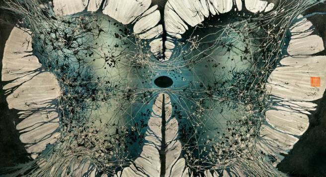
The banner for today’s post was sourced from Greg Dunn

Editorial: Putting 200 years into context

200

Here at the SoPD we understand and are deeply sympathetic to the frustration felt by the Parkinson’s community regarding the idea of ‘200 years and still no cure’.

As research scientists, we are in the trench everyday – fighting the good fight – trying to find ways of alleviating this terrible condition. And some of us are also in the clinics, interacting with sufferers and their families, listening to their stories and trying to help. While we do not deal directly with the day-to-day trials of living with Parkinson’s disease, we are keenly aware of many of the issues and are fully invested in trying to correct this condition.

972px-Paralysis_agitans_(1907,_after_St._Leger)

Source: Wikipedia

We do feel, however, that it is important to put some context into that ‘200 years’ time point that we are observing this week. It is too easy for people to think “wow, 200 years and still no cure?”

In our previous post – made in collaboration with Prof Frank Church of the Journey with Parkinson’s blog – we listed the major historical milestones and discoveries made in the Parkinson’s disease field during the last 200 years.

The most striking feature of that time line, however, is how just little actually happened during the first 100 years.

In fact for most of that period, Parkinson’s disease wasn’t even called ‘Parkinson’s disease’.

Of the 48 events that we covered on that time line, 37 of them have occurred in the last 50 years (26 since 2000).

Taking this line of thought one step further, 2017 is also the 20 year anniversary of the discovery of alpha synuclein‘s association with Parkinson’s disease. And what a remarkable 20 years that has been. In 1997, a group of researcher at the National institute of Health led by Robert Nussbaum reported the first genetic mutation in the alpha synuclein gene that infers vulnerability to Parkinson’s disease.

Since then, we have:

  • identified multiple additional mutations within that same gene that increase the risk of developing Parkinson’s disease.
  • determined which forms of alpha synuclein are toxic.
  • identified alpha synuclein as an important component of Lewy bodies – the dense clusters of protein found in the Parkinsonian brain.
  • discovered numerous methods by which alpha synuclein can be passed between cells – potentially aiding in the spread of Parkinson’s disease.
  • developed and validated models of Parkinson’s disease based on manipulations of alpha synuclein (including numerous genetically engineered mice, viral over-expression models, etc).
  • identified alpha synuclein in the lining of the gut of people with Parkinson’s disease and this has aided us in developing new theories as to how the condition may start.
  • set up and run numerous clinical trials targeting alpha synuclein (and we eagerly await the results of those trials).
  • published over 6200 scientific papers (don’t believe me? Click here) – that’s over 300 publications per year!

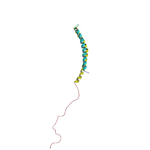
PBB_Protein_SNCA_image

Alpha synuclein protein. Source: Wikipedia

And the truly amazing part? All of these particular achievements are only dealing with just the one gene: alpha synuclein.

Since the identification of the alpha synuclein mutations, we have subsequently discovered genetic mutations in over 20 other genes that increase the risk of developing Parkinson’s disease. And we have conducted the same activities/experiments for most of those genes as we have for alpha synuclein.

For example, in 2004 we discovered that people with genetic mutations in a gene called glucocerebrosidase (or GBA) had an increased risk of developing Parkinson’s disease. In 2016, just 12 years after that discovery we have started a clinical trial designed specifically for those people (Click here for more on this).

wwwnew2_0

Source: Parkinson’s UK

We here at the SoPD are fully supportive of campaigns like #WeWontWait, and this post was not written (nor meant to be taken) as an excuse response to the ‘200 years and no cure’ frustration. I can understand how it may be read that way, but I did not know how else to write it. And I thought it needed to be written.

The point of this entire post is that those 200 years need to be put into context.

And while all of these words aren’t going to make life easier for someone living with Parkinson’s to deal with their situation, in addition to raising awareness this week I think it is important for the Parkinson’s community to also understand just how far we have come, and how fast we are currently progressing.

The question can be asked: will this be the last major anniversary we acknowledge with regards to Parkinson’s disease?

I sincerely think that there is cause to hope that it is.


Let me finish with a personal note:

I have a good friend – let’s call him Matt.

As a young boy, Matt remembers his grandfather having Parkinson’s disease. He remembers growing up watching the trials and tribulations that the old man went through with the condition. There were basically no treatment options when Matt’s grandfather was diagnosed and little in the way of support for the family. His grandfather’s body simply froze up as the disease progressed. L-dopa probably only became available to Matt’s grandfather during the latter stages of the disease.

Four years ago Matt’s father was diagnosed with Parkinson’s disease.

Thanks to scientific advances, however, Matt’s dad now has a wide range of treatment options on the medication side of things. The disease can be managed so that he can still play his golf and enjoy his retirement – in a way that his own father never could. He also has numerous surgical options once those medications lose their effectiveness (eg. deep brain stimulation, Pallidotomy, etc). The chances are very likely that Matt’s father will pass on by natural causes before he requires many of those additional options.

This is the progress that we have made.

But there is still a lot of work to be done of course.

During a lunch shortly after his father’s diagnosis, Matt looked squarely across the table at me. Me, the Parkinson’s researcher. All of the usual jovial nature was missing from his face and he simply muttered the words ‘hurry up’.

Whether he was speaking for his father, himself or his own young kids, I understood where his words were coming from and the sentiment.

And, as this post and the previous post point out, we are hurrying up.


The banner for today’s post was sourced from BMO

Milestones in Parkinson’s disease research and discovery

Self-Reflected-in-violets

FrankFor today’s post, we have teamed up with Prof Frank Church from the Journey with Parkinson’s blog to bring readers an ‘Introduction to the historical timeline on Parkinson’s disease’.

The idea for this project started as a conversation between Frank and his partner Barbara during a recent weekend at the beach in North Carolina.

Frank said: “Wouldn’t it be cool to publish a Parkinson’s historical timeline for Parkinson’s awareness month?”

However, to complete this project Frank felt it necessary to bring in some extra help in the form of a Parkinson’s expert.

And when everyone else said they were too busy, Frank contacted us.

Truly flattered, we immediately said yes. And the rest is history.


We are happy to present the milestones in Parkinson’s disease research and discover, though we do apologise to the clinicians, scientists, health-care specialists, and their projects that were not cited here but we limited the timeline to ~50 notations.

Below there are six panels outlining different stages of the history of Parkinson’s disease, and under each of them we have briefly described each of the events in the panel.

We hope you like it.

1817-1919- Milestones in Parkinson’s Disease Research and Discovery (Part 1a: Historical):

Slide1

First description of Parkinson’s disease

In 1811, Mr James Parkinson of no. 1 Hoxton Square (London) published a 66 page booklet called an ‘An Essay on the Shaking Palsy’. At the date of printing, it sold for 3 shillings (approx. £9 or US$12). The booklet was the first complete description of a condition that James called ‘Paralysis agitans’ or shaking palsy. In his booklet, he discusses the history of tremor and distinguishes this new condition from other diseases. He then describes three of his own patients and three people who he saw in the street.

The naming of Parkinson’s disease

Widely considered the ‘Father of modern neurology’, the importance of Jean-Martin Charcot’s contribution to modern medicine is rarely in doubt. From Sigmund Freud to William James (one of the founding fathers of Psychology), Charcot taught many of the great names in the early field of neurology. Between 1868 and 1881, Charcot focused much of his attention on the ‘paralysis agitans’. Charcot rejected the label ‘Paralysis agitans’, however, suggesting that it was misleading in that patients were not markedly weak and do not necessarily have tremor. Rather than Paralysis Agitans, Charcot suggested that Maladie de Parkinson (or Parkinson’s disease) would be a more appropriate name, bestowing credit to the man who first described the condition. And thus 70 years after passing away, James Parkinson was immortalized with the disease named after him.

The further clinical characterisation of Parkinson’s disease

British neurologist Sir William Gowers published a two-volume text called the Manual of Diseases of the Nervous System (1886, 1888). In this book he described his personal experience with 80 people with Parkinson’s disease in the 1880s. He also identified the subtle male predominance of the disorder and provided illustrations of the characteristic posture. In his treatment of Parkinson’s tremor, Gower used hyoscyamine, hemlock, and hemp (cannabis) as effective agents for temporary tremor abatement.

The discovery of the chemical dopamine

In the Parkinsonian brain there is a severe reduction in the chemical dopamine. This chemical was first synthesised in 1910 by George Barger and James Ewens at the Wellcome labs in London, England.

The discovery of Lewy bodies

One of the cardinal features of Parkinson’s disease in the brain is the presence of Lewy bodies – circular clusters of protein. In 1912, German neurologist Friedrich Lewy, just two years out of medical school and still in his first year as Director of the Neuropsychiatric Laboratory at the University of Breslau (now Wroclaw, Poland) Medical School discovered these ‘spherical inclusions’ in the brains of a people who had died with Parkinson’s disease.

The importance of the substantia nigra in Parkinson’s disease

The first brain structure to be associated with Parkinson’s disease was the substantia nigra. This region lies in an area called the midbrain and contains the majority of the dopamine neurons in the human brain. It was in 1919 that a Russian graduate student working in Paris, named Konstantin Tretiakoff, first demonstrated that the substantia nigra was associated with Parkinson’s disease. Tretiakoff also noticed circular clusters in the brains he examined and named them ‘corps de Lewy’ (or Lewy bodies) after the German neurologist Friedrich Lewy who first discovered them.

1953-1968- Milestones in Parkinson’s Disease Research and Discovery (Part 1b: Historical):

Slide2

The first complete pathologic analysis of the Parkinsonian brain

The most complete pathologic analysis of Parkinson’s disease with a description of the main sites of damage was performed in 1953 by Joseph Godwin Greenfield and Frances Bosanquet.

The discovery of a functional role for dopamine in the brain

Until the late 1950s, the chemical dopamine was widely considered an intermediate in the production of another chemical called norepinephrine. That is to say, it had no function and was simply an ingredient in the recipe for norepinephrine. Then in 1958, Swedish scientist Arvid Carlsson discovered that dopamine acts as a neurotransmitter – a discovery that won Carlsson the 2000 Nobel prize for Physiology or Medicine.

The founding of the Parkinson’s Disease Foundation

In 1957, a nonprofit organisation called the Parkinson’s Disease Foundation was founded by William Black. It was committed to finding a cure for Parkinson’s Disease. Since its founding in 1957, PDF has funded more than $115 million worth of scientific research in Parkinson’s disease.

The discovery of the loss of dopamine in the brain of people with Parkinson’s disease

In 1960, Herbert Ehringer and Oleh Hornykiewicz demonstrated that the chemical dopamine was severely reduced in brains of people who had died with Parkinson’s disease.

The first clinical trials of Levodopa

Knowing that dopamine can not enter the brain and armed with the knowledge that the chemical L-dopa was the natural ingredient in the production of dopamine, Oleh Hornykiewicz & Walther Birkmayer began injecting people with Parkinson’s disease with L-dopa in 1961. The short term response to the drug was dramatic: “Bed-ridden patients who were unable to sit up, patients who could not stand up when seated, and patients who when standing could not start walking performed all these activities with ease after L-dopa. They walked around with normal associated movements and they could even run and jump.” (Birkmayer and Hornykiewicz 1961).

The first internationally-used rating system for Parkinson’s disease

In 1967, Melvin Yahr and Margaret Hoehn published a rating system for Parkinson’s disease in the journal Neurology. It involves 5 stages, ranging from unilateral symptoms but no functional disability (stage 1) to confinement to wheel chair (stage 5). Since then, a modified Hoehn and Yahr scale has been proposed with the addition of stages 1.5 and 2.5 in order to help better describe the intermediate periods of the disease.

Perfecting the use of L-dopa as a treatment for Parkinson’s disease

In 1968, Greek-American scientist George Cotzias reported dramatic effects on people with Parkinson’s disease using oral L-dopa. The results were published in the New England Journal of Medicine. and L-dopa becomes a therapeutic reality with the Food and Drug Administration (FDA) approving the drug for use in Parkinson’s disease in 1970. Cotzias and his colleagues were also the first to describe L-dopa–induced dyskinesias.

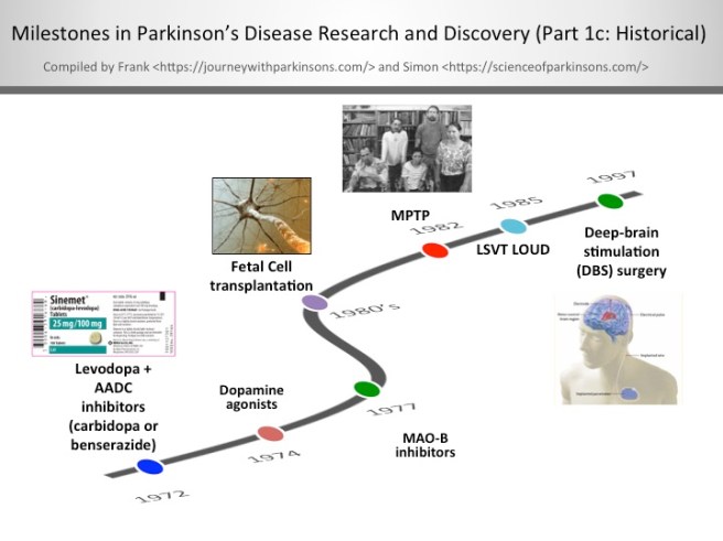
1972-1997- Milestones in Parkinson’s Disease Research and Discovery (Part 1c: Historical):

Slide3

Levodopa + AADC inhibitors (carbidopa or benserazide)

When given alone levodopa is broken down to dopamine in the bloodstream, which leads to some detrimental side effects.  By including an aromatic amino acid decarboxylase (AADC) inhibitor with levodopa allows the levodopa to get to the blood-brain barrier in greater amounts for better utilisation by the neurons. In the U.S., the AADC inhibitor of choice is carbidopa and in other countries it’s benserazide.

The discovery of dopamine agonists

Dopamine agonists are ‘mimics’ of dopamine that pass through the blood brain barrier to interact with target dopamine receptors. Since the mid-1970’s, dopamine agonists are often the first medication given most people to treat their Parkinson’s; furthermore, they can be used in conjunction with levodopa/carbidopa. The most commonly prescribed dopamine agonists in the U.S. are Ropinirole (Requip®), Pramipexole (Mirapex®), and Rotigotine (Neupro® patch). There are some challenging side effects of dopamine agonists including compulsive behaviour (e.g., gambling and hypersexuality),  orthostatic hypotension, and hallucination.

The clinical use of MAO-B inhibitors

In the late-1970’s, monoamine oxidase-B (MAO-B) inhibitors were created to block an enzyme in the brain that breaks down levodopa. MAO-B inhibitors have a modest effect in suppressing the symptoms of Parkinson’s.  Thus, one of the functions of MAO-B inhibitors is to prolong the half-life of levodopa to facilitate its use in the brain.  Very recently in clinical trials, it’s been shown that MAO-B inhibitors have some neuroprotective effect when used long-term.  The most widely used MAO-B inhibitors in the U.S. include Rasagiline (Azilect) and Selegiline (Eldepryl and Zelpar); MAO-B inhibitors may reduce “off” time and extend “on” time of levodopa.

Fetal Cell transplantation

After successful preclinical experiments in rodents, a team of researchers in Sweden, led by Anders Bjorklund and Olle Lindvall, began the first clinical trials of fetal cell transplantation for Parkinson’s disease. These studies involved taking embryonic dopamine cells and injecting them into the brains of people with Parkinson’s disease. The cells then matured and replaced the cells that had been lost during the progression of the disease.

The discovery of MPTP

In July of 1982, Dr. J. William Langston of the Santa Clara Valley Medical Center in San Jose (California) was confronted with a group of heroin addicts who were completely immobile. A quick investigation demonstrated that the ‘frozen addicts’ had injected themselves with a synthetic heroin that had not been prepared correctly. The heroin contained a chemical called MPTP, which when injected into the body rapidly kills dopamine cells. This discovery provided the research community with a new tool for modelling Parkinson’s disease.

1997-2006- Milestones in Parkinson’s Disease Research and Discovery (Part 1d: Historical):

Slide4

Alpha synuclein becomes the first gene associated with familial cases of Parkinson’s disease and its protein is found in Lewy bodies

In 1997, a group of researchers at the National institute of Health led by Robert Nussbaum reported the first genetic aberration linked to Parkinson’s disease. They had analysed DNA from a large Italian family and some Greek familial cases of Parkinson’s disease, and they

The gene Parkin becomes the first gene associated with juvenile Parkinson’s disease

The gene Parkin provides the instructions for producing a protein that is involved with removing rubbish from within a cell. In 1998, a group of Japanese scientists identified mutations in this gene that resulted in affected individuals being vulnerable to developing a very young onset (juvenile) version of Parkinson’s disease.

The first use of PET scan brain imaging for Parkinson’s disease

Using the injection of a small amount of radioactive material (known as a tracer), the level of dopamine present in an area of the brain called the striatum could be determined in a live human being. Given that amount of dopamine in the striatum decreases over time in Parkinson’s disease, this method of brain scanning represented a useful diagnostic aid and method of potentially tracking the condition.

The launch of Michael J Fox Foundation

In 1991, actor Michael J Fox was diagnosed with young-onset Parkinson’s disease at 29 years of age. Upon disclosing his condition in 1998, he committed himself to the campaign for increased Parkinson’s research. Founded on the 31st October, 2000, the Michael J Fox Foundation has funded more than $700 million in Parkinson’s disease research, representing one of the largest non-governmental sources of funding for Parkinson’s disease.

The Braak Staging of Parkinson’s pathology

In 2003, German neuroanatomist Heiko Braak and colleagues presented a new theory of how Parkinson’s disease spreads based on the postmortem analysis of hundreds of brains from people who had died with Parkinson’s disease. Braak proposed a 6 stage theory, involving the disease spreading from the brain stem (at the top of the spinal cord) up into the brain and finally into the cortex.

The gene DJ1 is linked to early onset PD

DJ1 (also known as PARK7) is a protein that inhibits the aggregation of Parkinson’s disease-associated protein alpha synuclein. In 2003, researchers discovered mutations in the DJ1 gene that made people vulnerable to a early-onset form of Parkinson’s disease.

The first GDNF clinical trial indicates neuroprotection in people with Parkinson’s disease

A small open-label clinical study involving the direct delivery of the chemical Glial cell-derived neurotrophic factor (GDNF) into the brains of people with Parkinson’s disease indicated that neuroprotection. The subjects involved in the study exhibited positive responses to the treatment and postmortem analysis of one subjects brain indicated improvements in the brain.

The genes Pink1 and LRRK2 are associated with early onset PD

Early onset Parkinson’s is defined by age of onset between 20 and 40 years of age, and it accounts for <10% of all patients with Parkinson’s.  Genetic studies are finding a causal association for Parkinson’s with five genes: alpha synuclein (SNCA), parkin (PARK2), PTEN-induced putative kinase 1 (PINK1), DJ-1 (PARK7), and Leucine-rich repeat kinase 2 (LRRK2). However it happens, and at whatever age it occurs, there is no doubt that genetics and environment combine together to contribute to the development of Parkinson’s.

The discovery of induced pluripotent stem (IPS) cells

In 2006, Japanese researchers demonstrated that it was possible to take skin cells and genetically reverse engineer them into a more primitive state – similar to that of a stem cell. This amazing achievement involved a fully mature cell being taken back to a more immature state, allowing it to be subsequently differentiated into any type of cell. This research resulted in the discoverer, Shinya Yamanaka being awarded the 2012 Nobel prize for Physiology or Medicine.

2007-2016- Milestones in Parkinson’s Disease Research and Discovery (Part 1e: Historical):

Slide5

The introduction of the MDS-UPDRS revised rating scale

The Movement Disorder Society (MDS) unified Parkinson’s disease rating scale (UPDRS) was introduced in 2007 to address two limitations of the previous scaling system, namely a lack of consistency among subscales and the low emphasis on the non-motor features. It is now the most commonly used scale in the clinical study of Parkinson’s disease.

The discovery of Lewy bodies in transplanted dopamine cells

Postmortem analysis of the brains of people with Parkinson’s disease who had fetal cell transplantation surgery in the 1980-1990s demonstrated that Lewy bodies are present in the transplanted dopamine cells. This discovery (made by three independent research groups) suggests that Parkinson’s disease can spread from unhealthy cells to healthy cells. This finding indicates a ‘prion-like’ spread of the condition.

SNCA, MAPT and LRRK2 are risk genes for idiopathic Parkinson’s disease

Our understanding of the genetics of Parkinson’s is rapidly expanding. There is recent evidence of multiple genes linked to an increase the risk of idiopathic Parkinson’s. Interestingly, microtubule-associated protein tau (MAPT) is involved in microtubule assembly and stabilization, and it can complex with alpha synuclein (SNCA).  Future therapies are focusing on  the reduction and clearance of alpha synuclein and inhibition of Lrrk2 kinase activity.

IPS derived dopamine neurons from people with Parkinson’s disease

The ability to generate dopamine cells from skin cells derived from a person with Parkinson’s disease represents not only a tremendous research tool, but also opens the door to more personalized treatments of suffers. Induced pluripotent stem (IPS) cells have opened new doors for researchers and now that we can generate dopamine cells from people with Parkinson’s disease exciting opportunities are suddenly possible.

Neuroprotective effect of exercise in rodent Parkinson’s disease models

Exercise has been shown to be both neuroprotective and neurorestorative in animal models of Parkinson’s. Exercise promotes an anti-inflammatory microenvironment in the mouse/rat brain (this is but one example of the physiological influence of exercise in the brain), which helps to reduce dopaminergic cell death.  Taking note of these extensive and convincing model system results, many human studies studying exercise in Parkinson’s are now also finding positive benefits from strenuous and regular exercise to better manage the complications of Parkinson’s.

Transeuro cell transplantation trial begins

In 2010, a European research consortium began a clinical study with the principal objective of developing an efficient and safe treatment methodology fetal cell transplantation in people with Parkinson’s disease. The trial is ongoing and the subjects will be followed up long term to determine if the transplantation can slow or reverse the features of Parkinson’s disease.

Successful preclinical testing of dopamine neurons from embryonic stem cells

Scientists in Sweden and New York have successfully generated dopamine neurons from human embryonic stem cells that can be successfully transplanted into animal models of Parkinson’s disease. Not only do the cells survive, but they also correct the motor deficits that the animals exhibit. Efforts are now being made to begin clinical trials in 2018.

Microbiome of the gut influences Parkinson’s disease

Several research groups have found the Parkinson’s disease-associated protein alpha synuclein in the lining of the gut, suggesting that the intestinal system may be one of the starting points for Parkinson’s disease. In 2016, researchers found that the bacteria in the stomachs of people with Parkinson’s disease is different to normal healthy individuals. In addition, experiments in mice indicated that the bacteria in the gut can influence the healthy of the brain, providing further evidence supporting a role for the gut in the development of Parkinson’s disease.

2016-2017- Milestones in Parkinson’s Disease Research and Discovery (Part 2: Clinical trials either recently completed or in progress)

Slide6

Safety, Tolerability and Efficacy Assessment of Dynacirc (Isradipine) for PD (STEADY-PD) III trial

Isradipine is a calcium-channel blocker approved for  treating high blood pressure; however, Isradipine is not approved for treating Parkinson’s. In animal models, Isradipine has been shown to slow the progression of PD by protecting dopaminergic neurons.  This study is enrolling newly diagnosed PD patients not yet in need of symptomatic therapy. Participants will be randomly assigned Isradipine or given a placebo.

Treatment of Parkinson’s Psychosis with Nuplazid

Approximately 50% of the people with Parkinson’s develop psychotic tendencies. Treatment of their psychosis can be relatively difficult. However, a new drug named Nuplazid was recently approved by the FDA specifically designed to treat Parkinson’s psychosis.

Opicapone (COMT Inhibitor) as Adjunct to Levodopa Therapy in Patients With Parkinson Disease and Motor Fluctuations

Catechol-O-methyl transferase (COMT) inhibitors prolong the effect of levodopa by blocking its metabolism. COMT inhibitors are used primarily to help with the problem of the ‘wearing-off’ phenomenon associated with levodopa. Opicapone is a novel, once-daily, potent third-generation COMT inhibitor.  It appears to be safer than existing COMT drugs. If approved by the FDA, Opicapone is planned for use in patients with Parkinson’s taking with levodopa who experience wearing-off issues.

Nilotinib (Tasigna® by Novartis) indicates positive results in phase I trial.

Nilotinib is a drug used in the treatment of leukemia. In 2015, it demonstrated beneficial effects in a small phase I clinical trial of Parkinson’s disease. Researchers believe that the drug activates the disposal system of cells, thereby helping to make cells healthier. A phase II trial of this drug to determine how effective it is in Parkinson’s disease is now underway.

ISCO cell transplantation trial begins

International Stem Cell Corporation is currently conducting a phase I clinical cell transplantation trial at a hospital in Melbourne, Australia. The company is transplanting human parthenogenetic stem cells-derived neural stem cells into the brains of people with Parkinson’s disease. The participants will be assessed over 12 months to determine whether the cells are safe for use in humans.

Neuropore’s alpha-synuclein stabilizer (NPT200-11) passes phase I trial

Neuropore Therapies is a biotech company testing a compound (NPT200-11) that inhibits and stablises the activity of the Parkinson’s disease-associated protein alpha synuclein. This alpha-synuclein inhibitor has been shown to be safe and well tolerated in humans in a phase I clinical trial and the company is now developing a phase II trial.

mGluR4 PAM  (PXT002331) well tolerated in phase I trial

Prexton Therapeutics recently announced positive phase I clinical trial results for their lead drug, PXT002331, which is the first drug of its kind to be tested in Parkinson’s disease. PXT002331 is a mGluR4 PAM – this is a class of drug that reduces the level of inhibition in the brain. In Parkinson’s disease there is an increase in inhibition in the brain, resulting in difficulties with initiating movements. Phase II clinical trials to determine efficacy are now underway.

Initial results of Bristol GDNF trial indicate no effect

Following remarkable results in a small phase I clinical study, the recent history of the neuroprotective chemical GDNF has been less than stellar. A subsequent phase II trial demonstrated no difference between GDNF and a placebo control, and now a second phase II trial in the UK city of Bristol has reported initial results also indicating no effect. Given the initial excitement that surrounded GDNF, this result has been difficult to digest. Additional drugs that behave in a similar fashion to GDNF are now being tested in the clinic.

Immunotherapies proves safe in phase I trials (AFFiRis & Prothena)

Immunotherapy is a treatment approach which strengthens the body’s own immune system. Several companies (particularly ‘AFFiRis’ in Austria and ‘Prothena’ in the USA) are now conducting clinical trials using treatments that encourage the immune system to target the Parkinson’s disease-associated protein alpha synuclein. Both companies have reported positive phase I results indicating the treatments are well tolerable in humans, and phase II trials are now underway.

Living Cell Technologies Limited continue Phase II trial of NTCELL

A New Zealand company called Living Cell Technologies Limited have been given permission to continue their phase II clincial trial of their product NTCELL, which is a tiny capsule that contains cells which release supportive nutrients when implanted in the brain. The implanted participants will be blindly assessed for 26 weeks, and if the study is successful, the company will “apply for provisional consent to treat paying patients in New Zealand…in 2017”.

MAO-B inhibitors shown to be neuroprotective.

MAO-B inhibitors block/slow the break down of the chemical dopamine. Their use in Parkinson’s disease allows for more dopamine to be present in the brain. Recently, several longitudinal studies have indicated that this class of drugs may also be having a neuroprotective effect.

Inhalable form of L-dopa

Many people with Parkinson’s disease have issues with swallowing. This makes taking their medication in pill form problematic. Luckily, a new inhalable form of L-dopa will shortly become available following recent positive Phase III clinical trial results, which demonstrated a statistically significant improvements in motor function for people with Parkinson’s disease during OFF periods.

Exenatide trial results expected

Exenatide is a drug that is used in the treatment of diabetes. It has also demonstrated beneficial effects in preclinical models of Parkinson’s disease, as well as an open-label clinical study over a 14 month period. Interestingly, in a two year follow-up study of that clinical trial – conducted 12 months after the patients stopped receiving Exenatide – the researchers found that patients previously exposed to Exenatide demonstrated significant improvements compared to how they were at the start of the study. There is currently a placebo-controlled, double blind phase II clinical trial being conducted and the results should be reported before the end of 2017.


A personal reflection

As I suggested at the start of this post, this endeavour was entirely Frank’s idea – full credit belongs with him. I was more than happy to help him out with it though as I thought it was a very worthy project. During this 200 year anniversary, I believe it is very important to acknowledge just how far we have come in our understanding of Parkinson’s disease since James first put pen to paper and described the six cases he had seen in London.

And Frank’s idea perfectly captures this.


The banner for today’s post was sourced from Greg Dunn (we are big fans!)

Hepatitis – Parkinson’s goes viral?

maxresdefault

Last week a new piece of Parkinson’s disease research has been widely discussed in the media.

It involves Hepatitis – the viral version of it at least.

In today’s post we will review the research and discuss what it may mean for Parkinson’s disease.


Fig2_v1c

A lewy body (brown with a black arrow) inside a cell. Source: Cure Dementia

A definitive diagnosis of Parkinson’s disease can only be made at the postmortem stage with an examination of the brain. Until that moment, all cases of Parkinson’s disease are ‘suspected’.

Critical to that postmortem diagnosis is the presence of circular shaped, dense clusters of proteins, called Lewy bodies (see the image above for a good example).

What causes Lewy bodies? We don’t know, but many people have theories.

This is Friedrich Heinrich Lewy (1885-1950).

DrLewy

Friedrich Lewy. Source: Lewy Body Society

As you can probably guess, Friedrich was the first to discover the ‘Lewy body’. His finding came by examining the brains of 85 people who died with Parkinson’s disease between 1908 – 1923.

In 1931, Friedrich Lewy read a paper at the International Congress of Neurology in Bern. During that talk he noted the similarities between the circular inclusions (called ‘negri bodies’) in the brains of people who suffered from rabies and his own Lewy bodies (observed in Parkinson’s disease).

rabies

A Negri body in a cell affected by rabies (arrow). Source: Nethealthbook

Given the similarities, Lewy proposed a viral cause for Parkinson’s disease.

Now, the idea that Parkinson’s disease could have a viral component has existed for a long time – even before Lewy made his conclusion. As we have previous mentioned, theories of viral causes for Parkinson’s have been circulating ever since the 1918 flu pandemic (Click here to read our post on this topic).

vonecomo-parkinson

An example of post-encephalitic Parkinsonism. Source: Baillement

About the same time as the influenza virus was causing havoc around the world, another condition began to appear called ‘encephalitis lethargica‘ (also known as post-encephalitic Parkinsonism). This disease left many of the victims in a statue-like condition, both motionless and speechless – similar to Parkinson’s disease. Initially, it was assumed that the influenza virus was the causal factor, but more recent research has left us not so sure anymore.

Since then there, however, has been additional bits of evidence suggesting a viral role in Parkinson’s disease. Such as this report:

H1N1

Title: Highly pathogenic H5N1 influenza virus can enter the central nervous system and induce neuroinflammation and neurodegeneration.
Author: Jang H, Boltz D, Sturm-Ramirez K, Shepherd KR, Jiao Y, Webster R, Smeyne RJ.
Journal: Proc Natl Acad Sci U S A. 2009 Aug 18;106(33):14063-8.
PMID: 19667183

The researchers in this study found that when they injected the highly infectious H5N1 influenza virus into mice, the virus progressed from the periphery (outside the brain) into the brain itself, where it induced Parkinson’s disease-like symptoms. The virus also caused a significant increase in the accumulation of the Parkinson’s associated protein Alpha Synuclein. Importantly, they witnessed the loss of dopamine neurons in the midbrain of the mice 60 days after resolution of the infection – that cell loss resembling what is observed in the brains of people with Parkinson’s disease.

The Parkinson’s associated protein alpha synuclein has also recently demonstrated anti-viral properties:

Beckham

Title: Alpha-Synuclein Expression Restricts RNA Viral Infections in the Brain.
Authors: Beatman EL, Massey A, Shives KD, Burrack KS, Chamanian M, Morrison TE, Beckham JD.
Journal: J Virol. 2015 Dec 30;90(6):2767-82. doi: 10.1128/JVI.02949-15.
PMID: 26719256               (This article is OPEN ACCESS if you would like to read it)

David Beckham (not the football player) and his research colleagues introduced West nile virus to brain cells grown in cell culture and they observed an increase in alpha synuclein production. They also found that the brains of people with West nile infections had increased levels of alpha synuclein.

The researchers then injected West Nile virus into both normal mice and genetically engineered mice (which produced no alpha synuclein) and they found that the genetically engineered mice which produced no alpha synuclein died quicker than the normal mice. They reported that there was an almost 10x increase in viral production in the genetically engineered mice. This suggested to them that alpha synuclein may be playing a role in protecting cells from viral infections.

Interesting, but what about this new data involving Hepatitis?

Yes, indeed. Let’s move on.

Wait a minute, what is Hepatitis exactly?

The name Hepatitis comes from the Greek: Hepat – liver; and itis – inflammation, burning sensation. Thus – as the label suggests – Hepatitis is inflammation of liver tissue.

Progress-of-Liver-Damage

Hepatitis and the liver. Source: HealthandLovepage

It can be caused by infectious agents (such as viruses, bacteria, and parasites), metabolic changes (induced by drugs and alcohol), or autoimmune/genetic causes (involving a genetic predisposition).

The most common cause of hepatitis is viral.

There are five main types of viral hepatitis (labelled A, B, C, D, and E). Hepatitis A and E are mainly spread by contaminated food and water. Both hepatitis B and hepatitis C are commonly spread through infected blood (though Hepatitis B is mainly sexually transmitted). Curiously, Hepatitis D can only infect people already infected with hepatitis B.

Hepatitis A, B, and D are preventable via the use of immunisation. A vaccine for hepatitis E has been developed and is licensed in China, but is not yet available elsewhere

Hepatitis C, however, is different.

There is currently no vaccine for it, mainly because the virus is highly variable between strains and the virus mutates very quickly, making an effective vaccine a difficult task. A number of vaccines under development (Click here for more on this).

What is known about Hepatitis C and the brain?

Quite a bit.

Similar to HIV (which we discussed in a previous post), the hepatitis C virus (HCV) enters the brain via infected blood-derived macrophage cells. In the brain, it is hosted by microglial cells, which results in altered functioning of those microglial cells. This causes problems for neuronal cells – including dopamine neurons. For example, people infected with HCV have reduced dopamine transmission, based on brain imaging studies (Click here and here for more on this result).

Have there been connections between hepatitis C virus and Parkinson’s disease before?

Yes.

Dopatitle


Title: Hepatitis C virus infection: a risk factor for Parkinson’s disease.
Authors: Wu WY, Kang KH, Chen SL, Chiu SY, Yen AM, Fann JC, Su CW, Liu HC, Lee CZ, Fu WM, Chen HH, Liou HH.
Journal: J Viral Hepat. 2015 Oct;22(10):784-91.
PMID: 25608223

The researchers in this study used data collected from a community-based screening program in north Taiwan which involved 62,276 people. The World Health Organisation (WHO) estimates that the prevalence of hepatitis C viral infection worldwide is approximately 2.2–3%, representing 130–170 million people. Taiwan is a high risk area for hepatitis, with antibodies for hepatitis viruses in Taiwan present in 4.4% in the general population (Source).

The researchers found that the significant association between hepatitis C viral infections and Parkinson’s disease – that is to say, a previous infection of hepatitis C increased the risk of developing Parkinson’s disease (by 40%). The researchers then looked at what the hepatitis C and B viral infections do to dopamine neurons growing in cell culture. They found that hepatitis C virus induced 60% dopaminergic cell death, while hepatitis B had no effect.

This study was followed up a few months later, by a second study suggesting an association between Hepatitis C virus and Parkinson’s disease:

Hep title

Title: Hepatitis C virus infection as a risk factor for Parkinson disease: A nationwide cohort study.
Authors: Tsai HH, Liou HH, Muo CH, Lee CZ, Yen RF, Kao CH.
Journal: Neurology. 2016 Mar 1;86(9):840-6.
PMID: 26701382

The researchers in this study wanted to investigate whether hepatitis C could be a risk factor for Parkinson’s disease. They did this by analyzing data from 2000-2010 drawn again from the Taiwan National Health Insurance Research Database.

The database included 49,967 people with either hepatitis B, hepatitis C or both, in addition to 199,868 people without hepatitis. During the 12 year period, 270 participants who had a history of hepatitis developed Parkinson’s disease (120 still had hepatitis C). This compared with 1,060 participants who were free of hepatitis, but went on to develop Parkinson’s disease.

When the researchers controlled for potentially confounding factors (such as age, sex, etc), the researchers found participants with hepatitis C had a 30% greater risk of developing Parkinson’s disease than the controls.

So if this has been demonstrated, why is this new study last week so important?

Good question.

The answer is very simple: This study is not based on statistics from Taiwan – this new study has found the same result from a new population.

HEP TITLE

Title: Viral hepatitis and Parkinson disease: A national record-linkage study.
Authors: Pakpoor J, Noyce A, Goldacre R, Selkihova M, Mullin S, Schrag A, Lees A, Goldacre M.
Journal: Neurology. 2017 Mar 29. [Epub ahead of print]
PMID: 28356465

These researchers used the English National Hospital Episode Statistics database and linked it to mortality data collected from 1999 till 2011. They too have found a strong association between hepatitis C and Parkinson’s disease (standardized rate ratio 1.51, 95% CI 1.18–1.9).

Curiously (and different from the previous studies), the researchers in this study also found a strong association for hepatitis B and Parkinson’s disease (standardized rate ratio 1.76, 95% CI 1.28–2.37). And these associations appear to be specific to Hepatitis B and C, as the investigators did not find any association between autoimmune hepatitis, chronic hepatitis, or HIV.

One important caveat with this new study, however, is that the authors could not
control for lifestyle factors (such as smoking or alcohol consumption). In addition, their system of linking medical records may underestimate the numbers of patients with
Parkinson’s disease as it would not take into account people with Parkinson’s disease who do not seek medical advice or those who are misdiagnosed (given a wrong diagnosis – it does happen!).

Regardless of these cautionary notes, the results still add to the accumulating evidence of an association between the virus that causes Hepatitis and the neurodegenerative condition of Parkinson’s disease.

But what about those people with Parkinson’s disease who have never had Hepatitis?

Yeah, this is a good question.

But there is a rather uncomfortable answer to it.

Here’s the rub: “Approximately 70%–80% of people with acute Hepatitis C do not have any symptoms” (Source: Centre for Disease Control). That is to say, the majority of people infected with the Hepatitis C virus will not be aware that they are infected. Some of those people who are infected may think that they have a case of the flu (HCV symptoms include fever, fatigue, loss of appetite,…), while others will simply not display any symptoms at all.

So many people with Parkinson’s disease may have had HCV, but never been aware of it.

And this is the really difficult part of researching the causal elements of Parkinson’s disease.

The responsible agent may actually leave little or no sign that they were ever present. For a long time, people have suggested that Parkinson’s disease is caused by a thief in the night – some agent that comes in, causes a problem and disappears without detection.

Perhaps Hepatitis is that thief.

But hang on a second, 60–70% of HCV infected people will go on to develop chronic liver disease (Source). Do people with Parkinson’s disease have liver issue?

Umm, well actually, in some cases: yes.

There have been studies of liver function in Parkinson’s disease where abnormalities have been found (Click here for more on this). And dopamine cell dysfunction has been seen in people with cirrhosis issues (Click here for more on this). In fact, the prevalence of Parkinsonism in people with cirrhosis has been estimated to be as high as 20% (and Click here for more on that).

So what are we saying? Hepatitis causes Parkinson’s disease???

No, we are not saying that.

Proving causality is the hardest task in science.

In addition, there have been a few studies in the past that have looked at viral infections as the cause of Parkinson’s disease that found strong associations with other viruses. For example this study:

Title: Infections as a risk factor for Parkinson’s disease: a case-control study.
Authors: Vlajinac H, Dzoljic E, Maksimovic J, Marinkovic J, Sipetic S, Kostic V.
Journal: Int J Neurosci. 2013 May;123(5):329-32.
PMID: 23270425

In this study, the researchers found that Parkinson’s Disease was also significantly associated to mumps, scarlet fever, influenza, and whooping cough as well as herpes simplex 1 infections. They found no association between Parkinson’s disease and Tuberculosis, measles or chickenpox though.

This result raises the tantalizing possibility that other viruses may also be involved with the onset of Parkinson’s disease (it should be added though that this study was based on only 110 people with Parkinson’s (compared with 220 controls) in one particular geographical location (Belgrade, Serbia)).

So different viruses may cause Parkinson’s disease?

We are not saying that either, but we would like to see more research on this topic.

And the situation may actually be more complicated than we think.

Recently, it has been reported that previous infection with flaviviruses (such as dengue) actually enhances the effect of Zika virus infect (Click here to read more on this). That is to say, a prior infection by one particular virus may exacerbate the infection of another virus. It could be that a previous infection by one virus increases that chance that a later infection by another virus – a particular combination of viral infections – may result in Parkinsonian symptoms (we are simply speculating here). 

Add to this complicated situation, the sheer number of unknown viruses. It is estimated that there are a minimum of 320,000 mammalian viruses still awaiting discovery (Click here for the source of this statistic), thus it is possible that additional unknown viruses may be involved with disease initiation for conditions like Parkinson’s disease.

A gang of unknown thieves in the night perhaps?

So what does it all mean?

Summing up: last week a new study was published that supported previous results that Hepatitis C viral infections could increase the risk of developing Parkinson’s disease. The results are important because they replicate previous findings from a different population of people.

The findings do not immediately mean that people with Hepatitis C are going to develop Parkinson’s disease, but it does suggest that they may be more vulnerable. The findings also suggest that more research is needed on the role of viral/infectious agents in the development of Parkinson’s disease.

We would certainly like to see more research in this area.


The banner for today’s post was sourced from Youtube

James: The man behind the disease (Part 1)

jamesp-signature

As part of Parkinson’s awareness month, and in observation of the 200 year anniversary of the first description of Parkinson’s disease, today we begin a four part set of posts looking at the man who made that first observation: James Parkinson.

Each week we will present various aspects about the man and his life.

Much of the material presented here has been replicated from our sister site Searching 4 James – a (much neglected) website celebrating the man and documenting the search for his likeness. To date, no portrait or image of the man has ever been found.

L0068467 The Villager's Friend and Physician

Source: Wellcome Images.

In her excellent book “James Parkinson, 1755-1824: From Apothecary to General Practitioner“, Shirley Roberts wrote that other sources have proposed that the man standing in the middle of the image above, talking to the villagers, is James Parkinson. The image appeared in James’ book ‘The Villager’s friend and physician’ (published in 1800), but (and I think you’ll agree) it does not give us much to work with.

Unfortunately Shirley Roberts made no reference to the sources of the proposal, but it is as close as we get to a likeness of the man, as he died before the first photographs were taken and there is no recorded painting of him.


Most people think of James Parkinson as a medical practitioner given his association with the disease that bears his name. But this singular association doesn’t really do the man credit. His contributions to medicine went well beyond the first description of ‘Parkinson’s disease’ – for example, James also gave the western world our first description of gout – a form of inflammatory arthritis that he and his father both suffered.

In addition, James was a ‘rockstar’ to the geological community, producing one of the most well regarded series of textbooks on the subject at the time. He was a political radical who wrote many pamphlets under the pseudonym “Old Hubert” and his associations with other radicals almost got him ‘transported’ (shipped out to the colonies). He was also a social reformist, calling for parliamentary reforms and universal suffrage. And his religious devotion made him a prominent figure within his church.

In short, he was a very interesting chap, who lived in (and had an impact on) interesting times.

THE WORLD OF JAMES

Before discussing the man himself, we must consider the world that James Parkinson was born into and the era he lived through. It provides us with the context within which we can fully appreciate the contributions that he made (including those beyond medicine).

James Parkinson was born on the 11th April 1755.

In the grand scale of things, the mid 1700’s was the peak of the little ice age, the middle of the age of enlightenment, and (critically) the start of the industrial revolution. The world was:

  • Pre USA (1776)
  • Pre French Revolution (1789)
  • Pre public electricity supply (1881)
  • Pre Napoleon (1769)
  • Pre Darwin (1809)

In London, King George II was on the English throne (soon to be replaced by George III), and Westminster bridge had just been finished (1750). The population of the city was approx. 700,000, but most of them lived in terrible conditions.

seutter_1750_london_view

A view of London (1750). Source: Historic Cities

James was born into a world where 74% of children born in London failed to reach the age of five. The medical world still practised humoral medicine (black bile, yellow bile, phlegm, and blood). Diseases were believed to be caused by an accumulation of “poisons” in the body, cured by bleeding, enemas, and sweating or blistering. The medical profession was:

  • Pre Ed Jenner’s vaccine for smallpox (1796)
  • Pre Rene Laennec’s stethoscope (1816)
  • Pre nitrous oxide (1800) or ether anaesthesia (1846)
  • Pre germ theory (Ignaz Semmelweis, 1847)
  • Pre Joseph Lister’s anti-septic surgery (1863).

Amputations were by far the most frequent surgeries, but the survival rate of the procedure was only 40% (and remember, there was no anaesthesia).

James Parkinson was born at no. 1 Hoxton Square in the liberty of Hoxton in Shoreditch, Middlesex. He would live all but the last 2 years of his life at that address.

In 1755, Hoxton was simply a scattering of houses, orchards and market gardens that lay approximately half a mile from one of the north-east gates of the walls of London. During the 17-1800s, Hoxton Square was considered a very fashionable area and young James would have grown up surrounded by open, reasonably well to do areas.

The maps below were made shortly before James was born, and it suggests open spaces, gardens, orchards and fields surrounding Hoxton.

London-1746

London in 1746 (Shoreditch is indicated by the black square)
Source: John Roque’s Map

Shoreditch

A map of Hoxton in 1746 – no 1. Hoxton Square (red arrow)
and St Leonard Church (blue arrow) are indicated.

James was born at the onset of the industrial revolution and with London prospering there was an enormous increase in the number of inhabitants. As more and more of London’s real estate became dedicated to business purposes, the inhabitants began spilling out into the surrounding areas. With transportation still limited to foot and horse, the people who worked in London needed to stay close to their place of employment, thus areas like Hoxton began to fill up rapidly. In 1788, there were 34,700 people living in Hoxton (in 5730 houses), which grew to 109,200 people in 1851 (in 15,433 houses).

Thus, during James’ life, Hoxton went through a radical transition. The large homes, orchards and gardens of his youth gave way to factories and over-crowding. And as a result, the ‘Parkinson and Son’ practise that he ran with his father (and later his own son) changed from serving a middle class clientele to dealing predominantly with the working class. With the prosperity of the time, there came a new trend of philanthropy, giving rise to the building of hospitals and mental asylums (‘madhouses’). James was the medical attendant for one of these madhouses, Holly House (Hoxton road, Hoxton).

The maps below were made in 1830 (shortly after James died – 1824) and indicate tremendous growth and expansion in London and the Hoxton area with the loss of much of the open spaces.

Greenwood

GREENWOOD MAP OF LONDON 1830 – Hoxton is indicated by the black square;
Tower of London (black arrow) and Westminster Abbey (red arrow) are also labelled – source: here

JP-Hoxton

 A map of Hoxton in 1830 – no 1. Hoxton Square (red arrow), St Leonard Church (blue arrow) and Holly House (Magenta arrow) are indicated.


THE FORMATIVE YEARS

James was baptised on the 29th of April 1755 in St Leonard’s church (Shoreditch) – the same church where he attended weekly services, got married, baptised his own children (and married some of them), and where he was eventually buried. The details of the baptism are recorded in the parish register, and read simply: James son of John and Mary Parkinson. Hoxton Square, Born 11th. Baptised 29th inst.

St Leonards

St Leonard’s church (1827) – Source

St Leonard’s church formed one of the key pillars of James’ life, and he could readily view the spire of the church one just block away from no.1 Hoxton Square.

The Parkinson family never owned the house at no. 1 Hoxton Square, which was owned by one Joshua Jenning. The building they lived in is gone now, but it was still standing in 1910 when Prof Leonard George Rowntree, a lecturer at Johns Hopkins Medical School (Baltimore), visited it and described it as:

“The house is a plain old three story building facing the east, on the northwest corner of Hoxton Square. Behind the main building and connected with it is a smaller two-story one with a central door opening into the little side street. This apparently was Parkinson’s office. Behind this again is another smaller building which may have served as a laboratory, as a library, or perhaps as a museum. Leading up to the deeply set, black, massive looking front door are a stone walk and deeply worn stone steps. The house is only a few feet back from the street and before it stands an old iron fence.

Uninteresting though the exterior is, upon entering this building one is impressed at the large size of the rooms and with the evidences of the prosperity of other days. We see in almost every room great carved open fire-places of elaborate design, and between some rooms large connecting arches. The deep panelling of walls and ceiling which was formerly so much in vogue is well preserved in some of the rooms on the second floor. One is surprised to find such an interesting interior behind such an uninviting exterior”  

(Rowntree, 1912)

url

An image of no.1 Hoxton Square – Source

James was the eldest surviving child of John (an apothecary and surgeon) and Mary Parkinson. James had two sisters who survived to adulthood, Margaret Townley Parkinson (born 3rd August,1759) and Mary Sedgewood (born 11th January, 1763).

Little is known about the formative years of James Parkinsons. From his own writings, we know that he had a solid education in Latin and Greek as well as chemistry, biology and mathematics. James was fortunate to grow up in a ‘comfortable, cultured home’ with ‘a medical atmosphere’. But a thriving literary, scientific, and religious atmosphere also existed in Hoxton square. No fewer than fifteen residents of the Square are biographized in the Dictionary of National Biography – a distinction not shared by any other London Square from that time. Nothing is known about where James received his education. His name does not appear on the registry of scholars of the well known public schools of London, such as St Paul’s, the charterhouse, Christ’s Hospital, Merchants Taylors – all of which were within walking distance of Hoxton Square. Private home schooling was very popular during this time. James certainly did not attend Cambridge or Oxford University.

At age 16, James began his training to be an apothecary. In accordance with an antiquated Elizabethan Act of Parliament, in order to become a surgeon a young man had to serve an apprenticeship of seven years. James was apprenticed to his father, but 20 years later he wrote that “no apprenticeship should be advisable except to a hospital”. James was extremely critical of the traditional methods used in the teaching of medicine at the time:

“The first four or five years are almost entirely appropriated to the compounding of medicines; the art of which,with every habit of necessary exactness, might just as well be obtained in as many months. The remaining years of his apprenticeship bring with them the acquisition of the art of bleeding, of dressing a blister, and, for the completion of the climax – of exhibiting an enema”  Parkinson, J. p32 (1800)

To further his training, James became one of the first medical students of the London Hospital Medical College (Whitechapel Road), founded by William Blizzard – surgeon of the Hospital. The college register records that he entered for training on Feb 20th 1776 when he was in his twentieth year. He was a ‘hospital pupil’ (or dresser) under Richard Grindall, FRS, at that time assistant surgeon. James remained for 6 months, but after this training he still felt ‘miserably ignorant’.

NPG D12199; Richard Grindall by William Daniell, after  George Dance

Richard Grindall (1716-1795) – by William Daniell, (21 Aug 1793)  – Source

On 1st April 1784, James was examined and granted the grand diploma of the Company of Surgeons. He then joined his father in a practice, called “Parkinson and Son” (that practice was to last through 4 generations – approx. 80 years). Unfortunately, John Parkinson died only 6 years later, and James was left to manage the practice single-handedly. James was fortunate to take over his father’s prosperous practise as he noted that ‘a physician seldom obtains bread by his profession until he has no teeth left to eat it’. The clientele requiring the services of Parkinson and son, however, would change dramatically during James’s life. Parkinson’s and son’s evolved from a upper-middle class practise to an almost entirely working class practise by the time James passed on.

It says a great deal about the man that he did not move away from the community as it evolved (as many early inhabitants of Hoxton Square did).

On 21st May 1783, James married Mary Dale in St Leonard’s Church, by special license which was the custom of the upper and middle classes of that period. He was 25 and she was 23 years old. James’s friend Wakelin Welch Jr of Lympstone (Devon) acted as his best man (many years later, James’ book ‘Organic Remains of a Former World’ was dedicated to Welch).

According to the Family Pursuits website, Mary Dale (daughter of John Dale and Mary Hardy) was born 2nd September, 1757 in Shoreditch, Middlesex. Her family lived lived in Charles Square, Hoxton. Mary’s grandfather, Francis Dale (1650-1716), was an apothecary in Hoxton Old Town. He had three sons: Francis (also an apothecary), Thomas (1699-1750), and John (a silk merchant and Mary’s father). Her family not only had a medical history, but also geological. Mary’s grand uncle, Samuel Dale (1659-1739) was a keen botanist and one of the first to describe the fossils in the cliffs of Harwich (Essex).


d88e38cf

Samuel Dale (1659-1739) – Source: The Essex Field Club

Thus the marriage was most likely a good fit for James. Mary Parkinson would live a long life, dying on 28 March 1838 of typhus fever (Gardner-Thorpe, 2013). Together with James, she had six children, key amongst them was John William Keys Parkinson (born 11th July, 1785) who apprenticed to his father and would later become the ‘Son’ in ‘Parkinson and Son’ (and ultimately John’s son James Keys Parkinson would follow in this process).


In the next post of this series, we will look at James’ early years as a physician and his foray into political radicalism.

ADHD and Parkinson’s disease

maxresdefault

The chemical dopamine plays a critical role in Parkinson’s disease.

It is also involved with the condition Attention deficit hyperactivity disorder, and recently researchers have been looking at whether there are any links between the two.

In today’s post we will look at what Attention deficit hyperactivity disorder is, how it relates to Parkinson’s disease, and what new research means for the community.


o-ADHD-facebook

Source: Huffington Post

We really have little idea about how Parkinson’s disease actually develops.

It could be kicked off by a virus or environmental factors or genetics…or perhaps a combination of these. We really don’t know, and it could vary from person to person.

There is a lot of speculation, however, as to what additional conditions could make one susceptible to Parkinson’s disease, even those conditions with early developmental onsets, such as autism (which we have previously written about – click here to see that post).

Recently researchers in Germany have asked if there is any connections between Parkinson’s and ADHD?

What is ADHD?

Attention deficit hyperactivity disorder (ADHD) is a neurodevelopmental condition that begins in childhood and persists into adulthood in 60% of affected individuals.

It is classically characterised in the media by hyperactive children who struggle to concentrate and stay focused on what they are doing. They are often treated with drugs such as Methylphenidate (also known as ritalin). Methylphenidate acts by blocking a protein called the dopamine transporter, which is involved with reabsorbing the chemical dopamine back into the cell after it has performed it’s function.

1280px-Ritalin

Ritalin. Source: Wikipedia

So are there any connections between ADHD and Parkinson’s disease?

This is an interesting question.

While there have been no reported findings of increased (or decreased) frequency of Parkinson’s disease in people with ADHD (to our knowledge), there are actually several bits of evidence suggesting a connection between the two conditions, such as abnormalities in the substantia nigra:

substantia

Title: Structural abnormality of the substantia nigra in children with attention-deficit hyperactivity disorder
Authors: Romanos M, Weise D, Schliesser M, Schecklmann M, Löffler J, Warnke A, Gerlach M, Classen J, Mehler-Wex C.
Journal: J Psychiatry Neurosci. 2010 Jan;35(1):55-8.
PMID: 20040247               (This article is OPEN ACCESS if you would like to read it)

The substantia nigra is a structure in the brain where the dopamine neurons reside. In Parkinson’s disease, the dopamine neurons of the substantia nigra start to degenerate – 50% are lost by the time a person is diagnosed with the condition.

In this study, the researchers used a technique called echogenicity to examine the substantia nigra of 22 children with ADHD and 22 healthy controls. Echogenicity is the ‘ability to bounce an echo’. This sort of assessment measures the return of an ultrasound signal that is aimed at a structure.

The researchers found that the ADHD subjects had a larger substantia nigra area than the healthy controls – which apparently indicates dopamine dysfunction. This finding is similar to results that have been observed in Parkinson’s disease (Click here to read more regarding that study).


Another connection between the two conditions was recent research has shown that genetic variations in the PARK2 gene (also known as Parkin) contribute to the genetic susceptibility to ADHD.

Parkin title

Title: Genome-wide analysis of rare copy number variations reveals PARK2 as a candidate gene for attention-deficit/hyperactivity disorder.
Authors: Jarick I, Volckmar AL, Pütter C, Pechlivanis S, Nguyen TT, Dauvermann MR, Beck S, Albayrak Ö, Scherag S, Gilsbach S, Cichon S, Hoffmann P, Degenhardt F, Nöthen MM, Schreiber S, Wichmann HE, Jöckel KH, Heinrich J, Tiesler CM, Faraone SV, Walitza S, Sinzig J, Freitag C, Meyer J, Herpertz-Dahlmann B, Lehmkuhl G, Renner TJ, Warnke A, Romanos M, Lesch KP, Reif A, Schimmelmann BG, Hebebrand J, Scherag A, Hinney A.
Journal: Mol Psychiatry. 2014 Jan;19(1):115-21.
PMID: 23164820         (This article is OPEN ACCESS if you would like to read it)

There are about 20 genes that have been associated with Parkinson’s disease, and they are referred to as the PARK genes. Approximately 10-20% of people with Parkinson’s disease have a genetic variation in one or more of these PARK genes (we have discussed these before – click here to read that post). PARK2 is a gene called Parkin. Mutations in Parkin can result in an early-onset form of Parkinson’s disease. The Parkin gene produces a protein which plays an important role in removing old or sick mitochondria (we discussed this in our previous post – click here to read that post).

In this report, the researchers conducted a genetic sequencing study on 489 young subjects with ADHD (average age 11 years old) and 1285 control individuals. They replicated the study with a similar sized population of people affected by ADHD and control subjects, and in both studies they found that certain deletions and replications in the Parkin gene influences susceptibility to ADHD – two of the genetic variations were found in 335 of the ADHD cases and none in 2026 healthy controls (from both sets of studies).

So there are are some interesting possible connections between  ADHD and Parkinson’s disease.

And what has the recent research from the German scientists found?

In this study, the researchers have looked at additional genetic variations that have been suggested to infer susceptibility to ADHD.

adhd

Title: No genetic association between attention-deficit/hyperactivity disorder (ADHD) and Parkinson’s disease in nine ADHD candidate SNPs
Authors: Geissler JM; International Parkinson Disease Genomics Consortium members., Romanos M, Gerlach M, Berg D, Schulte C.
Journal: Atten Defic Hyperact Disord. 2017 Feb 7. doi: 10.1007/s12402-017-0219-8. [Epub ahead of print]
PMID: 28176268

The researchers analysed nine genetic variations in seven genes:

  • one variant in the gene synaptosomal-associated protein, 25kDa1 (SNAP25)
  • one variant in the gene dopamine transporter (DAT; also known as SLC6A3)
  • one variant in the gene dopamine receptor D4 (DRD4)
  • one variant in the gene serotonin receptor 1B (HTR1B)
  • three mutations in cadherin 13 (CDH13)
  • one mutation located within the gene tryptophan hydroxylase 2 (TPH2)
  • one mutation located within the gene noradrenaline transporter (SLC6A2)

These genetic variations were assessed in 5333 cases of Parkinson’s disease and 12,019 healthy controls. The researchers found no association between any of the genetic variants and Parkinson’s disease. This finding lead the investigators to conclude that these genetic alterations associated with ADHD do not play a substantial role in increasing the risk of developing Parkinson’s disease.

Have ADHD medications ever been tested in Parkinson’s disease?

Yes.

Given the association of both ADHD and Parkinson’s disease with altered dopamine processing in the brain, a clinical trial of ritalin in Parkinson’s disease was set up and run in 2006 (Click here to read more about that trial). The results of the trial were published in 2007:


Ritalin title

Title: Effects of methylphenidate on response to oral levodopa: a double-blind clinical trial.
Authors: Nutt JG, Carter JH, Carlson NE.
Journal: Arch Neurol. 2007 Mar;64(3):319-23.
PMID: 17353373       (This article is OPEN ACCESS if you would like to read it)

In this study, the researchers recruited 12 people with Parkinson’s disease and examined their response to 0.4 mg/kg of ritalin – given 3 times per day – in conjunction with their normal anti-Parkinsonian medication (L-dopa). They then tested the subjects with either ritalin or a placebo control and failed to find any clinically significant augmentation of L-dopa treatment from the co-administration of ritalin.

What does it all mean?

So summing up: both Attention deficit hyperactivity disorder (ADHD) and Parkinson’s disease are associated with changes in the processing of the brain chemical dopamine. There are loose connections between the two conditions, but nothing definitive.

It will be interesting to follow up some of the individuals affected by ADHD, to determine if they ultimately go on to develop Parkinson’s (particularly those with Parkin mutations/genetic variants). But until then, the connection between these two conditions is speculative at best.


The banner for today’s post was sourced from Youtube

Resveratrol: From the folks who brought you Nilotinib


vc_spotlightsonoma_breaker_winegrapes_stock_rf_525141953_1280x640

Recently the results of a small clinical study looking at Resveratrol in Alzheimer’s disease were published. Resveratrol has long been touted as a miracle ingredient in red wine, and has shown potential in animal models of Parkinson’s disease, but it has never been clinically tested.

Is it time for a clinical trial?

In today’s post we will review the new clinical results and discuss what they could mean for Parkinson’s disease.


maxresdefault

From chemical to wine – Resveratrol. Source: Youtube

In 2006, there was a research article published in the prestigious journal Nature about a chemical called resveratrol that improved the health and survival of mice on a high-calorie diet (Click here for the press release).

Wine2
Title: Resveratrol improves health and survival of mice on a high-calorie diet.
Authors: Baur JA, Pearson KJ, Price NL, Jamieson HA, Lerin C, Kalra A, Prabhu VV, Allard JS, Lopez-Lluch G, Lewis K, Pistell PJ, Poosala S, Becker KG, Boss O, Gwinn D, Wang M, Ramaswamy S, Fishbein KW, Spencer RG, Lakatta EG, Le Couteur D, Shaw RJ, Navas P, Puigserver P, Ingram DK, de Cabo R, Sinclair DA.
Journal: Nature. 2006 Nov 16;444(7117):337-42.
PMID: 17086191          (This article is OPEN ACCESS if you would like to read it)

In this study, the investigators placed middle-aged (one-year-old) mice on either a standard diet or a high-calorie diet (with 60% of calories coming from fat). The mice were maintained on this diet for the remainder of their lives. Some of the high-calorie diet mice were also placed on resveratrol (20mg/kg per day).

After 6 months of this treatment, the researchers found that resveratrol increased survival of the mice and insulin sensitivity. Resveratrol treatment also improved mitochondria activity and motor performance in the mice. They saw a clear trend towards increased survival and insulin sensitivity.

The report caused a quite a bit of excitement – suddenly there was the possibility that we could eat anything we wanted and this amazing chemical would safe us from any negative consequences.

Grape

Source: Nature

That report was proceeded by numerous studies demonstrating that resveratrol could extend the life-span of various micro-organisms, and it was achieving this by activating a family of genes called sirtuins (specifically Sir1 and Sir2) (Click herehere and here for more on this).

Subsequent to these reports, there have been numerous scientific publications suggesting that resveratrol is capable of all manner of biological miracles.

Wow! So what is resveratrol?

grapes

Do you prefer your wine in pill form? Source: Patagonia

Resveratrol is a chemical that belongs to a group of compounds called polyphenols. They are believed to act like antioxidants. Numerous plants produce polyphenols in response to injury or when the plant is under attack by pathogens (microbial infections).

Fruit are a particularly good source of resveratrol, particularly the skins of grapes, blueberries, raspberries, mulberries and lingonberries. One issue with fruit as a source of resveratrol, however, is that tests in rodents have shown that less than 5% of the oral dose was observed as free resveratrol in blood plasma (Source). This has lead to the extremely popular idea of taking resveratrol in the form of wine, in the hope that it could have higher bioavailability compared to resveratrol in pill form. Red wines have the highest levels of Resveratrol in their skins (particularly Mabec, Petite Sirah, St. Laurent, and pinot noir). This is because red wine is fermented with grape skins longer than is white wine, thus red wine contains more resveratrol.


EDITOR’S NOTE: Sorry to rain on the parade, but it is important to note here that red wine actually contains only small amounts of resveratrol – less than 3-6 mg per bottle of red wine (750ml). Thus, one would need to drink a great deal of red wine per day to get enough resveratrol (the beneficial effects observed in the mouse study described above required 20mg/kg of resveratrol per day. For a person weighting 80kg, this would equate to 1.6g per day or approximately 250 750ml bottles). 

We would like to suggest that consuming red wine would NOT be the most efficient way of absorbing resveratrol. And obviously we DO NOT recommend any readers attempt to drink 250 bottles per day (if that is even possible). 

The recommended daily dose of resveratrol should not exceed 250 mg per day over the long term (Source). Resveratrol might increase the risk of bleeding in people with bleeding disorders. And we recommend discussing any change in treatment regimes with your doctor before starting.


So what did they find in the Alzheimer’s clinical study?

Well, the report we will look at today is actually a follow-on to published results from a phase 2/safety clinical trial that were reported in 2015:

trial.jpg

Title: A randomized, double-blind, placebo-controlled trial of resveratrol for Alzheimer disease.
Authors: Turner RS, Thomas RG, Craft S, van Dyck CH, Mintzer J, Reynolds BA, Brewer JB, Rissman RA, Raman R, Aisen PS; Alzheimer’s Disease Cooperative Study.
Title: Neurology. 2015 Oct 20;85(16):1383-91.
PMID: 26362286          (This article is OPEN ACCESS if you would like to read it)

The researchers behind the study are associated with the Georgetown research group that conducted the initial Nilotinib clinical study in Parkinson’s disease (Click here for our post on this).

The investigators conducted a randomized, placebo-controlled, double-blind, multi-center phase 2 trial of resveratrol in individuals with mild to moderate Alzheimer disease. The study lasted 52 weeks and involved 119 individuals who were randomly assigned to either placebo or resveratrol 500 mg orally daily treatment.

EDITOR’S NOTE: We appreciate that is daily dose exceeds the recommended daily dose mentioned above, but it is important to remember that the participants involved in this study were being closely monitored by the study investigators.

Brain imaging and samples of cerebrospinal fluid (the liquid within which the brain sits) were collected at the start of the study and after completion of treatment.

The most important result of the study was that resveratrol was safe and well-tolerated. The most common side effect was feeling nausea and diarrhea in approximately 42% of individuals taking resveratrol (curiously 33% of the participants blindly taking the placebo reported the same thing). There was also a weight loss effect between the groups, with the placebo group gaining 0.5kg on average, while the resveratrol treated group lost 1kg on average.

The second important take home message is that resveratrol crossed the blood–brain barrier in humans. The blood brain barrier prevents many compounds from having any effect in the brain, but it does not stop resveratrol.

The investigators initially found no effects of resveratrol treatment in various Alzheimer’s markers in the cerebrospinal fluid. Not did they see any effect in brain scans, cognitive testing, or glucose/insulin metabolism. The authors were cautious about their conclusions based on these results, however, as the study was statistically underpowered (that is to say, there were not enough participants in the various groups) to detect clinical benefits. They recommended a larger study to determine whether resveratrol is actually beneficial.

While exploring the idea of a larger study, the researchers have re-analysed some of the data, and that brings us to the report we want to review today:

moussa

Title: Resveratrol regulates neuro-inflammation and induces adaptive immunity in Alzheimer’s disease.
Authors: Moussa C, Hebron M, Huang X, Ahn J, Rissman RA, Aisen PS, Turner RS.
Journal: J Neuroinflammation. 2017 Jan 3;14(1):1. doi: 10.1186/s12974-016-0779-0.
PMID: 28086917       (This article is OPEN ACCESS if you would like to read it)

In this report, the investigators conducted a retrospective study re-examining the cerebrospinal fluid and blood plasma samples from a subset of subjects involved in the clinical study described above. In this study, they only looked at the subjects who started with very low levels in the cerebrospinal fluid of a protein called Aβ42.

Amyloid beta (or Aβ) is the bad boy/trouble maker of Alzheimer’s disease; considered to be critically involved in the disease. A fragment of this protein (called Aβ42) begin clustering in the brains of people with Alzheimer’s disease and as a result, low levels of Aβ42 in cerebrospinal fluid have been associated with increased risk of Alzheimer’s disease and considered a possible biomarker of the condition (Click here to read more on this).

The resveratrol study investigators collected all of the data from subjects with cerebrospinal fluid levels of Aβ42 less than 600 ng/ml at the start of the study. This selection criteria gave them 19 resveratrol-treated and 19 placebo-treated subjects.

In this subset re-analysis study, resveratrol treatment appears to have slowed the decline in cognitive test scores (the mini-mental status examination), as well as benefiting activities of daily living scores and cerebrospinal fluid levels of Aβ42.

One of the most striking results from this study is the significant decrease observed in the cerebrospinal fluid levels of a protein called Matrix metallopeptidase 9 (or MMP9) after resveratrol treatment. MMP9 is slowly emerged as an important player in several neurodegenerative conditions, including Parkinson’s disease (Click here to read more on this). Thus the decline observed is very interesting.

This re-analysis indicates beneficial effects in some cases of Alzheimer’s as a result of taking resveratrol over 52 weeks. The researchers concluded that the findings of this re-analysis support the idea of a larger follow-up study of resveratrol in people with Alzheimer’s disease.

Ok, but what research has been done on resveratrol in Parkinson’s disease?

Yes, good question.

One of the earliest studies looking at resveratrol in Parkinson’s disease was this one:

Reserv
Title: Neuroprotective effect of resveratrol on 6-OHDA-induced Parkinson’s disease in rats.
Authors: Jin F, Wu Q, Lu YF, Gong QH, Shi JS.
Journal: Eur J Pharmacol. 2008 Dec 14;600(1-3):78-82.
PMID: 18940189

In this study, the researchers used a classical rodent model of Parkinson’s disease (using the neurotoxin 6-OHDA). One week after inducing Parkinson’s disease, the investigators gave the animals either a placebo or resveratrol (at doses of 10, 20 or 40 mg/kg). This treatment regime was given daily for 10 weeks and the animals were examined behaviourally during that time.

The researchers found that resveratrol improved motor performance in the treated animals, with them demonstrating significant results as early as 2 weeks after starting treatment. Resveratrol also reduced signs of cell death in the brain. The investigators concluded that resveratrol exerts a neuroprotective effect in this model of Parkinson’s disease.

Similar results have been seen in other rodent models of Parkinson’s disease (Click here and here to read more).

Subsequent studies have also looked at what effect resveratrol could be having on the Parkinson’s disease associated protein alpha synuclein, such as this report:

PD-title

Title: Effect of resveratrol on mitochondrial function: implications in parkin-associated familiarParkinson’s disease.
Authors: Ferretta A, Gaballo A, Tanzarella P, Piccoli C, Capitanio N, Nico B, Annese T, Di Paola M, Dell’aquila C, De Mari M, Ferranini E, Bonifati V, Pacelli C, Cocco T.
Journal: Biochim Biophys Acta. 2014 Jul;1842(7):902-15.
PMID: 24582596                     (This article is OPEN ACCESS if you would like to read it)


In this study, the investigators collected skin cells from people with PARK2 associated Parkinson’s disease.

What is PARK2 associated Parkinson’s disease?

There are about 20 genes that have been associated with Parkinson’s disease, and they are referred to as the PARK genes. Approximately 10-20% of people with Parkinson’s disease have a genetic variation in one or more of these PARK genes (we have discussed these before – click here to read that post).

PARK2 is a gene called Parkin. Mutations in Parkin can result in an early-onset form of Parkinson’s disease. The Parkin gene produces a protein which plays an important role in removing old or sick mitochondria.

Hang on a second. Remind me again: what are mitochondria?

We have previously written about mitochondria (click here to read that post). Mitochondria are the power house of each cell. They keep the lights on. Without them, the lights go out and the cell dies.

Mitochondria

Mitochondria and their location in the cell. Source: NCBI

You may remember from high school biology class that mitochondria are bean-shaped objects within the cell. They convert energy from food into Adenosine Triphosphate (or ATP). ATP is the fuel which cells run on. Given their critical role in energy supply, mitochondria are plentiful and highly organised within the cell, being moved around to wherever they are needed.

Another Parkinson’s associated protein, Pink1 (which we have discussed before – click here to read that post), binds to dysfunctional mitochondria and then grabs Parkin protein which signals for the mitochondria to be disposed of. This process is an essential part of the cell’s garbage disposal system.

Park2 mutations associated with early onset Parkinson disease cause the old/sick mitochondria are not disposed of correctly and they simply pile up making the cell sick. The researchers that collected the skin cells from people with PARK2 associated Parkinson’s disease found that resveratrol treatment partially rescued the mitochondrial defects in the cells. The results obtained from these skin cells derived from people with early-onset Parkinson’s disease suggest that resveratrol may have potential clinical application.

Thus it would be interesting (and perhaps time) to design a clinical study to test resveratrol in people with PARK2 associated Parkinson’s disease.

So why don’t we have a clinical trial?

Resveratrol is a chemical that falls into the basket of un-patentable drugs. This means that big drug companies are not interested in testing it in an expensive series of clinical trials because they can not guarantee that they will make any money on their investment.

There was, however, a company set up in 2004 by the researchers behind the original resveratrol Nature journal report (discussed at the top of this post). That company was called “Sirtris Pharmaceuticals”.

e4d4a0ddab6419c9de2bd8ca4f199e0c

Source: Crunchbase

Sirtris identified compounds that could activate the sirtuins family of genes, and they began testing them. They eventually found a compound called SRT501 which they proposed was more stable and 4 times more potent than resveratrol. The company went public in 2007, and was subsequently bought by the pharmaceutical company GlaxoSmithKline in 2008 for $720 million.

Sirtris_rm

Source: Xconomy

From there, however, the story for SRT501… goes a little off track.

In 2010, GlaxoSmithKline stopped any further development of SRT501, and it is believed that this decision was due to renal problems. Earlier that year the company had suspended a Phase 2 trial of SRT501 in a type of cancer (multiple myeloma) because some participants in the trial developed kidney failure (Click here to read more).

Then in 2013, GlaxoSmithKline shut down Sirtris Pharmaceuticals completely, but indicated that they would be following up on many of Sirtris’s other sirtuins-activating compounds (Click here to read more on this).

Whether any of those compounds are going to be tested on Parkinson’s disease is yet to be determined.

What we do know is that the Michael J Fox foundation funded a study in this area in 2008 (Click here to read more on this), but we are yet to see the results of that research.

We’ll let you know when we hear of anything.

So what does it all mean?

Summing up: Resveratrol is a chemical found in the skin of grapes and berries, which has been shown to display positive properties in models of neurodegeneration. A recent double blind phase II efficacy trial suggests that resveratrol may be having positive benefits in Alzheimer’s disease.

Preclinical research suggests that resveratrol treatment could also have beneficial effects in Parkinson’s disease. It would be interesting to see what effect resveratrol would have on Parkinson’s disease in a clinical study.

Perhaps we should have a chat to the good folks at ‘CliniCrowd‘ who are investigating Mannitol for Parkinson’s disease (Click here to read more about this). Maybe they would be interested in resveratrol for Parkinson’s disease.


ONE LAST EDITOR’S NOTE: Under absolutely no circumstances should anyone reading this material consider it medical advice. The material provided here is for educational purposes only. Before considering or attempting any change in your treatment regime, PLEASE consult with your doctor or neurologist. SoPD can not be held responsible for actions taken based on the information provided here. 


The banner for today’s post was sourced from VisitCalifornia