A new theory of Parkinson’s disease

emc2

The great American baseball legend, Yogi Berra, once said: “In theory, there is no difference between theory and practice. But in practice, there is.”

Silly as it reads, there is a great deal of truth to that statement.

In science, we very quickly chase after a particular theory as soon as a little bit of evidence is produced that supports it. Gradually, these theories become our basic understanding of a situation, until someone points out the holes in the theory and we have to revise it.

A new theory of Parkinson’s disease has recently been proposed. In today’s post we will review what the theory is suggesting and what evidence there is to support it.


150119_sipress-2005-05-23

“I still say it’s only a theory”. Source: NewYorker

In the age of ‘alternative facts’, it is always important to remember that we don’t know as much as we think we do. In fact, much of our modern world still relies on a kind of faith rather than actual ‘facts’. For example, we take a particular type of medicine, because it has worked for some people in the past, not because it will definitely make us better.

And the same applies to our understanding of neurodegenerative conditions, like Parkinson’s disease. Based on all the evidence we have collected thus far, we have theories of how Parkinson’s disease may be progressing. But there are always exceptions to the rule, and these force us to refine or reconsider our theories.

Recently a refinement to our theory of Parkinson’s disease has been suggested.

Who has suggested it?

This is Prof Ole Isacson.

maxresdefault

Source: Crunchbase

He’s a dude.

He is is a Professor of Neurology at Harvard Medical School, and Chief Scientific Officer of the Neuroscience Research Unit and Senior Vice President at the pharmaceutical company Pfizer.

And this is Dr Simone Engelender.

8678381191

Source: Rappaport

She’s awesome as well.

She is Associate Professor of Molecular Pharmacology at the Rappaport Family Institute for Research in the Medical Sciences in Haifa, Israel.

Together they have proposed a new theory of Parkinson’s disease that has the research community talking:

trends
Title: The Threshold Theory for Parkinson’s Disease.
Authors: Engelender S, Isacson O.
Journal: Trends Neurosci. 2017 Jan;40(1):4-14.
PMID: 27894611

The new theory proposes that Parkinson’s disease may actually be a ‘systemic condition’ (that is, affecting cells everywhere at the same time), but the clinical features – such as motor issues – only appear as certain thresholds are passed in the affected populations of neurons in the brain.

What does that mean?

Wait a minute. Let’s start at the beginning.

Before discussing what the new theory suggests, shall we first have a look at what the old theories proposed?

Ok, what did the old theory propose?

This is Prof Heiko Braak:

heiko-braak-01

Source – Memim.com

He’s pretty cool too. Nice guy.

Many years ago, Prof Braak – a German neuroanatomist – sat down and examined hundreds of postmortem brains from people with Parkinson’s disease.

He had collected brains from people at different stages of Parkinson’s disease – from just after being diagnosed to having had the condition for decades – and he was looking for any kind of pattern that might explain where and how the disease starts. His research led to what is referred to as the “Braak stages of Parkinson’s disease” – a six step explanation of how the disease spreads up from the brain stem and into the rest of the brain (Click here to read more about this).

nrneurol.2012.80-f1

The Braak stages of PD. Source: Nature

Braak’s results also led him to propose that Parkinson’s disease may actually begin in the brain stem (which connects the brain to the spinal cord) and the disease slowly works it’s way up into the brain.

That is the ‘ascending’ theory of Parkinson’s disease.

This idea has been further adapted by Braak and others with the discovery of Parkinson’s disease features in the gut (we have discussed this in previous posts – Click here and here to read those posts).

But how does the disease actually spread?

Good question.

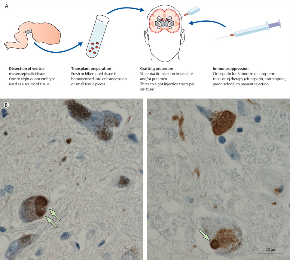
The spread of the condition is believed to be due to the protein alpha synclein being passed between cells in some manner. This idea stemmed from the analysis of the brains of people with Parkinson’s disease who received cell transplantation therapy in the 1980-90’s. After those people passed away (due to natural causes), their brains were analysed and it was discovered that some of the cells in the transplants (1-5%) have Lewy bodies in them (Lewy bodies are one of the hallmarks of Parkinson’s disease, dense circular clusters of proteins including alpha synuclein). This suggests that the disease is passed on to the healthy transplanted cells in some way.

gr3

Photos of neurons from the post-mortem brains of people with Parkinson’s that received transplants. White arrows in the images above indicate lewy bodies inside transplanted cells. Source: The Lancet

So the research community has been working with the idea of an ‘ascending’ theory of Parkinson’s disease, and the spreading of the condition via the passing of alpha synuclein from cell to cell. And this theory has been fine,…

Why do I feel like there’s a ‘but’ coming?

Because there is a ‘but’ coming.

And it’s a big BUT.

But as Prof Isacson and Dr Engelender point out there are some holes in this theory.

Some big holes.

For example, in a 2008 study of 71 postmortem brains from people with Parkinson’s disease, 47% of the cases did not fit the predicted ‘Braak theory’ spread of alpha synuclein, and 7% of those cases did not have any cell loss in the dorsal motor nucleus (one of the first sites of damage in the Braak theory – Click here to read more).

Ok, so the theory is not perfect…what are Prof Isacson and Dr Engelender proposing instead?

They suggest that alpha synuclein accumulation starts at about the same time in nerve cells throughout the body, but the different groups of nerve cell differ in how much toxicity they can handle.

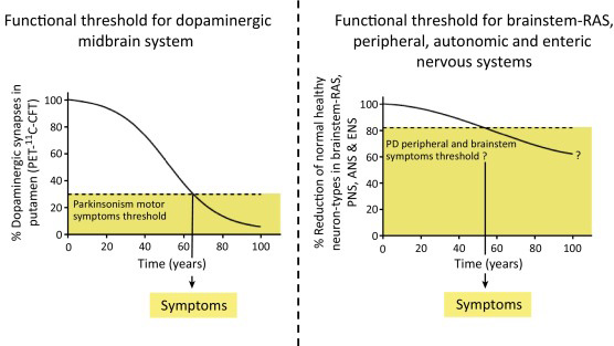
Some of these groups of cells can handle a lot (and more than half of the cells need to be lost before clinical features begin to appear), while others have a lower ‘threshold’ (only a few cells need to die before symptoms appear).

Prof Isacson and Dr Engelender argue that the nerve cells around the gut, for example, have a lower reserve (or total number), and, therefore, symptoms related to the gut become more obvious sooner as those cells die off or become less efficient. This lower threshold is in contrast to the more well known cell loss of the dopamine producing neurons in the midbrain, where approximately 50-70 percent of the dopamine neurons disappear before the classical motor features of Parkinson’s start to appear. Their theory suggests that this part of the brain has a larger reserve, and thus higher threshold.

Hence the reason why this is being called the ‘threshold theory’.

1-s2.0-S016622361630145X-gr1

Some groups of cells may have a higher threshold in Parkinson’s disease. Source: Cell

Some cells may have a low threshold and only require a few cells to be lost before the clinical features associated with those cells begin to appear. These symptoms would obviously appear earlier than those features associated with a high threshold population of cells, which required substantial loss before symptoms appear.

This idea would explain differing results seen in research findings regarding, for example, vagotomies (the cutting of the vagus nerve to the gut – click here to read more about this). This new theory would suggest that the procedure might not have any impact at all on lowering the risk of Parkinson’s disease.

Both scientists insist that searching for treatments that slow or block the aggregation of alpha synuclein is still necessary.

“Instead of studying how proteins move from one neuron to another and searching for compounds that prevent the ‘spread’ of aggregated alpha-synuclein, we need to study why alpha-synuclein accumulates within neurons and how these neurons die in the disease, and search for compounds that prevent the general neuronal dysfunction,” – Dr Engelender

(Source: Science Daily)

So are there any problems with this new theory?

The new theory is a very interesting idea and deserves consideration. It solves some of the problems with the “ascending theory” discussed further above. But it also faces some of the same problems that the ascending theory has to deal with.

For example, in one large autopsy study which investigated 904 brains, the investigators blindly collected all of the brains that had alpha synuclein present in the groups of neurons that are affected in Parkinson’s disease (eg. the dorsal motor nucleus of vagus, substantia nigra, and/or basal forebrain nuclei.). They found that alpha synuclein was observed in 11.3% (or 106 cases). But when the researchers then looked at the clinical notes associated with those cases, only 32 (30%) had been diagnosed with a neurodegenerative disorder. The rest had demonstrated no clinical features.

Another study found that 8.3% of the aged control brains had alpha synuclein present in them. In addition, the presence of alpha synuclein is not specific to Parkinson’s disease – approximately 50% of people who die with Alzheimer’s disease have been found to have Lewy bodies. These results suggest that alpha synuclein aggregation can be present in both healthy and diseased brains. But if this is so, what role is alpha synuclein playing in Parkinson’s disease?

(You see the sort of problems we are dealing with in research when trying to come up with a theory of how something complicated is actually working?)

What does it all mean?

The central job of a scientist is to test hypotheses.

A hypothesis is a true or false statement (for example, hypothesis: the sun will come up tomorrow – easy to test as the sun either will or won’t come up; the statement is either true or false). In building one hypothesis on top of another hypothesis, we develop theories about how the world around us works.

Sometimes our hypotheses can unwittingly take us in a particular direction, depending on different variables. The danger in this process (one which must be met with discipline and control procedures) is that one can start to look for results that support a hypothesis or theory. It is a very human characteristic to become blind to any evidence to the contrary.

A new theory of Parkinson’s disease has been proposed. It suggests that rather than the condition starting in one location and progressively moving higher into the brain, Parkinson’s disease may actually start everywhere and it is the varying levels of tolerance between different types of cells that determines which cells die first.

It is certainly a new take of the available evidence and the research community is considering it. It will be interesting to see what kind of feedback results from this article, and we will post updates on that feedback as they become available.


The banner for today’s post was sourced from Sott

A yeast model of Parkinson’s disease

yeast-cell

When I say the word ‘yeast’, you might think of making bread or beer.

One does not automatically think of Parkinson’s disease.

But yeast has actually been incredibly useful in enhancing our understanding of the genetics of Parkinson’s disease, and may well now provide us with novel treatments for the condition. In today’s post we will discuss how yeast research is leading the way for Parkinson’s disease.


bn-qp674_obit_l_gr_20161103124141

Prof Susan Lindquist. Source: WallStreetJournal

It was with sadness that we heard about the passing of Prof Susan Lindquist in October last year. She was truly a pioneer in the field of molecular biology. In addition to advancing our understanding of gene functioning in degenerative diseases like Parkinson’s disease and Alzheimer’s, she also started a company, Yumanity, which is currently testing new drugs to tackle these conditions. Hopefully her legacy will have enormous impact for the millions of people around the world struggling with these conditions.

And that legacy all started with a bold (some even called it ‘crazy’) idea.

It involved yeast.

What is yeast?

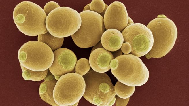
Quite possibly the earliest domesticated species, yeast is a single-celled microorganism, traditionally classified as a member of the fungus kingdom. The evolutionary lineage of yeast dates back hundreds of millions of years old and there are at least 1500 species of yeast (Source: Wikipedia).

cell

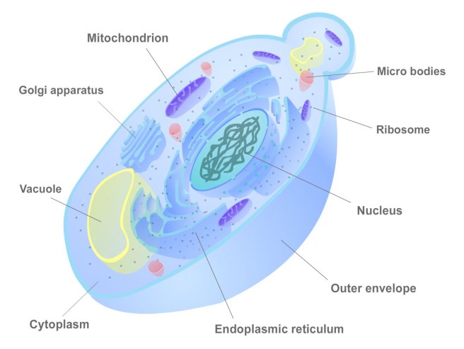
The cellular structure of yeast. Source: Biocourseware

More importantly, yeast is one of the most centrally important model organisms used in modern biological research, representing one of the most thoroughly researched organisms in the world.

We know more about the biology of yeast than we do about ourselves!

And this statement is made further evident as researchers use yeast to produce the world’s first synthetic organism (an organism for which the genome has been designed or engineered). By the end of 2017, the Synthetic Yeast 2.0 consortium plans to have produced a new form of yeast in which all 16 chromosomes will have been made in the lab (for more on this, read this STAT article).

Why do scientists like studying yeast?

The main reason is that yeast cells are very similar to human cells, but they grow a lot faster (human cells on average divide a rate of about once every 12 hours, while yeast cells divide every two hours). Yeast is similar to human cells in that it has all of the eukaryote structures, including a nucleus, cytoplasm, and mitochondria (eukaryote meaning a cell with a nucleus).

Yeast has played a fundamental role in many major scientific discoveries since the early 1900s. In 1907, German scientist Edward Buchner won the Nobel Prize in Chemistry for research involving yeast extract and fermentation. Ninety one years later (2006), Roger D. Kornberg won the same prize for his work on DNA transcription using yeast.

Yeast was the first eukaryote to have its genome (DNA) fully sequenced (in 1996). Yeast is literally leading the way in biological research.

So what has this got to do with Parkinson’s disease?

This is where Prof Susan Lindquist comes into the story.

maxresdefault

Source: Youtube

In the early 2000s, she suggested the idea of using yeast to look at neurodegenerative conditions like Parkinson’s disease. It was a wild concept. Talking to the New York Times in 2007, she said “Even people in my laboratory thought we were crazy to try to study neurodegenerative diseases with a yeast cell. It’s not a neuron”.

But they persevered and in 2003 they published this research report in the journal Science:

yeast

Title: Yeast genes that enhance the toxicity of a mutant huntingtin fragment or alpha-synuclein.
Authors: Willingham S, Outeiro TF, DeVit MJ, Lindquist SL, Muchowski PJ.
Journal: Science. 2003; 302(5651):1769-72.
PMID: 14657499

In this study, the researchers conducted a genome-wide screens of mutant genes in yeast to identify genes that enhanced the toxic effects of the mutant huntingtin gene or alpha-synuclein protein. That is to say, they randomly mutated (made un-operational) just one gene per yeast cell, and these yeast cells either had the mutant huntingtin gene (which causes the neurodegenerative condition of Huntington’s disease) or too much alpha synuclein (which causes protein clumping in the yeast cells). Using this approach, they could determine which genes were responsible for increasing the negative effects of the mutant huntington gene or the alpha synuclein protein.

Of the 4850 yeast genes that the researcher mutated (and can we just point out that that is A LOT of work!!!), 52 were identified that exaggerated the affect of the mutant huntingtin gene and 86 increased sensitivity to alpha-synuclein (curiously, only one mutant gene resulted in increased sensitivity to both).

When they looked at the known functions of 86 genes that were increasing the sensitivity to alpha synuclein, the researchers found that most of them were involved in the processes of lipid metabolism (the synthesis and degradation of lipids) and vesicle-mediated transport (this occurs at the tips of neural branches – where alpha synuclein is located). Alpha synuclein is known to be involved with lipid metabolism (Click here and here for more on this). This reenforced the belief with the researchers that yeast could be used to assess disease relevant pathways – this study gave them the ‘proof of concept’.

In addition, the majority of the genes had human ‘orthologs’ (genes in different species that evolved from a common ancestral gene), meaning that the findings of yeast studies could potentially be translated to higher order creatures, like humans. The researchers concluded that they had found cell autonomous genes that are relevant to Parkinson’s disease.

With the publication of this work, people in Prof Lindquist’s lab were probably thinking the idea wasn’t so crazy anymore.

And this first publication led to many more, such as this report which was published in the same journal in 2006:

Linds
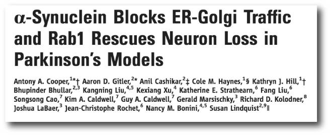
Title: Alpha-synuclein blocks ER-Golgi traffic and Rab1 rescues neuron loss in Parkinson’s models.
Authors: Cooper AA, Gitler AD, Cashikar A, Haynes CM, Hill KJ, Bhullar B, Liu K, Xu K, Strathearn KE, Liu F, Cao S, Caldwell KA, Caldwell GA, Marsischky G, Kolodner RD, Labaer J, Rochet JC, Bonini NM, Lindquist S.
Journal: Science. 2006 Jul 21;313(5785):324-8.
PMID: 16794039      (This article is OPEN ACCESS if you would like to read it)

In this study, the researchers doubled the amount of alpha synuclein that yeast cells produce and they observed that the cell stopped growing and started to die. They then looked at the earliest cellular events following theover production of alpha synuclein and they noticed that there was a blockage in the endoplasmic reticulum (ER)-to-Golgi vesicular trafficking.

Yes, I know what you are going to ask: What is ER-to-Golgi vesicular trafficking?

The endoplasmic reticulum (or ER) is a network of tubules connected to the nucleus. It is involved in the production of proteins and lipids, which are then transported to the Golgi apparatus which then modifies them, sorts them and and packs them into small bags called vesicles. These vesicles can then be taken to the cell membrane where the proteins are released to do their jobs.

ergolgi

The ER to Golgi pathway. Source: Welkescience

Now the fact that too much alpha synuclein was blocking this pathway was interesting, but the researchers wanted to go further. They conducted another genome-wide screens of genes in yeast to identify genes that could rescue this blockage – BUT this time, instead of mutating genes, the researchers randomly over produced one protein (and there was 3000 of them!!!) in each cell. Because the overproduction of alpha synuclein kills the yeast cells, all the researchers had to do was wait until the end of the experiment and determine which protein was over produced in the surviving cells.

And this led them to a protein called RAB1.

RAB1 is a protein that is critical to the transportation of proteins in cells. The researchers next tested the ability of RAB1 to rescue dopamine cells in Drosophila (flies), Caenorhabditis elegans (microscopic worms), and cell culture models of Parkinson’s disease and they found that it was able to rescue the cells in all three cases. While this result was very interesting, it also provided full validation of the approach that Prof Lindquist and her colleagues were taking using yeast cells to find new therapies for neurodegenerative conditions, like Parkinson’s disease.

But Prof Lindquist and her colleagues didn’t stop there.

Over the next decade numerous research reports were published taking advantage of this approach, including these two reports which appeared back-to-back in the journal Science:

lindq1

Title: Identification and rescue of α-synuclein toxicity in Parkinson patient-derived neurons.
Authors: Chung CY, Khurana V, Auluck PK, Tardiff DF, Mazzulli JR, Soldner F, Baru V, Lou Y, Freyzon Y, Cho S, Mungenast AE, Muffat J, Mitalipova M, Pluth MD, Jui NT, Schüle B, Lippard SJ, Tsai LH, Krainc D, Buchwald SL, Jaenisch R, Lindquist S.
Journal: Science. 2013 Nov 22;342(6161):983-7.
PMID: 24158904

And:

lindq2

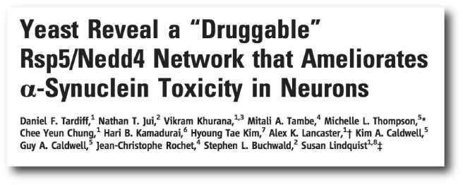
Title: Yeast reveal a “druggable” Rsp5/Nedd4 network that ameliorates α-synuclein toxicity in neurons.
Authors: Tardiff DF, Jui NT, Khurana V, Tambe MA, Thompson ML, Chung CY, Kamadurai HB, Kim HT, Lancaster AK, Caldwell KA, Caldwell GA, Rochet JC, Buchwald SL, Lindquist S.
Journal: Science. 2013 Nov 22;342(6161):979-83.
PMID: 24158909

In these reports, Prof Lindquist and colleagues tested whether some of the proteins that had come up in their various screens could actually have positive benefits in human cells, specifically induced pluripotent stem (iPS) cells (which we have discussed before – click here to read that post). The researchers grew brains cells (neurons and glial cells) from the iPS cells derived from people who suffered from Parkinson’s disease with dementia. These cells exhibited a number of features that indicated that they were not healthy. From their yeast screens, the researchers identified a protein called Nedd4 that reversed the pathologic features in these neurons.

Nedd4 is an E3 ubiquitin-protein ligase, which is a protein involved in the removal of old or damaged proteins from a cell. It is part of the garbage disposal process. Importantly, Nedd4 has been shown to label alpha synuclein for disposal (Click here to read more about this) and it is also present in Lewy Bodies – the circular clusters of proteins present in the brains of people with Parkinson’s disease. Importantly, it is a ‘druggable’ target. Nedd4 has been considered a therapeutic target for cancer (Click here to read more on this).

More recently (and following the passing of Prof Lindquist) the group has published two research reports back-to-back in the journal Cell Systems:

yeast-3

Title: Genome-Scale Networks Link Neurodegenerative Disease Genes to α-Synuclein through Specific Molecular Pathways.
Authors: Khurana V, Peng J, Chung CY, Auluck PK, Fanning S, Tardiff DF, Bartels T, Koeva M, Eichhorn SW, Benyamini H, Lou Y, Nutter-Upham A, Baru V, Freyzon Y, Tuncbag N, Costanzo M, San Luis BJ, Schöndorf DC, Barrasa MI, Ehsani S, Sanjana N, Zhong Q, Gasser T, Bartel DP, Vidal M, Deleidi M, Boone C, Fraenkel E, Berger B, Lindquist S.
Journal: Cell Syst. 2017 Jan 25. pii: S2405-4712(16)30445-8.
PMID: 28131822        (This article is OPEN ACCESS if you would like to read it)

And:

chung2

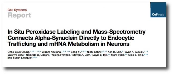
Title: In Situ Peroxidase Labeling and Mass-Spectrometry Connects Alpha-Synuclein Directly to Endocytic Trafficking and mRNA Metabolism in Neurons.
Authors: Chung CY, Khurana V, Yi S, Sahni N, Loh KH, Auluck PK, Baru V, Udeshi ND, Freyzon Y, Carr SA, Hill DE, Vidal M, Ting AY, Lindquist S.
Journal: Cell Syst. 2017 Jan 25. pii: S2405-4712(17)30002-9.
PMID: 28131823

In these reports, Prof Lindquist and colleagues systematically mapped out molecular pathways underlying the toxic effects of alpha-synuclein. They applied their yeast derived 332 genes that impact alpha-synuclein toxicity, and linked them to multiple Parkinson’s-associated genes and druggable targets, using software called TransposeNet.

1-s2.0-S2405471216304458-fx1

Source: Cell

The investigators then validated some of the connections in human iPS cells derived from people with Parkinson’s disease, confirming that some of the Parkinson’s disease-related genetic interactions observed in yeast are ‘conserved’ (that is maintained across evolution) to humans. And these findings fully vindicated Prof Lindquist’s ‘crazy’ idea of using yeast cells to investigate neurodegenerative disease.

In the second report, the researchers identified 225 proteins in close physical proximity to alpha-synuclein in neurons using a new technique (called ascorbate peroxidase (APEX) labeling – let’s just say it’s complicated, but if you’d like to read more about it, Click here). Many of those 225 proteins were well known to the researchers being involved with activities in vesicles and synaptic terminals, where alpha synuclein is often found. But the researchers also found microtubule-associated proteins (including tau) rubbing shoulders with alpha-synuclein, as well as proteins involved with mRNA binding, processing, and translation (which they were not expecting). Thus, not only has Prof Lindquist’s yeast model of Parkinson’s disease provided us with novel therapeutic targets, but also opened new avenues of research related to alpha synuclein functioning.


And there is now a ‘yeast’ biotech company?

It is called Yumanity.

yumanity-screenshot2

Yumanity. Source: ScientificAmerican

Started in December 2014, with $45 million in funding, Yumanity is focused on determining new targets for neurodegenerative conditions in yeast cells, testing those new targets in human cells, and then moving towards clinical trials with the best candidates. To date, they have not announced any clinical trial candidates, but they working on compounds that are targeting the NEDD4 pathway in Parkinson disease (discussed above). We will be watching this company with great interest.

So what does it all mean?

Back in the early 2000s, Prof Lindquist and her team asked a simple but strange question:

Can we use our knowledge of yeast genetics to study neurodegeneration?

We now know that the answer is ‘yes’. The small single cell organism that most of us associate with baking and beer, shares enough genetics with us that we can use it as an assay for investigating molecular pathways involved with diseases of the brain. And in the not so distant future, this simple little organism may be providing us with new treatments and therapies for those diseases.

As we suggested at the start of this post, Prof Lindquist has left an amazing legacy. If Yumanity can move a new drug into clinical trials for Parkinson’s disease, it will only further strengthen that legacy.

Susan Lee Lindquist – June 5, 1949 to October 27, 2016


The banner for today’s post was sourced from NewEuropeans

The Journal of Parkinson’s disease – special issue

JPD_logo_editors

Our policy at the SoPD is not to advertise or endorse commercial products or services. This is to avoid any ethical or conflict of interest situations.

Every now and then, however, we see something that we believe will be of interest and value to the Parkinson’s community…aaand we bend our policy rule book.


Today the Journal of Parkinson’s disease released a “200 years of Parkinson’s disease” OPEN ACCESS special issue of their journal which highlights some of the major discoveries in the field of Parkinson’s disease research.

Critically, the articles provide insights into how the discoveries were made, and they are written by some of the biggest names in the Parkinson’s research community (many of whom were actually there when the discoveries were made).

The issue has articles dealing with topics including:

Click here to see all of the articles in this special issue.

We fully recommend readers take advantage of this OPEN ACCESS issue and learn about how some of these great discoveries were made.

Happy reading.


Full disclosure: The Journal of Parkinson’s disease is a product of IOS Press. The SoPD has not been approached by or made any offers to IOS Press or anyone at the Journal of Parkinson’s disease. We merely thought that the material in this particular OPEN ACCESS issue would be of interest to our readers.


The banner for today’s post was sourced from the Journal of Parkinson’s disease

The red headed mice of Boston

redhair

Recently scientists have found a possible link in the curious relationship of red hair, melanoma and Parkinson’s disease.

It involves red headed mice (not a typo – you read that correctly).

In today’s post we will discuss the new research and explain what it means for Parkinson’s disease.


ginger

Red or ginger hair. Source: theLocal

We have previously discussed the curious association between red hair and Parkinson’s disease (Click here for that post).

We have also previously discussed the curious association between melanoma and Parkinson’s disease (Click here for that post).

Melanoma

Melanoma. Source: Wikipedia

Basically, people with red hair are more vulnerable to Parkinson’s disease that dark haired people, and people with a history of melanoma (skin cancer) are more likely to develop Parkinson’s disease than people with no history.

And given that people with red hair are generally more vulnerable to melanoma that dark haired people, you can understand why scientists have recently been very interested in this curious triangle of seemingly unrelated biological features.

Recently, however, scientists in Boston (USA) have provided evidence that the genetic mutation which causes red hair and increases the risk of melanoma, might also make the brain more vulnerable to Parkinson’s disease.

Red hair is caused by a genetic mutation?

Before we answer this question: the word ‘mutation’ carries a negative connotation thanks to it’s use in popular media and films. In biology, researchers prefer to use the word genetic ‘variation’. And EVERYONE has variations. They are what makes each of us unique. A father will pass on many of his own genetic variations to his son, but there will also be 50-100 spontaneous variations. And this is how, red hair can sometimes pop up in a family with little history of it.

Ok, so red hair is caused by a genetic variation?

Yes.

Red hair, which occurs naturally in 1–2% of the general population (though there are some regional/geographical variation), results from one of several genetic variations. Approximately 80% of people with red hair have a variation in a gene called melanocortin-1 receptor (or MC1R). Another gene associated with red hair is called HCL2 – ‘Hair colour 2’.

So what did the researchers find?

red

Title: The melanoma-linked “redhead” MC1R influences dopaminergic neuron survival.
Authors: Chen X, Chen H, Cai W, Maguire M, Ya B, Zuo F, Logan R, Li H, Robinson K, Vanderburg CR, Yu Y, Wang Y, Fisher DE, Schwarzschild MA.
Journal: Ann Neurol. 2016 Dec 26. doi: 10.1002/ana.24852. [Epub ahead of print]
PMID: 28019657

In their study, the researchers have investigated mice that carry a mutation of the MC1R gene (thus inactivating the gene – and yes, these mice have red/ginger fur!). They noticed that the mice displayed a progressive decline in their locomotor activity, moving around significantly less than non-red furred control mice at 8 months of age. The MC1R mutant mice also displayed a reduction in the number of dopamine producing neurons in the brain, when compared to the non-red furred controls (dopamine a chemical in the brain that helps to regulate movement).

The MC1R mutant mice were more vulnerable to toxin induced models of Parkinson’s disease (both 6OHDA and MPTP), but (most interestingly) when the researchers used a substance that binds to MC1R and initiates a response (an MC1R agonist called BMS-470539) they found that this treatment improved the survival of the dopamine producing cells in the brain.

The researchers are now seeking to further understand how the loss of MC1R renders the dopamine cells more vulnerable, and follow up the finding that MC1R agonists are neuroprotective.

Has there ever been any other evidence to suggest that MC1R is neuroprotective?

No. To our knowledge this is the first evidence that targeting MC1R could be a novel therapeutic strategy in a brain related condition (there has been some evidence of MC1R activation having beneficial effects in other parts of the body – click here for more on this).

And there are some indications as to how this positive effect could be working:

nurr1-2
Title: Melanocortin-1 receptor signaling markedly induces the expression of the NR4A nuclear receptor subgroup in melanocytic cells.
Authors: Smith AG, Luk N, Newton RA, Roberts DW, Sturm RA, Muscat GE.
Journal: J Biol Chem. 2008 May 2;283(18):12564-70.
PMID: 18292087

In this study, the researchers found that activating MC1R increases the levels of a protein called NR4A2 (or Nurr1). Nurr1 is a protein involved in the development and maintenance of dopamine producing neurons, and numerous recent studies have suggested that it is neuroprotective for these cells as well (Click here to read more on this).

So what does it all mean?

For some time there has been a curious link between people with red hair, melanoma and Parkinson’s disease. Now researchers in Boston have provided new evidence that the link exists, but they have also highlighted a new pathway via which novel therapies for Parkinson’s disease might be researched and developed.  Not a bad day at the office.


The banner for today’s post was sourced from Fancy mice

Phase II trial launched for Nilotinib

DSK_4634s

Big news today from Georgetown University with the announcement that they will be starting a phase II trial for the cancer drug Nilotinib.

Click here to read the press release.

In this post we will discuss what has happened thus far and what the new trial will involve.


gt

Georgetown University (Washington DC). Source: Wallpapercave

In October 2015, researchers from Georgetown University announced the results of a small clinical trial at the Society for Neuroscience conference in Chicago.

It is no understatement to say that the results of that study got the Parkinson’s community very excited.

The study (see the abstract here) was a small clinical trial (12 subjects; 6 month study) that was aiming to determine the safety and efficacy of a cancer drug, Nilotinib (Tasigna® by Novartis), in advanced Parkinson’s Disease and Lewy body dementia patients. In addition to checking the safety of the drug, the researchers also tested cognition, motor skills and non-motor function in these patients and found 10 of the 12 patients reported meaningful clinical improvements.

In their presentation at the conference in Chicago, the investigators reported that one individual who had been confined to a wheelchair was able to walk again; while three others who could not talk before the study began were able to hold conversations. They suggested that participants who were still in the early stages of the disease responded best, as did those who had been diagnosed with Lewy body dementia.

The study involved the cancer drug Nilotinib.

What is Nilotinib?

Nilotinib (pronounced ‘nil-ot-in-ib’ and also known by its brand name Tasigna) is a small-molecule tyrosine kinase inhibitor, that has been approved for the treatment of imatinib-resistant chronic myelogenous leukemia (CML). That is to say, it is a drug that can be used to treat a type of leukemia when the other drugs have failed. It was approved for this treating cancer by the FDA in 2007.

How does Nilotinib work?

The researchers behind the study suggest that Nilotinib works by turning on autophagy – the “garbage disposal machinery” inside each neuron. Autophagy is a process that clears waste and toxic proteins from inside cells, preventing them from accumulating and possibly causing the death of the cell.

Print

The process of autophagy. Source: Wormbook

Waste material inside a cell is collected in membranes that form sacs (called vesicles). These vesicles then bind to another sac (called a lysosome) which contains enzymes that will breakdown and degrade the waste material.

The investigators believe that nilotinib may be helping in Parkinson’s disease, by clearing away the waste building up in cells – allowing the remaining cells to function more efficiently.

This is great, so what happened in 2016?

That’s a great question.

First, the results of the study being published (Click here to read those results). Second, the U.S. Food and Drug Administration (FDA) reviewed Georgetown’s investigational new drug application (IND) for nilotinib in Parkinson’s disease, and they informed the Georgetown University investigators that a new clinical trial could proceed.

But after that, there were whispers of issues and problems behind the scenes.

Back in August we wrote a post about the Phase II trial being delayed due to disagreements about the design of the study (Read that post by clicking here). Two separate research groups emerged from those disagreements (Georgetown University researchers themselves and a consortium including the Michael J Fox Foundation). Click here for the STAT website article outlining the background of the issues, and click here for the Michael J Fox Foundation statement regarding the situation. The Georgetown University team have a lot of leverage in this situation as they control the patent side of things (Click here to see the patent).

We are not sure what has happened since August, but the Georgetown University team has now announced that they are going to go ahead with a phase II trial to look at safety and efficacy of nilotinib in Parkinson’s disease.

What do we know about the new trial?

At the moment the details are basic:

The design of the study involves two parts:

In the first part of the study, one third of the participants receiving a low dose (150mg) of nilotinib, another third receiving a higher dose (300mg) of nilotinib and the final third will receive a placebo drug (a drug that has no bioactive effect to act as a control against the other two groups). The outcomes will be assessed clinically at six and 12 months by investigators who are blind to the treatment of each subject. These results will be compared to clinical assessments made at the start of the trial. (We are not sure if brain imaging – for example, a DATscan – will be included in the assessment, but it would be useful)

In the second part of the study, there will be a one-year open-label extension trial, in which all participants will be randomized given either the low dose (150mg) or high dose (300mg) of nilotinib. This extension is planned to start upon the completion of the first part (the placebo-controlled trial) to evaluate nilotinib’s long-term effects. (We are a little confused by this study design with regards to efficacy, but determining the safety issues of using nilotinib long term is important to establish).

We are not clear on how many subjects will be involved in the study or what the criteria for eligibility will be. All we can suggest is that if you are interested in finding out more about this new study, you can sign up here to receive more information as it becomes available.

 – – – – – – – – – – – – – –

Summing up, this is welcomed news for the Parkinson’s community as we will finally be able to determine if nilotinib is having positive effects in Parkinson’s disease. There have been some concerns raised that the effects of the drug in the first clinical study may have been the result of removing additional Parkinsonian treatments during the study (Click here for more on this). This new study will hopefully help to clarify things.

And fingers crossed provide us with a useful new treatment for Parkinson’s disease.


The banner for today’s post was sourced from William-Jon

New kiwi research in Parkinson’s disease

0f6b3c3205fbdc30c9216c205d1bc039

I really didn’t expect to be writing about Parkinson’s research being conducted in New Zealand again so quickly, but yesterday a new study was published which has a few people excited.

It presents evidence of how the disease may be spreading… using cells collected from people with Parkinson’s disease.

In today’s post we will review the study and discuss what it means for Parkinson’s disease.


022217_ts_zealandia_main_free

The South Island of NZ from orbit. Source: Sciencenews

We may have mentioned the protein Alpha synuclein once or twice on this blog.

For anyone familiar with the biology of Parkinson’s disease, alpha synuclein is a major player. It is either public enermy no.1 in the underlying pathology of this condition or else it is the ultimate ‘fall guy’, left standing in the crime scene holding the bloody knife.

Remind me, what is alpha synuclein?

Alpha synuclein is an extremely abundant protein in our brains – making up about 1% of all the proteins floating around in each neuron (one of the main types of cell in the brain).

In healthy brain cells, normal alpha synuclein is typically found just inside the surface of the membrane surrounding the cell body and in the tips of the branches extending from the cell (in structures called presynaptic terminals which are critical to passing messages between neurons).

And why is alpha synuclein important in Parkinson’s disease?

Genetic mutations account for 10-20% of the cases in Parkinson’s disease.

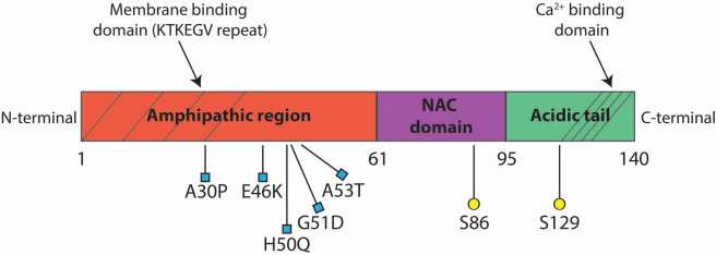
Five mutations in the alpha-synuclein gene have been identified which are associated with increased risk of Parkinson’s disease (A53T, A30P, E46K, H50Q, and G51D – these are coordinates for locations on the alpha synuclein gene). Rare duplication or triplication of the gene have also been associated with  Parkinson’s disease.

biomolecules-05-00865-g001

The structure of alpha synuclein protein – blue squares are mutations. Source: Mdpi

So genetically, alpha synuclein is associated with Parkinson’s disease. But it is also involved at the protein level.

In brains of many people with Parkinson’s disease, there are circular clumps of alpha synuclein (and other proteins) that collect inside cells. These clumps are called Lewy bodies. They are particularly abundant in areas of the brain that have suffered cell loss.

Fig2_v1c

A lewy body (brown with a black arrow) inside a cell. Source: Cure Dementia

No one has ever seen the process of Lewy body formation, so all we can do is speculate about how these aggregates develop. Currently there is a lot of evidence supporting the idea that alpha synuclein can be passed between cells. Once inside the new cell, the alpha synuclein helps to seed the formation of new Lewy bodies, and this is how the disease is believed to progress.

Mechanism of syunuclein propagation and fibrillization

The passing of alpha synuclein between brain cells. Source: Nature

Exactly how alpha synuclein is being passed between cells is the topic of much research at the moment. There are many theories and some results implicating methods such as direct penetration, or via a particular receptor. Perhaps even by a small package called an exosome being passed between cells (see image above).

How this occurs in the Parkinson’s disease brain, however, is unknown.

And this (almost) brings us to the kiwi scientists.

Last years, a group of Swiss scientists demonstrated that alpha synuclein could be passed between cells via ‘nanotubes’ – tiny tubes connecting between cells. The outlined their observations and results in this article:

switzerland
Title: Tunneling nanotubes spread fibrillar α-synuclein by intercellular trafficking of lysosomes.
Authors: Abounit S, Bousset L, Loria F, Zhu S, de Chaumont F, Pieri L, Olivo-Marin JC, Melki R, Zurzolo C.
Journal: EMBO J. 2016 Oct 4;35(19):2120-2138.
PMID: 27550960

The researchers who conducted this study were interested in tunneling nanotubes.

Yes, I know, ‘What are tunneling nanotubes?’

Tunneling nanotubes (also known as Membrane nanotubes or cytoneme are long protrusions extending from one cell membrane to another, allowing the two cells to share their contents. They can extend for long distances, sometimes over 100 μm – 0.1mm, but that’s a long way in the world of cells!

nanotubes

Tunneling nanotubes (arrows). Source: Wikipedia (and PLOSONE)

Previous studies had demonstrated that tunneling nanotubes can pass different infectious agents (HIV for example – click here to read more on this), supporting the idea that these structures could be a general conduit by certain diseases could be spreading.

nanotubes

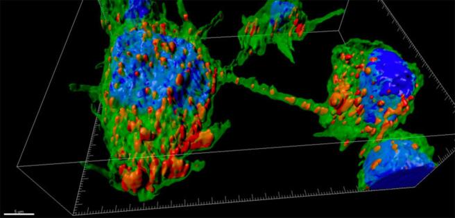
A tunneling nanotube between two cells. Source: Pasteur

In their study the Swiss researchers found that alpha synuclein could be transferred between brain cells (grown in culture) via tunneling nanotubes. In addition, following that process of transfer, the alpha synuclein was able to induce the aggregation (or clumping) of the alpha synuclein in recipient cells.

A particularly interesting finding was that alpha synuclein appeared to encourage the appearance of tunneling nanotubes (there were more tunneling nanotubes apparent when cells produced more alpha synuclein). And the alpha synuclein that was being transferred was being passed on in ‘lysosomal vesicles’ – these are the rubbish bags of the cell (lysosomal vesicles are used to take proteins away for degradation).

Paints a rather insidious picture of the ‘ultimate fall guy’ huh!

And that image was made worse by the results published by the kiwis last night:

maurice

Title: α-synuclein transfer through tunneling nanotubes occurs in SH-SY5Y cells and primary brain pericytes from Parkinson’s disease patients
Authors: Dieriks BV, Park TI, Fourie C, Faull RL, Dragunow M, Curtis MA.
Journal: Scientific Reports, 7, Article number: 42984
PMID: 28230073                    (This article is OPEN ACCESS if you would like to read it)

In their study, the New Zealand scientists extended the Swiss research by looking at cells collected from people with Parkinson’s disease. The researchers took human brain pericytes, which were derived from the postmortem brains of people who died with Parkinson’s disease.

And before you ask: pericytes are cells that wrap around the cells lining small blood vessels. They are important to the development of new blood vessels and maintaining the structural integrity of microvasculature.

aa-pericit4ub6b

A pericyte (blue) hugging a blood vessel (red). Source: Xvivo

Pericytes contain alpha synuclein precipitates like those seen in neurons, and the kiwi scientists demonstrated that pericytes too can transfer alpha synuclein via tunneling nanotubes to neighbouring cells – representing a non-neuronal method of transport.

They also found that the transfer through the tunneling nanotubes can be very rapid – within 30 seconds – and the transferred alpha synuclein can hang around for more than 72 hours, suggesting that it is difficult for the receiving cell to dispose of. The researchers did note that the transfer through tunneling nanotubes occurred only in small subset of cells, but that this could explain the slow progression of Parkinson’s disease over time.

What does it all mean?

In order for us to truly tackle Parkinson’s disease and bring it under control, we need to know how this slowly progressing neurodegenerative condition is spreading. Some researchers in New Zealand have provided evidence involving cells collected from people with Parkinson’s disease that indicates one method by which the disease could be passed from one cell to another.

Tiny tunnels between cells, allowing material to be shared, could explain how the disease slowly progresses. The scientists observed the Parkinson’s associated protein alpha synuclein being passed between cells and then hanging around for more than a few days.

This method of transfer was made more interesting because the New Zealand researchers reported that non-neuronal cells (Pericytes, collected from people with Parkinson’s disease) could also form tunneling nanotubes. This observation raises questions as to what role non-neuronal cells could be playing in Parkinson’s disease.

This line of questions will obviously be followed up in future research, as will efforts to determine if tunneling nanotubes are actually present in the human brain or simply biological oddities present only in the culture dish. Demonstrating nanotubes in the brain will be difficult, but it would provide us with solid evidence that this method of disease transfer could be a bonafide cause of disease spread.

We watch with interest for further work in this area.


FULL DISCLOSURE: The author of this blog is a kiwi… and proud of it. He is familiar with the researchers who have conducted this research, but has had no communication with them regarding the publishing of this post. He simply thought that the results of their study would be of interest to the Parkinson’s community.


The banner for today’s post was sourced from Pinterest

HIV and Parkinson’s disease

hiv-aids-definition2


I was recently made aware of an interesting fact:

Approximately 5% of people with Human immunodeficiency virus (HIV) infections develop Parkinson’s disease-like features.

Why is this?

In today’s post we will try to understand what is going on, and what it may mean for Parkinson’s disease.


hiv-budding-colo2r

HIV (in green) budding (being released) from a blood cell (lymphocyte). Source: Wikipedia

Ok, let’s start at the beginning:

What is HIV?

Human immunodeficiency virus (or HIV) – as the name suggests – is the virus.

It causes the infection which gives rise to Acquired Immune Deficiency Syndrome (or AIDS). AIDS is a progressive failure of the immune system – the body loses its ability to fight infections. Without treatment, average survival period after infection with HIV is between 9 – 12 years.

HIV can be spread by the transfer of bodily fluids, such as blood and semen. The World Health Organisation (WHO) has estimated that approximately 36.9 million people worldwide were living with HIV/AIDS at the end of 2014 (that is equivalent to the entire population of Canada!).

hi-virion-structure_en-svg

The structure of the HIV virus. Source: Wikipedia

Does HIV affect the brain?

Yes.

At postmortem examinations, less than 10% of the brains from HIV infected individuals are histologically normal (Source).

HIV is a member of the lentivirus family of viruses, which readily infect immune cells (such as blood cells). HIV can also infect other types of cells though, including those in the brain. HIV will usually enter the central nervous system within the first month following infection. It enters the brain via infected blood cells which come into contact with brain ‘immune system/helper’ cells such as microglia and macrophages at the blood-brain-barrier.

f1-large

How HIV enters the brain. Source: Disease Models and Mechanisms

HIV can also infect astrocytes (albeit at a lower frequency than microglia and macrophages), by direct cell-cell contact with infected T cells (blood cells) at the blood-brain-barrier (No. 1 in the image above). After infecting astrocytes, there is dysfunction in the astrocyte and it will no longer be so supportive to the local neurons (No. 2 in the image above). Once inside the brain, HIV-infected macrophages will allow for infection of other macrophages and microglia (No. 3 in the image above), and all together these HIV-infected astrocytes and microglia will cause damage to neurons by releasing viral proteins (two in particular, called Tat and gp120) and additional nasty chemicals which are bad for the neurons (No. 4 in the image above). Finally, as the disease progresses, the protective layer of the blood-brain-barrier becomes compromised and HIV-infected T cells eventually enter the brain and they cause damage to neurons by releasing pro-inflammatory chemicals (making the environment harsh for neurons).

There is remarkably little evidence of HIV actually infecting neurons (Click here for a review on this), so any cell loss in the brain that is associated with HIV does not result from neurons themselves being infected. This may be due to the fact that neurons do not have the HIV receptors (such as CD4) on their cell membrane. Similarly, oligodendrocytes (a supporting cell) does not appear to be easily infected by HIV. The bulk of the infected cells in the brain appear to be of the microglial, macrophage and astrocytes. And without these supporting cells doing their jobs in a normal fashion, it is easy to see how neurons can start dying off.

The severity, characteristics and distribution of HIV-induced injury in the brain varies greatly between affected individuals. It is most likely associated with the viral load (or the number of viral particles) in the brain, which can vary from a few thousand to more than a million copies per mL.

Do HIV-infected people show any signs of the virus entering the brain?

For the majority of people infected with HIV, this entry of the virus into the nervous system is neurologically asymptomatic (meaning they will not notice it), except for the occasional mild headache (for more on this read this review). As a result of the HIV virus entering the brain, however, many infected individuals will suffer from a specific set of neurological disorders, collectively called the AIDS dementia complex (ADC) (also known as HIV-associated cognitive/motor complex, or simply HIV dementia).

So how does HIV infection result in Parkinson’s disease-like features?

As we have suggested in the introduction to this post, on rare occasions (approximately 5% of cases), HIV-infected patients may present an illness virtually identical to Parkinson’s disease. More commonly, people with HIV will exhibit an increased sensitivity to dopamine receptor-blocking agents, such as drugs with a low potential for inducing Parkinsonism, (for example prochlorperazine and metoclopropamide).

The exact mechanism by which HIV infection results in Parkinson’s disease-like features is the subject of debate, but what is clear is that the basal ganglia (a structure involved in Parkinson’s disease) faces the brunt of the HIV infection in the brain. HIV-infected microglia and macrophage are most prominent in the basal ganglia when compared to other brain regions (Click here and here for more on this), and the basal ganglia is where the chemical dopamine from the midbrain is being released.

In addition, there are other changes in the brains of HIV infected people which may aid in the appearance of Parkinsonian features:


viraltitle

Title: Increased frequency of alpha-synuclein in the substantia nigra in human immunodeficiency virusinfection.
Authors: Khanlou N, Moore DJ, Chana G, Cherner M, Lazzaretto D, Dawes S, Grant I, Masliah E, Everall IP; HNRC Group.
Journal: J Neurovirol. 2009 Apr;15(2):131-8.
PMID: 19115126       (This article is OPEN ACCESS if you would like to read it)

The researchers in this study used staining techniques to look at the amount of alpha synuclein – the Parkinson’s associated protein – in slices of brain tissue taken from postmortem autopsies of 73 HIV+ individuals aged between 50 and 76 years of age.

The presence of alpha synuclein in the substantia nigra (an area of the brain affected by Parkinson’s disease) was a lot higher in the HIV+ brains when compared with healthy control samples (16% of the HIV+ brains had high levels of alpha synclein vs 0% for the healthy brains).

Interestingly, nearly all of the brains analysed (35 out of 36 HIV+ brains) had high levels of the Alzheimer’s disease associated protein, beta amyloid (which again raises the question of whether beta amyloid could be playing a defensive role in infections – see our previous post on this). Also interesting, was that there was no correlation between these proteins being present and the age of the person at death – that is to say, older brains did not have more of these proteins when compared with younger brains.

There are also additional ways in which HIV could be causing Parkinson’s-like features, such as:

  • HIV has been shown to affect the protein levels of Parkinson’s disease associated proteins, such as DJ1 and Lrrk2 (Click here and here to read more on this).
  • HIV can, in some cases, increase the level of Dopamine transporter, which would reduce the levels of free floating dopamine in the brain (Click here to read more about this).

How is HIV treated?

aidspills

Treating HIV. Source: NPR

There is currently no cure for HIV infection.

There are, however, treatments which help to slow the virus down. These are called Anti-retroviral drugs (HIV is a retrovirus). There are different kinds of anti-retroviral drugs, which act at different stages of the HIV life cycle. Combinations of several anti-retroviral drugs (generally three or four) is known as ‘Highly Active Anti-Retroviral Therapy'(or HAART).

hiv-drug-classes-svg

Mechanism by which four classes of anti-retroviral drugs work against HIV. Source: Wikipedia

As the schematic image above highlights, there are many ways to slow down the HIV virus. For example, you can prevent it from attaching to a cell and fusing with the cell membrane (fusion inhibitors). By treating HIV infected people with multiple medications attacking different parts of the HIV life cycle, the virus has been slowed down.

Does HAART treatments for HIV help with these Parkinson’s-like features?

In some cases, the answer appears to be yes.

There are numerous case studies in the literature which demonstrate the alleviation of HIV-associated Parkinsonian symptoms with HAART, such as this report:

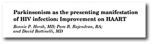
hersh

Title: Parkinsonism as the presenting manifestation of HIV infection: improvement on HAART.
Authors: Hersh BP, Rajendran PR, Battinelli D.
Journal: Neurology. 2001 Jan 23;56(2):278-9.
PMID: 11160977

In this study the researchers described the case of a 37 year old man who developed Parkinson’s like features in the setting of an HIV infection, which were resolved after 1 year of HAART.

Over a period of 4 months, the man developed co-ordination issue, clumsiness and an irregular tremor in his right hand (there was, however, no resting tremor). He noted a generalised slowness and exhibited a tendency towards decreased right arm swinging. He also developed dystonia in the right hand/arm. Following L-dopa treatment (25/100; one tablet 3x per day) there was improvement in balance & co-ordination, speech, facial expression, and the tremor (L-dopa does appear to improve most cases of HIV-associated Parkinson’s-like features).

Six months after first displaying these Parkinsonian features (and two month after initiating L-dopa treatment), the subject was placed on HAART treatment. Four months later, he discontinued L-dopa treatment and 12 months after starting the HAART regime his Parkinsonian features were largely resolved.

More case studies of HAART alleviating HIV-associated Parkinsonisms can be found by clicking here and here.

What does this mean for Parkinson’s disease?

This post was written for the research community rather than people with Parkinson’s disease. I thought the fact that some people with HIV can start to have Parkinson’s like features was an interesting curiosity and wanted to share/spread the information.

Having said that, this post raises some really interesting questions, such as if a virus like HIV can have this effect on the brain, could other viruses be having similar effects? Could some cases of Parkinson’s disease simply be the result of a viral infection? Either multiple hits from a particular virus or different viruses each taking a varying toll over the course of a life time.

This idea would explain many of the curious features of Parkinson’s disease, such as:

  • the asymmetry of the symptoms (people with Parkinson’s usually have the disease starting on one side of the body.
  • the fact that some cells in the brain are more vulnerable to the disease than others (perhaps they are more receptive to a particular virus).
  • the protein clusterings in the cells (Lewy bodies may be defensive efforts against viral infections).

As we have previous mentioned, theories of viral causes for Parkinson’s have been circulating ever since the 1918 flu pandemic (Click here to read our previous post on this topic). About the same time as the influenza virus was causing havoc around the world, another condition began to appear called ‘encephalitis lethargica‘. This disease left many of the victims in a statue-like condition, both motionless and speechless – similar to Parkinson’s disease. Initially, it was assumed that the influenza virus was the causal factor, but more recent research has left us not so sure anymore.

The point is, however, perhaps it is time for us to re-examine the possibility of a viral agent being involved in the development of Parkinson’s disease.

There is new technology that allows us to determine the viral history of each individual from a simple blood test (Click here for more on this), so it would be interesting to compare blood samples from people with Parkinson’s disease with healthy controls to determine any differences.

In addition to the overall question of a viral role in Parkinson’s disease, there also remains the question of why only a small fraction of people with HIV are affected by Parkinsonisms. It could be interesting to genetically screen those people with HIV that exhibit Parkinsonisms and compare them with people with HIV that do not. Do those affected individuals have recognised Parkinson’s related genetic mutations? Or do they have novel genetic variations that could tell us more about Parkinson’s disease?

Food for thought. Would be happy to hear others thoughts.


The banner for today’s post was sourced from AidsServices

PARIS is always a good idea

bzn8qhy

Audrey Hepburn was taking about the city when she uttered the words that title this post, but today we will be talking about the protein that bears the same name: PARIS.

Last week new research was published which demonstrated that in the absence of Parkin and Pink1 protein, the protein PARIS builds up and becomes toxic for cells.

Today’s post will review that research and we’ll discuss what it all means for Parkinson’s disease.


paris

No label required. A magnificent city. Source: HathawaysofHaworth

Today’s post has nothing to do with the city of Paris, but it is always nice to have photos of this European capital gracing the page.

We have recently discussed the Parkinson’s associated proteins Pink1 and Parkin (click here for that post). Today we will be revisiting these proteins as we discuss another protein that they interact with: PARIS (specifically PARIS1).

What is PARIS?

PARIS (aka TBC1D2 or TBC1 Domain Family Member 2) is a GTPase-activating protein.

What does that mean?

Getting a signal from outside of a cell into the interior is a complicated affair. There are numerous ways to do it, but one of the most common involves ‘G-proteins‘. These are involved with transmitting a signal from the outside of a cell into the interior, and when inside the cell G-proteins act as molecular switches.

G-proteins are located inside the cell membrane and are activated by G-protein-coupled receptors. When a signaling molecule binds to the G-protein-coupled receptor on the outside of the cell membrane, the portion of the receptor inside the cell activates the G-protein which then starts of a chain of events resulting in the signal being passed on.

11_07bgprotcoupledrec-l

Source: Bio1151

The role of GTPase-Activating Proteins in this process is to turn the G protein’s activity off. In step 4 of the image above, a GTPase-Activating Protein (which is not shown) binds to the G-protein and terminate the activity of the signalling event – returning it to an inactive state.

Thus, GTPase-Activating Proteins – like PARIS – are important regulators of signalling inside the cell.

What do we know about PARIS1 in Parkinson’s disease?

So a few years ago, a group of researchers led by Prof Ted Dawson at John Hopkins School of Medicine published this study:

cell

Title: PARIS (ZNF746) repression of PGC-1α contributes to neurodegeneration in Parkinson’s disease.
Authors: Shin JH, Ko HS, Kang H, Lee Y, Lee YI, Pletinkova O, Troconso JC, Dawson VL, Dawson TM.
Journal: Cell. 2011 Mar 4;144(5):689-702.
PMID: 21376232        (This article is OPEN ACCESS if you would like to read it)

In this study, the researchers noticed that the protein PARIS was accumulating in cells that did not have the Parkinson’s associated protein, Parkin. In those cells, the Parkin gene was mutated so that the Parkin protein was not produced properly. The researchers discovered that Parkin was important for labelling old PARIS protein for disposal. Thus in the absence of Parkin, PARIS protein would not be disposed of and simply piled up.

This build up of PARIS resulted in the loss of dopamine neurons in mice that did not produce Parkin. When the researchers re-introduced normal Parkin protein, the researchers were able to rescue the cell loss. Interestingly, the researchers also found that over production of PARIS in normal mice resulted in cell loss which could be rescued by a similar over production of Parkin.

When they looked in postmortem human brains, the researchers found that levels of PARIS protein were more than two times higher in regions affected by Parkinson’s disease (the striatum and the substantia nigra) of people with sporadic Parkinson’s disease when compared to healthy controls. Interestingly, this increase was only seen with PARIS protein, and not PARIS RNA (where the scientists saw no different with control samples), suggesting a build up of PARIS protein in the Parkinsonian brain.

The investigators concluded that this meant PARIS was could be playing a role in the cell loss associated with Parkinson’s disease.

They followed up this research a few years later with this publication:

parkins

Title: Parkin loss leads to PARIS-dependent declines in mitochondrial mass and respiration.
Authors: Stevens DA, Lee Y, Kang HC, Lee BD, Lee YI, Bower A, Jiang H, Kang SU, Andrabi SA, Dawson VL, Shin JH, Dawson TM.
Journal: Proc Natl Acad Sci U S A. 2015 Sep 15;112(37):11696-701.
PMID: 26324925     (This article is OPEN ACCESS if you would like to read it)

In this study, the same researchers found that when they remove the Parkin protein from the brains of adult mice there would be a decrease in the size and number of mitochondria. We have previous discussed mitochondria – the power stations of the cell – and their loss is bad news for a cell (click here to read more on mitochondria).

The researchers next demonstrated that this loss of mitochondria could reversed by removing PARIS protein from the Parkin mutant mice, and this prevented the loss of dopamine neurons. They also showed that the loss of mitochondria (and loss of dopamine neurons) could be caused by over production of PARIS in normal mice.

These results pointed towards an important role for both Parkin and PARIS in the maintenance of healthy mitochondria.

So what new research has been published about PARIS1?

This study was published last week:

dawson

Title: PINK1 Primes Parkin-Mediated Ubiquitination of PARIS in Dopaminergic Neuronal Survival.
Authors: Lee Y, Stevens DA, Kang SU, Jiang H, Lee YI, Ko HS, Scarffe LA, Umanah GE, Kang H, Ham S, Kam TI, Allen K, Brahmachari S, Kim JW, Neifert S, Yun SP, Fiesel FC, Springer W, Dawson VL, Shin JH, Dawson TM.
Journal: Cell Rep. 2017 Jan 24;18(4):918-932.
PMID: 28122242       (This article is OPEN ACCESS if you would like to read it)

In their study the researchers found that Parkin is not the only Parkinson’s associated protein in the PARIS story.

We have previously talked about the protein Pink1 (click here to read more on) – and yes, you would be forgiven if you start to think that all Parkinson’s related proteins start with the latter ‘P’. Pink1 grabs Parkin and causes it to bind to dysfunctional mitochondria. Parkin then signals to the rest of the cell for that particular mitochondria to be disposed of. In this study, the researchers found that Pink1 also grabs PARIS and signals for Parkin to dispose of it. In the absence of Pink1, normal Parkin protein does not label old PARIS protein for disposal and PARIS starts to pile up.

The researchers then began manipulating the levels of Pink in the brains of mice and they observed PARIS-dependent cell loss – that is to say, in the absence of Pink1, cells died only when PARIS was present.

These findings suggest that therapies targeting PARIS could be used in people with Parkinson’s disease who are carrying either a Parkin or a Pink1 mutation (both very common in early onset Parkinson’s disease).

What does it all mean?

People with early onset Parkinson’s disease quite often have a genetic mutation in one of a small number of genes – Pink1 and Parkin being prominent amongst these genes. The researchers who conducted the study that we have reviewed today have identified a common mechanism by which both of these proteins could be acting in their roles in Parkinson’s disease: a protein called PARIS.

Currently there is no treatment (that we are aware of) that targets the PARIS protein – nothing in the clinic nor being experimentally tested. Obviously, however, PARIS represents a VERY interesting protein for further investigations. The Dawson lab has several patents on PARIS (Click here and here for more on these), so evidently people will be working on drug candidates that inhibit PARIS.

There is a naturally occurring inhibitor, a micro RNA cluster miR-17-92 (also known as oncomir-1), which reduces the production of PARIS protein by blocking PARIS RNA (Click here for more on this). Using this micro RNA to target PARIS will be very difficult (both activating/delivering the micro RNA and unknown off target effects).

We are assuming that Prof Dawson and colleagues are rapidly screening compounds to determine which can block or inhibit PARIS activity and we will eagerly wait to see the results of that work.

Watch this space.


The banner for today’s post was sourced from Wallpapercave


EDITORIAL NOTE: Yay, 100 posts!

A smartphone application for Parkinson’s disease

umotif

Here at the SoPD, we like our gadgets and new technology.

And we believe that there is enormous potential for people with Parkinson’s disease to benefit hugely even from some of the small technological advances that seem to be occurring on a day basis.

Today’s post will review a recent study that looked at tested the benefits of a smartphone application for people with Parkinson’s disease.


selfcare-infographic-english

A schematic illustrating the limited monitoring of Parkinson’s. Source: Riggare

On her great blog, Swedish engineer and ‘proud mother’ Sara Riggare posted the image above to illustrate the ridiculous current situation regarding the standard monitoring of Parkinson’s disease.

As the schematic perfectly illustrates, in 2014 Sara spent 8,765 hours conducting ‘self care’. That is, she was applying her own knowledge and experience to managing her Parkinson’s disease. For just one hour in that year was her Parkinson’s actually being monitored by a medical clinician (8,766 being the number of hours in a year).

This is actually a very serious problem – for not only the Parkinson’s community – but anyone suffering from a long-term medical condition. How are they to gage their current situation on a day to day basis when they have such infrequent visits to their medical specialist?

And this is where technology can help.

But, before we begin:

FULL DISCLOSURE NO.1: the author of this blog is an author in the study that will be discussed (#ThisIsNotShamelessSelfPromotion).

FULL DISCLOSURE NO.2: We here at the SoPD are in no way benefitting from mentioning the study here. The company behind the product, umotif, has not asked us nor been contacted by us regarding this post (in fact, they are completely unaware that we are posting this). We are writing this post simply because we thought that it would be of interest to the wider Parkinson’s community. And yes, as other technology comes to along, we will bring it to you attention by posting about it here.

With all of that out of the way, the study is AMAZING!

smart

Title: Using a smartphone based self-management platform to support medication adherence and clinical consultation in Parkinson’s disease: Results from the SMART-PD Randomised Controlled Trial v4.
Authors: Lakshminarayana R, Wang D, Burn D, Barker RA, Chaudhuri KR, Galtrey C, van Guzman N, Hellman B, Pal S, Stamford J, Steiger M, Stott SRW, Teo J, Barker RA, Wang E, Bloem BR, van der Eijk M, Rochester L, & Williams A
Journal: NPJ Parkinson’s disease (2017), 3, 2.
PMID: N/A          (This article is OPEN ACCESS if you would like to read it)

The company behind the application approached various clinical research groups around the UK and proposed to run a study of their new product. The software had many features (including information about medication, a reminder alarm for when medication should be taken, tests/games, and links to other resources online).

41531_2016_3_fig4_html

A programmable reminder system for medication. Source: Nature

The primary focus of the software, however, was a flower-petal shaped motif that the participants could manipulate to indicate how they were feeling.

umotif-app-screenshot-2

The umotif flower motif. Source: SalusDigital

Participants could drag each coloured petal in or out to indicate how they were feeling at a particular moment in time. The smaller the petal, the more lower the score. And each petal represented different aspects of daily life, for example the moon and stars (dark blue) petal allowed an indication of how one slept.

Each time the participant indicated their current status on the flower motif, the information was recorded and could be tracked over days, weeks, and months. This level of information allowed people to begin to see patterns in their own behaviour over time, with some people getting poorer sleep during the middle of the month for example. And different variables could be compared (such as sleep score with exercise score), providing users with a more dynamic idea of their situation over time.

41531_2016_3_fig5_html-1

Comparing scores between measures over time. Source: Nature

A total of 215 people with Parkinson’s disease were randomly assigned to either receive the application (106 subjects) or not (acting as a control subject; 109 subjects). Both groups were contacted by the investigators fours times during the 16 week trial and feedback was provided in addition to any changes in their treatment regimes.

72% of participants application group continued to use and engage with the application throughout the 16-week trial. By the end of the study, the application group demonstrated significantly improved adherence to their treatment regime when compared to the control group. Curiously, the application also significantly improved patients’ perception of quality of follow up consultations, demonstrating unexpected benefits.

And at the end of the study all of the control group participants in the study were allowed to begin using the application, while the application group continued to use it.

One interesting aspect of the study was the lack of interaction with technology by the target population. 180 people who were initially invited to take part in the study could not because they did not have smartphones (with iPhone/iPad or Android operating systems). So obviously there are opportunities for alternative approaches to this kind of tracking (other than a smartphone).

What does it all mean?

This smartphone application is a user friendly approach to tracking someone’s Parkinson’s over time, getting around the ‘lack of monitoring’ issue that concerns many in the community.

Umotif and Parkinson’s UK have kindly made this video about the study results so we might just sit back and let them explain what it all means and point out all of the benefits.

We are working on additional posts about wearable tech for Parkinson’s disease, which will be coming soon.

So stay tuned.


The banner for today’s post was sourced from ParkinsonsMovement

The Dogfish solution for Parkinson’s

Spiny dogfish 096

This week an interesting study was published in the scientific journal, Proceedings of the National Academy of SciencesIt involved our old friend, alpha synuclein – the aggregating protein that is associated with Parkinson’s disease – and the dogfish shark.

Not natural dance partners, I agree. But the findings of the study are very interesting.

In today’s post we will review the study and explain the connection between the protein and the shark.


spiny-dogfish-shark-622x363

Source: Discovery

Some people call them spiny dogfish.

Others call them Spurdogs. Or Mud shark. Or even Piked dogfish.

Call them what you will – in the scientific realm they are referred to as Squalus acanthias. They are one of the most common members of the Squalide (dogfish) family of sharks. In the wild, Squalus acanthias are found in shallow waters, but can be seen further offshore in more temperate latitudes. They are relatively harmless to humans, but they do have venom in their rear fin – when under attack, the dogfish shark will arch its back and pierce/poison its attacker (so beware!).

Interesting, but what is the connection with Parkinson’s disease?

Good question.

So here’s the thing about dogfish sharks: they are extremely hardy when it comes to infection.

They don’t really get sick all that often. And this is despite having a relatively “primitive” immune system (Click here to read more on this). A team led by Prof Michael Zasloff (of Georgetown University) discovered that a chemical called ‘Squalamine’ may be one of the reasons for this robustness.

What is Squalamine?

Squalamine is steroid with a wide range of antimicrobial activity. Steroids are used as a treatment for certain inflammatory conditions, but the research published this week suggests another property for Squalamine.

This is the research article that was published:

pnas-dobson

Title: A natural product inhibits the initiation of α-synuclein aggregation and suppresses its toxicity
Authors: Perni M, Galvagnion C, Maltsev A, Meisl G, Müller MB, Challa PK, Kirkegaard JB, Flagmeier P, Cohen SI, Cascella R, Chen SW, Limboker R, Sormanni P, Heller GT, Aprile FA, Cremades N, Cecchi C, Chiti F, Nollen EA, Knowles TP, Vendruscolo M, Bax A, Zasloff M, Dobson CM.
Journal: PNAS 2017; doi:10.1073/pnas.1610586114
PMID: 28096355             (this article is OPEN ACCESS if you would like to read it)

In this study, the researchers discovered that squalamine can actually block alpha synuclein from aggregating (that is clumping together). They treated human cells (that produce too much alpha synuclein, which ultimately kills them) in culture with squalamine and they observed an almost complete suppression of the toxic effect of alpha synuclein.

c_elegans

Caenorhabditis elegans – cute huh? Source: Nematode

The researchers next looked at the effects of squalamine in a microscopic worm called Caenorhabditis elegans . These tiny creatures are widely used in biology because they can be easily genetically manipulated and their nervous system is very simple and well mapped out (they have just 302 neurons and 56 glial cells!). The particular strain of Caenorhabditis elegans used in this current study produced enormous amounts of alpha synuclein, which results in muscle paralysis.

By treating the worms with squalamine, the researchers observed a dramatic reduction of alpha synuclein protein aggregating and an almost complete elimination of the muscle paralysis. In addition, they noted a reduction in the cellular damage caused by the aggregation of alpha synuclein. All in all, a pretty impression result! The researchers suggested that their findings indicate that “squalamine could be a means of therapeutic intervention in Parkinson’s disease”.

So is squalamine being tested in the clinic?

The answer is: Yes, but not for Parkinson’s disease.

There is currently a clinical trial for squalamine in people with neovascular age-related macular degeneration – a condition of the eye (click here for more information about that trial). This work is being carried out by a company called Ohr Pharmaceuticals and as far as we are aware all of their work is focused on eye treatments. Squalamine has also been tested in clinical studies of fungal infection of the scalp – tinea capitis – and appeared to be well tolerated (Click here for more information).

Regarding Parkinson’s disease, there is just one small problem:

Squalamine doesn’t cross the blood-brain barrier
(click here to read more on this)

The blood brain barrier is a membrane that covers and protects the brain. It limits what chemicals can enter (or leave) the brain. Squalamine is one chemical that the blood brain barrier won’t let into the brain.

But this is not the end of the world!

Prof Zasloff and colleagues have designed a drug very similar to Squalamine, which they have called MSI-1436 which is currently being tested. And the good news is that it can cross the blood brain barrier (Click here to read more on this). MSI-1436 appears to exhibit potent appetite suppression and anti-diabetic properties when injected in animals. MSI-1436 has been clinically tested (phase 1) for tolerance in diabetes with obesity (Click here to see the details of that trial), but that clinical trial was conducted in 2008-9 and the results are still not available. The company behind the trial, ‘Genaera Corp’, has since been shut down (Click here for more on this), and we are unaware of any follow up clinical work on this drug.

What does it all mean?

Well, the researchers in this study have found a chemical (squalamine) which is able to prevent alpha synuclein from aggregating – which is believed to be one the underlying processes in Parkinson’s disease. This means that we have another experimental therapy to add to the growing arsenal of potential future Parkinson’s disease treatments.

It is important to appreciate, however, that this is the first time this result has been shown and what we need to see now is independent replication of these results. This follow-up work will also need to involve squalamine being tested in a more advanced animal model of Parkinson’s disease (worms are cute and all, but there is only so much data we can get from them!). In addition, if squalamine (or MSI-1436) has a future in treating Parkinson’s disease, we will need to better investigate the weight-loss properties of this chemical as this would not be an ideal side effect for people with Parkinson’s disease.

As this research progresses on squalamine, we’ll report it here.

Watch this space.


UPDATE – 16th May, 2016

Wow! So this is all happening very fast.

Today, Enterin Inc. has just enrolled their first patient in the RASMET study: a Phase 1/2a randomised, controlled, multi-center clinical study evaluating synthetic squalamine in people with PD. The study will enrol 50 patients over a 9-to-12-month period (Click here for the press release).

We’ll continue to watch this space… things appear to be moving very quick here!


The banner for today’s post was sourced from X-ray Mag