Helicobacter pylori: Unwanted passengers?

helicobacter-pylori


Whether we like it or not, we house a great deal of microbes.

Many of these tiny creatures aid us in our daily living by conducting important functions. Some of these microbes, however, may not be helping us, getting a free ride, and potentially causing trouble.

In today’s post we will review recent research regarding one particular family of bacteria, Helicobacter pylori, and what they might be doing in relations to Parkinson’s disease.


yong

Source: ScienceFriday

In his magnificent book, I contain multitudes, science writer/journalist Ed Yong writes that we – every single one of us – release approximately 37 million bacteria per hour. By talking, breathing, touching, or simply being present in the world, we are losing and also picking up the little passengers everywhere we go.

Reminds me of that Pascal Mercier book “Night Train to Lisbon” – We leave something of ourselves behind when we leave a place,… I’m not sure if this is what he was referring to though.

Yong also points out that: 80% of the bacteria on your right thumb are different to the bacteria on your left thumb.

It’s a fascinating book (and no, I am not receiving any royalties for saying that).

12-microbes.w710.h473.2x

Microbes. Source: NYmag

We have discussed microbes several times on this blog, particularly in the context of the gut and its connection to Parkinson’s disease (Click here, here and here to read some of those posts). Today we are going to re-visit one particular type of microbe that we have also discussed in a previous postHelicobacter pylori.

helicobacter-pylori-1

Helicobacter pylori. Source: Helico

Continue reading “Helicobacter pylori: Unwanted passengers?”

Improving the SoPD blog 2017 – any thoughts/suggestions?

improve-yourself1

Every six months or so, I put up a post asking for feedback/thoughts/suggestions on the style/content of the site. Or requests for any special topics readers would like to read.

In this post, I also try to provide some insight as to how the website is going and what is happening behind the scenes. 

The whole point of this particular post is to provide an opportunity to you the reader to help improve the site – any and all suggestions are welcomed.


sotu201211

The State of the Blog address. Source: Tngop

So lets begin with where things are at present.

The state of the blog:

The blog has been running since the 9th September, 2015. There are currently 155 individual posts (64 this year) dealing with all manner of Parkinson’s disease research-related content (for the full list, please see the site map page).

I have had some readers ask about how much traffic is visiting the site on a regular basis and in the interest of full transparency blah-blah-blah: the site is currently receiving about 3,000 visitors per week. Curiously, Mondays receive the most views (approximately 21% of visitors), and 8pm is the busiest time of each day for the site (approximately 12% of views – is nothing on TV on Mondays nights?).

Continue reading “Improving the SoPD blog 2017 – any thoughts/suggestions?”

The omnigenics of Parkinson’s disease?

agarose-gel-electrophoresis-dna

One of the most common observations that people make when they attend a Parkinson’s disease support group meeting is the huge variety of symptoms between sufferers.

Some people affected by this condition are more tremor dominant, while others have more pronounced gait (or walking) issues. In addition, some people have an early onset version, while others has a very later onset. What could explain this wide range of features?

A group of Stanford researchers have recently proposed an interesting new idea regarding our understanding of genetics that could partly explain some of this variability. In todays post I speculate on whether their idea could be applied to Parkinson’s disease.


shutterstock_225119212

Source: Discover

Earlier this year an interesting study was published in the prestigious journal Nature on the topic of the genetics of height (yes height. Trust me, I’m going somewhere with this):

Nature
Title: Rare and low-frequency coding variants alter human adult height
Authors: Marouli E, Graff M, Medina-Gomez C, Lo KS, Wood AR, Kjaer TR, Fine RS, Lu Y, Schurmann C,………at least 200 additional authors have been deleted here in order to save some space…….EPIC-InterAct Consortium; CHD Exome+ Consortium; ExomeBP Consortium; T2D-Genes Consortium; GoT2D Genes Consortium; Global Lipids Genetics Consortium; ReproGen Consortium; MAGIC Investigators, Rotter JI, Boehnke M, Kathiresan S, McCarthy MI, Willer CJ, Stefansson K, Borecki IB, Liu DJ, North KE, Heard-Costa NL, Pers TH, Lindgren CM, Oxvig C, Kutalik Z, Rivadeneira F, Loos RJ, Frayling TM, Hirschhorn JN, Deloukas P, Lettre G.
Journal: Nature. 2017 Feb 9;542(7640):186-190.
PMID: 28146470

In this study, the researchers – who are part of the GIANT consortium – were analysing DNA collected from over 700,000 people and trying to determine what genetic differences could influence height.

euPZ0hG

Height is not important for music. Source: Imgur

Why study height?

Good question. There are several reasons:

Firstly, it is easy to accurately measure. Second, the researchers believed that if we can master the complex genetics of something simple like height maybe what we learn will give us a blueprint for how we should study more complex medical disorders that have thus far eluded our complete understanding.

Continue reading “The omnigenics of Parkinson’s disease?”

The other anniversary: 20 years of Alpha Synuclein

20_years1

On the 27th June, 1997, a research report was published in the prestigious scientific journal ‘Science’ that would change the world of Parkinson’s disease research forever.

And I am not exaggerating here.

The discovery that genetic variations in a gene called alpha synuclein could increase the risk of developing Parkinson’s disease opened up whole new areas of research and eventually led to ongoing clinical trials of potential therapeutic applications.

Todays post recounts the events surrounding the discovery, what has happened since, and we will discuss where things are heading in the future.


original-26772-1364707371-8

Source: listchallenge

It is fair to say that 1997 was an eventful year.

In world events, President Bill Clinton was entering his second term, Madeleine Albright became the first female Secretary of State for the USA, Tony Blair became the prime minister of the UK, and Great Britain handed back Hong Kong to China.

1997_Clinton_Inauguration_-_Swearing-in_Ceremony

#42 – Bill Clinton. Source: Wikipedia

In the world of entertainment, author J. K. Rowling’s debut novel “Harry Potter and the Philosopher’s Stone” was published by Bloomsbury, and Teletubbies, South Park, Ally McBeal, and Cold Feet (it’s a British thing) all appeared on TV for the first time, amusing and entertaining the various age groups associated with them.

south_park_still_h_2016

South Park. Source: Hollywoodreporter

Musically, rock band Blur released their popular hit song ‘Song 2‘ (released 7th April), “Bitter Sweet Symphony” by the Verve entered the UK charts at number 2 in June, and rapper Notorious B.I.G. was killed in a drive by shooting. Oh, and let’s not forget that “Tubthumping” (also known as “I Get Knocked Down”) by Chumbawamba was driving everybody nuts for its ubiquitous presence.

And at the cinemas, no one seemed to care about anything except a silly movie called Titanic.

PCTV-1770001166-hd

Titanic. Source: Hotspot

Feeling old yet?

Continue reading “The other anniversary: 20 years of Alpha Synuclein”

Cholesterol, statins, and Parkinson’s disease

Eraser deleting the word Cholesterol

A new research report looking at the use of cholesterol-reducing drugs and the risk of developing Parkinson’s disease has just been published in the scientific journal Movement disorders.

The results of that study have led to some pretty startling headlines in the media, which have subsequently led to some pretty startled people who are currently taking the medication called statins.

In todays post, we will look at what statins are, what the study found, and discuss what it means for our understanding of Parkinson’s disease.


hg475_blood-vessel-cholesterol_fs

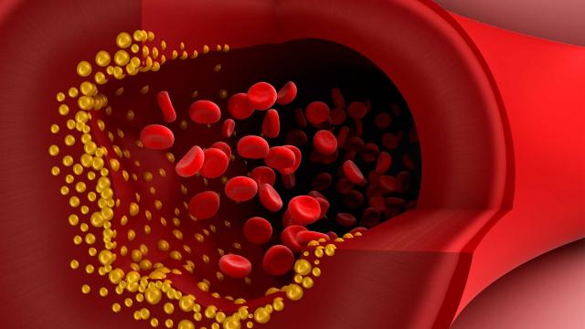
Cholesterol forming plaques (yellow) in the lining of arteries. Source: Healthguru

Cholesterol gets a lot of bad press.

Whether it’s high and low, the perfect balance of cholesterol in our blood seems to be critical to our overall health and sense of wellbeing. At least that is what we are constantly being told this by media and medical professionals alike.

But ask yourself this: Why? What exactly is cholesterol?

Good question. What is cholesterol?

Cholesterol (from the Greek ‘chole‘- bile and ‘stereos‘ – solid) is a waxy substance that is circulating our bodies. It is generated by the liver, but it is also found in many foods that we eat (for example, meats and egg yolks).

cholesterol-svg

The chemical structure of Cholesterol. Source: Wikipedia

Cholesterol falls into one of three major classes of lipids – those three classes of lipids being TriglyceridesPhospholipids and Steroids (cholesterol is a steroid). Lipids are major components of the cell membranes and thus very important. Given that the name ‘lipids’ comes from the Greek lipos meaning fat, people often think of lipids simply as fats, but fats more accurately fall into just one class of lipids (Triglycerides).

Like many fats though, cholesterol dose not dissolve in water. As a result, it is transported within the blood system encased in a protein structure called a lipoprotein.

Chylomicron.svg

The structure of a lipoprotein; the purple C inside represents cholesterol. Source: Wikipedia

Lipoproteins have a very simple classification system based on their density:

  • very low density lipoprotein (VLDL)
  • low density lipoprotein (LDL)
  • intermediate density lipoprotein (IDL)
  • high density lipoprotein (HDL).

Now understand that all of these different types of lipoproteins contain cholesterol, but they are carrying it to different locations and this is why some of these are referred to as good and bad.

The first three types of lipoproteins carry newly synthesised cholesterol from the liver to various parts of the body, and thus too much of this activity would be bad as it results in an over supply of cholesterol clogging up different areas, such as the arteries.

LDLs, in particular, carry a lot of cholesterol (with approximately 50% of their contents being cholesterol, compared to only 20-30% in the other lipoproteins), and this is why LDLs are often referred to as ‘bad cholesterol’. High levels of LDLs can result in atherosclerosis (or the build-up of fatty material inside your arteries).

Progressive and painless, atherosclerosis develops as cholesterol silently and slowly accumulates in the wall of the artery, in clumps that are called plaques. White blood cells stream in to digest the LDL cholesterol, but over many years the toxic mess of cholesterol and cells becomes an ever enlarging plaque. If the plaque ever ruptures, it could cause clotting which would lead to a heart attack or stroke.

ni2

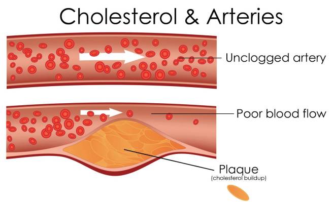
Source: MichelsonMedical

So yeah, some lipoproteins can be considered bad.

HDLs, on the other hand, collects cholesterol and other lipids from cells around the body and take them back to the liver. And this is why HDLs are sometimes referred to as “good cholesterol” because higher concentrations of HDLs are associated with lower rates of atherosclerosis progression (and hopefully regression).

But why is cholesterol important?

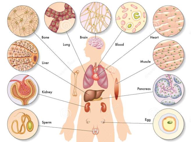
While cholesterol is usually associated with what is floating around in your bloodstream, it is also present (and very necessary) in every cell in your body. It helps to produce cell membranes, hormones, vitamin D, and the bile acids that help you digest fat.

It is particularly important for your brain, which contains approximately 25 percent of the cholesterol in your body. Numerous neurodegenerative conditions are associated with cholesterol disfunction (such as Alzheimer’s disease and Huntington’s disease – Click here for more on this). In addition, low levels of cholesterol is associated with violent behaviour (Click here to read more about this).

Are there any associations between cholesterol and Parkinson’s disease?

The associations between cholesterol and Parkinson’s disease is a topic of much debate. While there have been numerous studies investigating cholesterol levels in blood in people with Parkinson’s disease, the results have not been consistent (Click here for a good review on this topic).

Rather than looking at cholesterol directly, a lot of researchers have chosen to focus on the medication that is used to treat high levels of cholesterol – a class of drugs called statins.

Gao

Title: Prospective study of statin use and risk of Parkinson disease.
Authors: Gao X, Simon KC, Schwarzschild MA, Ascherio A.
Journal: Arch Neurol. 2012 Mar;69(3):380-4.
PMID: 22410446              (This article is OPEN ACCESS if you would like to read it)

In this study the researchers conduced a prospective study involving the medical details of 38 192 men and 90 874 women from two huge US databases: the Nurses’ Health Study (NHS) and the Health Professionals Follow-Up Study (HPFS).

NHS study was started in 1976 when 121,700 female registered nurses (aged 30 to 55 years) completed a mailed questionnaire. They provided an overview of their medical histories and health-related behaviours. The HPFS study was established in 1986, when 51,529 male health professionals (40 to 75 years) responded to a similar questionnaire. Both the NHS and the HPFS send out follow-up questionnaires every 2 years.

By analysing all of that data, the investigators found 644 cases of Parkinson’s disease (338 women and 306 men). They noticed that the risk of Parkinson’s disease was approximately 25% lower among people currently taking statins when compared to people not using statins. And this association was significant in statin users younger than 60 years of age (P = 0.02).

What are statins?

Also known as HMG-CoA reductase inhibitors, statins are a class of drug that inhibits/blocks an enzyme called 3-hydroxy-3-methylglutaryl coenzyme A (HMG-CoA) reductase.

HMG-CoA reductase is the key enzyme regulating the production of cholesterol from mevalonic acid in the liver. By blocking this process statins help lower the total amount of cholesterol available in your bloodstream.

photodune-13199316-generic-pack-of-statins-l

Source: Myelomacrowd

Statins are used to treat hypercholesterolemia (also called dyslipidemia) which is high levels of cholesterol in the blood. And they are one of the most widely prescribed classes of drugs currently available, with approximately 23 percent of adults in the US report using statin medications (Source).

Now, while the study above found an interesting association between statin use and a lower risk of Parkinson’s disease, the other research published on this topic has not been very consistent. In fact, a review in 2009 found a significant associations between statin use and lower risk of Parkinson’s disease was observed in only two out of five prospective studies (Click here to see that review).

New research published this week has attempted to clear up some of that inconsistency, by starting with a huge dataset and digging deep into the numbers.

So what new research has been published?

Statins

Title: Statins may facilitate Parkinson’s disease: Insight gained from a large, national claims database
Authors: Liu GD, Sterling NW, Kong L, Lewis MM, Mailman RB, Chen H, Leslie D, Huang X
Journal: Movement Disorder, 2017 Jun;32(6):913-917.
PMID: 28370314

Using the MarketScan Commercial Claims and Encounters database which catalogues the healthcare use and medical expenditures of more than 50 million employees and their family members each year, the researcher behind that study identified 30,343,035 individuals that fit their initial criteria (that being “all individuals in the database who had 1 year or more of continuous enrolment during January 1, 2008, to December 31, 2012, and were 40 years of age or older at any time during their enrolment”). From this group, the researcher found a total of 21,599 individuals who had been diagnosed with Parkinson’s disease.

In their initial analysis, the researchers found that Parkinson’s disease was positively associated with age, male gender, hypertension, coronary artery disease, and usage of cholesterol-lowering drugs (both statins and non-statins). The condition was negatively associated with hyperlipidemia (or high levels of cholesterol). This result suggests not only that people with higher levels of cholesterol have a reduced chance of developing Parkinson’s disease, but taking medication to lower cholesterol levels may actually increase ones risk of developing the condition.

One interesting finding in the data was the effect that different types of statins had on the association.

Statins can be classified into two basic groups: water soluble (or hydrophilic) and lipid soluble (or lipophilic) statins. Hydrophilic molecule have more favourable interactions with water than with oil, and vice versa for lipophilic molecules.

wataer_oil

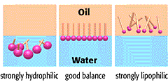
Hydrophilic vs lipophilic molecules. Source: Riken

Water soluble (Hydrophilic) statins include statins such as pravastatin and rosuvastatin; while all other available statins (eg. atorvastatin, cerivastatin, fluvastatin, lovastatin and simvastatin) are lipophilic.

In this new study, the researchers found that the association between statin use and increased risk of developing Parkinson’s disease was more pronounced for lipophilic statins (a statistically significant 58% increase – P < 0.0001), compared to hydrophilic statins (a non-significant 19% increase – P = 0.25). One possible explanation for this difference is that lipophilic statins (like simvastatin and atorvastatin) cross the blood-brain barrier more easily and may have more effect on the brain than hydrophilic ones.

The investigators also found that this association was most robust during the initial phase of statin treatment. That is to say, the researchers observed a 82% in risk of PD within 1 year of having started statin treatment, and only a 37% increase five years after starting statin treatment.; P < 0.0001). Given this finding, the investigators questioned whether statins may be playing a facilitatory role in the development of Parkinson’s disease – for example, statins may be “unmasking” the condition during its earliest stages.

So statins are bad then?

Can I answer this question with a diplomatic “I don’t know”?

It is difficult to really answer that question based on the results of just this one study. This is mostly because this new finding is in complete contrast to a lot of experimental research over the last few years which has shown statins to be neuroprotective in many models of Parkinson’s disease. Studies such as this one:

statins
Title: Simvastatin inhibits the activation of p21ras and prevents the loss of dopaminergic neurons in a mouse model of Parkinson’s disease.
Authors: Ghosh A, Roy A, Matras J, Brahmachari S, Gendelman HE, Pahan K.
Journal: J Neurosci. 2009 Oct 28;29(43):13543-56.
PMID: 19864567              (This study is OPEN ACCESS if you would like to read it)

In this study, the researchers found that two statins (pravastatin and simvastatin – one hydrophilic and one lipophilic, respectively) both exhibited the ability to suppress the response of helper cells in the brain (called microglial) in a neurotoxin model of Parkinson’s disease. This microglial suppression resulted in a significant neuroprotective effect on the dopamine neurons in these animals.

Another study found more Parkinson’s disease relevant effects from statin treatment:

Synau

TItle: Lovastatin ameliorates alpha-synuclein accumulation and oxidation in transgenic mouse models of alpha-synucleinopathies.
Authors: Koob AO, Ubhi K, Paulsson JF, Kelly J, Rockenstein E, Mante M, Adame A, Masliah E.
Journal: Exp Neurol. 2010 Feb;221(2):267-74.
PMID: 19944097            (This study is OPEN ACCESS if you would like to read it)

In this study, the researchers treated two different types of genetically engineered mice (both sets of mice produce very high levels of alpha synuclein – the protein closely associated with Parkinson’s disease) with a statin called lovastatin. In both groups of alpha synuclein producing mice, lovastatin treatment resulted in significant reductions in the levels of cholesterol in their blood when compared to the saline-treated control mice. The treated mice also demonstrated a significant reduction in levels of alpha synuclein clustering (or aggregation) in the brain than untreated mice, and this reduction in alpha synuclein accumulation was associated with a lessening of pathological damage in the brain.

So statins may not be all bad?

One thing many of these studies fail to do is differentiate between whether statins are causing the trouble (or benefit) directly or whether simply lowering cholesterol levels is having a negative impact. That is to say, do statins actually do something else? Other than lowering cholesterol levels, are statins having additional activities that could cause good or bad things to happen?


url

Source: Liverissues

The recently published study we are reviewing in this post suggested that non-statin cholesterol medication is also positively associated with developing Parkinson’s disease. Thus it may be that statins are not bad, but rather the lowering of cholesterol levels that is. This raises the question of whether high levels of cholesterol are delaying the onset of Parkinson’s disease, and one can only wonder what a cholesterol-based process might be able to tell us about the development of Parkinson’s disease.

If the findings of this latest study are convincingly replicated by other groups, however, we may need to reconsider the use of statins not in our day-to-day clinical practice. At the very least, we will need to predetermine which individuals may be more susceptible to developing Parkinson’s disease following the initiation of statin treatment. It would actually be very interesting to go back to the original data set of this new study and investigate what addition medical features were shared between the people that developed Parkinson’s disease after starting statin treatment. For example, were they all glucose intolerant? One would hope that the investigators are currently doing this.

Are Statins currently being tested in the clinic for Parkinson’s disease?

(Oh boy! Tough question) Yes, they are.

There is currently a nation wide study being conducted in the UK called PD STAT.

PDSTATLogo_Large

The study is being co-ordinated by the Plymouth Hospitals NHS Trust (Devon). For more information, please see their website or click here for the NHS Clinical trials gateway website.

Is this dangerous given the results of the new research study?

(Oh boy! Even tougher question!)

Again, we are asking this question based on the results of one recent study. Replication with independent databases is required before definitive conclusions can be made.

There have, however, been previous clinical studies of statins in neurodegenerative conditions and these drugs have not exhibited any negative effects (that I am aware of). In fact, a clinical trial for multiple sclerosis published in 2014 indicated some positive results for sufferers taking simvastatin:

MS-STAT
Title: Effect of high-dose simvastatin on brain atrophy and disability in secondary progressive multiple sclerosis (MS-STAT): a randomised, placebo-controlled, phase 2 trial.
Authors: Chataway J, Schuerer N, Alsanousi A, Chan D, MacManus D, Hunter K, Anderson V, Bangham CR, Clegg S, Nielsen C, Fox NC, Wilkie D, Nicholas JM, Calder VL, Greenwood J, Frost C, Nicholas R.
Journal: Lancet. 2014 Jun 28;383(9936):2213-21.
PMID: 24655729             (This article is OPEN ACCESS if you would like to read it)

In this double-blind clinical study (meaning that both the investigators and the subjects in the study were unaware of which treatment was being administered), 140 people with multiple sclerosis were randomly assigned to receive either the statin drug simvastatin (70 people; 40 mg per day for the first month and then 80 mg per day for the remainder of 18 months) or a placebo treatment (70 people).

Patients were seen at 1, 6, 12, and 24 months into the study, with telephone follow-up at months 3 and 18. MRI brain scans were also made at the start of the trial, and then again at 12 months and 25 months for comparative sake.

The results of the study indicate that high-dose simvastatin was well tolerated and reduced the rate of whole-brain shrinkage compared with the placebo treatment. The mean annualised shrinkage rate was significantly lower in patients in the simvastatin group. The researchers were very pleased with this result and are looking to conduct a larger phase III clinical trial.

Other studies have not demonstrated beneficial results from statin treatment, but they have also not observed a worsening of the disease conditions:

Alzh
Title: A randomized, double-blind, placebo-controlled trial of simvastatin to treat Alzheimer disease.
Authors:Sano M, Bell KL, Galasko D, Galvin JE, Thomas RG, van Dyck CH, Aisen PS.
Journal: Neurology. 2011 Aug 9;77(6):556-63.
PMID: 21795660            (This article is OPEN ACCESS if you would like to read it)

In this study, the investigators recruited a total of 406 individuals were mild to moderate Alzheimer’s disease, and they were randomly assigned to two groups: 204 to simvastatin (20 mg/day, for 6 weeks then 40 mg per day for the remainder of 18 months) and 202 to placebo control treatment. While Simvastatin displayed no beneficial effects on the progression of symptoms in treated individuals with mild to moderate Alzheimer’s disease (other than significantly lowering of cholesterol levels), the treatment also exhibited no effect on worsening the disease.


So what does it all mean?

Research investigating cholesterol and its association with Parkinson’s disease has been going on for a long time. This week a research report involving a huge database was published which indicated that using cholesterol reducing medication could significantly increase one’s risk of developing Parkinson’s disease.

These results do not mean that someone being administered statins is automatically going to develop Parkinson’s disease, but – if the results are replicated – it may need to be something that physicians should consider before prescribing this class of drug.

Whether ongoing clinical trials of statins and Parkinson’s disease should be reconsidered is a subject for debate well above my pay grade (and only if the current results are replicated independently). It could be that statin treatment (or lowering of cholesterol) may have an ‘unmasking’ effect in some individuals, but does this mean that any beneficial effects in other individuals should be discounted? If preclinical data is correct, for example, statins may reduce alpha synuclein clustering in some people which could be beneficial in Parkinson’s.

As we have said above, further research is required in this area before definitive conclusions can be made. This is particularly important given the inconsistencies of the previous research results in the statin and Parkinson’s disease field of investigation.


EDITORIAL NOTE: The information provided by the SoPD website is for information and educational purposes only. Under no circumstances should it ever be considered medical or actionable advice. It is provided by research scientists, not medical practitioners. Any actions taken – based on what has been read on the website – are the sole responsibility of the reader. Any actions being contemplated by readers should firstly be discussed with a qualified healthcare professional who is aware of your medical history. While some of the information discussed in this post may cause concern, please speak with your medical physician before attempting any change in an existing treatment regime.


The banner for today’s post was sourced from HarvardHealth

BioRxiv – open access preprints

si-bioRxiv

For the vast majority of the general population, science is consumed via mass media head lines and carefully edited summaries of the research.

The result of this simplified end product is an ignorance of the process that researchers need to deal with in order to get their research in the public domain.

As part of our efforts to educate the general public about the scientific research of Parkinson’s disease, it is necessary to also make them aware of that process, the issues associated with it, and how it is changing over time.

In todays post, we will look at how new research reports are being made available to the public domain before they are published.


ibiology-preprints-1024x576

Getting research into the public domain. Source: STAT

Every morning here at the SoPD, we look at what new research has entered the public domain over night and try to highlight some of the Parkinson’s disease relevant bits on our Twitter account (@ScienceofPD).

To the frustration of many of our followers, however, much of that research sits behind the pay-to-view walls of big publishing houses. One is allowed to read the abstract of the research report in most cases, but not the full report.

Given that charity money and tax payer dollars are paying for much of the research being conducted, and for the publication fee (approx. $1500 per report on average) to get the report into the journal, there is little debate as to the lack of public good in such a system. To make matter worse, many of the scientists doing the research can not access the published research reports, because their universities and research institutes can not afford the hefty access fees for all of the journals.

Problem-infographic3

Source: Libguides

To be fair, the large publishing houses have recognised that this is not a sustainable business model, and they have put forward the development of open-access web-based science journals, such as Nature communications, Scientific reports, and Cell reports. But the fees for publishing in these journals can in some cases be higher than the closed access publications.

This is crazy. What can we do about it?

Well, there have been efforts for some time to improve the situation.

Projects like the Public Library of Science (or PLOS) have been very popular and are now becoming a real force on the scientific publishing landscape (they recently celebrated their 10 year anniversary and during that time they have published more than 165,000 research articles). But they too have costs associated with maintaining their service and publications fees can still be significant.

url

Is there an easier way of making this research available?

So this is Prof Paul Ginsparg.

Ginsparg_at_Cornell_University

Source: Wikipedia

Looks like the mad scientist type right? Don’t be fooled. He’s awesome! Prof Ginsparg is a professor of Physics and Computing & Information Science at Cornell University.

Back in 1991, he started a repository of pre-print publications in the field of physics. The repository was named arXiv.org, and it allowed physics researchers to share and comment on each others research reports before they were actually published.

The site slowly became an overnight sensation.

The number of manuscripts deposited at arXiv passed the half-million mark on October 3, 2008, the million manuscript mark by the end of 2014 (with a submission rate of more than 8,000 manuscripts per month). The site currently has 1,257,315 manuscripts that are freely available to access. A future nobel prize winning bit of research is probably in there!

Now, by their very nature, and in a very general sense, biomedical researchers are a jealous bunch.

For many years they looked on with envy at the hive of activity going on at arXiv and wished that they had something like it themselves. And now they do! In November 2013, Cold Spring Harbor Laboratory in New York launched BioRxiv.

maxresdefault

Source: BioRxiv

And the website is very quickly becoming a popular destination: by April 21, 2017, >10,000 manuscript had been posted, at a current rate of over 800 manuscripts per month (Source).

Recently they got a huge nod of financial support from the Chan Zuckerberg Initiative – a foundation set up by Facebook founder Mark Zuckerberg and his wife Priscilla Chan to “advance human potential and promote equality in areas such as health, education, scientific research and energy” (Wikipedia).

5677ff2b-be2c-49e7-b0cb-7e33c18149dd-1467045359144Chan-Zuckerberg-Initiative-1

Source: ChangZuckerberg

In April of this year, the Chan Zuckerberg Initiative announced a partnership with Cold Spring Harbor Laboratory to help support the site (Click here to see the press release).

So what is bioRxiv?

bioRxiv is a free OPEN ACCESS service that allows researchers to submit draft copies of scientific papers — called preprints — for their colleagues to read and comment on before they are actually published in peer-reviewed scientific journals.

Here are two videos explaining the idea:

Sounds great right?

To demonstrate how the bioRxiv process works, we have selected an interesting manuscript from the database that we would like to review here on the SoPD.

This is the article:

HEMMER

Title: In Vivo Phenotyping Of Parkinson-Specific Stem Cells Reveals Increased a-Synuclein Levels But No Spreading
Authors: Hemmer K, Smits LM, Bolognin S, Schwamborn JC
Database: BioRxiv
DOI: https://doi.org/10.1101/140178
PMID: N/A                   (You can access the manuscript by clicking here)

In this study (which was posted on bioRxiv on the 19th May, 2017), the researchers have acquired skin cells from an 81 year old female with Parkinson’s disease who carries a mutation (G2019S) in the LRRK2 gene.

Mutations in the Leucine-rich repeat kinase 2 (or Lrrk2) gene are associated with an increased risk of developing Parkinson’s disease. The most common mutation of LRRK2 gene is G2019S, which is present in 5–6% of all familial cases of Parkinson’s disease, and is also present in 1–2% of all sporadic cases. We have previously discussed Lrrk2 (Click here to read that post).

image1

The structure of Lrrk2 and where various mutations lie. Source: Intech

The skin cells were transformed using a bit of biological magic in induced pluripotent stem (or IPS) cells. We have previously discussed IPS cells and how they are created (Click here to read that post). By changing a subjects skin cell into a stem cell, researchers can grow the cell into any type of cell and then investigate a particular disease on a very individualised basis (the future of personalised medicine don’t you know).

nature10761-f2.2

IPS cell options available to Parkinson’s disease. Source: Nature

Using this IPS cell with a mutation in the LRRK2 gene, the researchers behind todays manuscript next grew the cells in culture and encouraged the cells to become dopamine producing cells (these are some of the most vulnerable cells in Parkinson’s disease). The investigators had previously shown that neurons grown in culture from cells with the G2019S mutation in the LRRK2 gene have elevated levels of of the Parkinson’s disease protein alpha Synuclein (Click here to read that OPEN ACCESS paper).

In this present study, the investigators wanted to know if these cells would also have elevated levels of alpha synuclein when transplanted into the brain. Their results indicate that the cells did. Next, the investigators wanted to use this transplantation model to see if the high levels of alpha synuclein in the transplanted cells would lead to the protein being passed to neighbouring cells.

Why did they want to do that?

One of the current theories regarding the mechanisms underlying the progressive spread of Parkinson’s disease is that the protein alpha synuclein is lead culprit. Under normal conditions, alpha synuclein usually floats around as an individual protein (or monomer), but sometime it starts to cluster (or aggregate) with other monomers of alpha synuclein and these form what we call oligomers. These oligomers are believed to be a toxic form of alpha synuclein that is being passed from cell to cell. And it ‘seeds’ the disease in each cell it is passed on to (Click here for a very good OPEN ACCESS review of this topic).

Mechanism of syunuclein propagation and fibrillization

The passing of alpha synuclein between brain cells. Source: Nature

There have been postmortem analysis studies of the brains from people with Parkinson’s who have had cell transplantation therapy back in the 1990s. The analysis shows that some of the transplanted cells have evidence of toxic alpha synuclein in them – some of those cells have Lewy bodies in them, suggesting that the disease has been passed on to the healthy introduced cells from the diseased brain (Click here for the OPEN ACCESS research report about this).

In the current bioRxiv study, the investigators wanted to ask the reverse question:

Can unhealthy, toxic alpha synuclein producing cells cause the disease to spread into a healthy brain?

So after transplanted the Lrrk2 mutant cells into the brains of mice, they waited 11 weeks to see if the alpha synuclein would be passed on to the surrounding brain. According to their results, the unhealthy alpha synuclein did not transfer. They found no increase in levels of alpha synuclein in the cells surrounding the transplanted cells. The researchers concluded that within the parameters of their experiment, Parkinson’s disease-associated alpha synuclein spreading was not detected.

Interesting. When will this manuscript be published in a scientific journal?

We have no idea.

One sad truth of the old system of publication is: it may never be.

And this illustrates one of the beautiful features of bioRxiv.

This manuscript is probably going through the peer-review process at a particular scientific journal at the moment in order for it to be properly published. It is a process that will take several months. Independent reviewers will provide a critique of the work and either agree that it is ready for publication, suggest improvements that should be made before it can be published, or reject it outright due to possible flaws or general lack of impact (depending on the calibre of the journal – the big journals seem to only want sexy science). It is a brutal procedure and some manuscripts never actually survive it to get published, thus depriving the world of what should be freely available research results.

And this is where bioRxiv provides us with a useful forum to present scientific biological research that may never reach publication. Perhaps the researchers never actually intended to publish their findings, and just wanted to let the world know that someone had attempted the experiment and these are the results they got (there is a terrible bias in the world of research publishing to only publish positive results).

The point is: with bioRxiv we can have free access to the research before it is published and we do not have to wait for the slow peer-review process.

And there is definitely some public good in that.

EDITORS NOTE HERE: We are not suggesting for a second that the peer-review process should be done away with. The peer-review process is an essential and necessary aspect of scientific research, which helps to limit fraud and inaccuracies in the science being conducted.

What does it all mean?

This post may be boring for some of our regular readers, but it is important for everyone to understand that there are powerful forces at work in the background of scientific research that will determine the future of how information is disseminated to both the research community and general population. It is useful to be aware of these changes.

We hope that some of our readers will be bold/adventurous and have a look at some of what is on offer in the BioRxiv database. Maybe not now, but in the future. It will hopefully become a tremendous resource.

And we certainly encourage fellow researchers to use it (most of the big journals now accept preprint manuscripts being made available on sites like bioRxiv – click here to see a list of the journals that accept this practise) and some journals also allow authors to submit their manuscript directly to a journal’s submission system through bioRxiv via the bioRxiv to Journals (B2J) initiative (Click here for a list of the journals accepting this practise).

The times they are a changing…


The banner for today’s post was sourced from ScienceMag

Rotten eggs, Rotorua and Parkinson’s disease

fixedw_large_4x

Being a proud kiwi, I am happy to highlight and support any research coming out of New Zealand.

Recently a new commentary has been published suggesting that living in the NZ city of Rotorua (‘Roto-Vegas‘ to the locals) may decrease the risk of developing Parkinson’s disease.

In today’s post, we will review the research behind the idea and discuss what it could mean for people with neurodegenerative conditions, like Parkinson’s disease.


iStock_000060169360_new_zealand_champagne_pool_rotorua

The geothermal wonderlands of Rotorua. Source: Audleytravel

Rotorua is a small city in the central eastern area of the North Island of New Zealand (Aotearoa in the indigenous Māori language).

The name Rotorua comes from the Māori language (‘roto’ meaning lake and rua meaning ‘two’). The full Māori name for the spot is actually Te Rotorua-nui-a-Kahumatamomoe. The early Māori chief and explorer Ihenga named it after his uncle Kahumatamomoe. But given that it was the second major lake found in Aotearoa (after lake Taupo in the centre of the North Island), the name that stuck was Rotorua or ‘Second lake’.

TAM-BLOG-1

Maori culture. Source: TamakiMaoriVillage

Similar to lake Taupo, Rotorua is a caldera resulting from an ancient volcanic eruption (approximately 240,000 years ago). The lake that now fills it is about 22 km (14 mi) in diameter.

p-8709-gns

Lake Rotorua. Source: Teara

The volcano may have disappeared, but the surrounding region is still full of geothermal activity (bubbling mud pools and geysers), providing the region with abundant renewable power and making the city a very popular tourist destination.

FFP_150415-6431-Edit

Tourist playing with mud. Source: Rotoruanz

Before visiting the city, however, travellers should be warned that Rotorua’s other nicknames include “Sulphur City” and “Rotten-rua”, because of the smell that results from the geothermal activity.

And speaking from personal experience, the “rotten eggs” smell is prevalent.

Interesting, but what has this got to do with the science of Parkinson’s disease?

Well, the rotten egg smell is the result of hydrogen sulfide emissions, and recently it has been suggested that this pungent gas may be having positive benefits on people, particularly with regards to Parkinson’s disease.

This idea has been proposed by Dr Yusuf Cakmak at the University of Otago in a recent commentary:

Yusuf

Title: Rotorua, hydrogen sulphide and Parkinson’s disease-A possible beneficial link?
Author: Cakmak Y.
Journal: N Z Med J. 2017 May 12;130(1455):123-125.
PMID: 28494485

In his write up, Dr Cakmak points towards two studies that have been conducted on people from Rotorua. The first focused on examining whether there was any association between asthma and chronic obstructive pulmonary disease and exposure to hydrogen sulfide in Rotorua. By examining air samples and 1,204 participants, the investigators of that study no association (the report of that study is OPEN ACCESS and can be found by clicking here).

The second study is the more interesting of the pair:

roto

Title: Chronic ambient hydrogen sulfide exposure and cognitive function.
Authors: Reed BR, Crane J, Garrett N, Woods DL, Bates MN.
Journal: Neurotoxicol Teratol. 2014 Mar-Apr;42:68-76.
PMID: 24548790                 (This article is OPEN ACCESS if you would like to read it)

In this study, the investigators recruited 1,637 adults (aged 18-65 years) from Rotorua. They conducted neuropsychological tests on the subjects, measuring visual and verbal episodic memory, attention, fine motor skills, psychomotor speed and mood. The average amount of time the participants had lived in the Rotorua region was 18 years (ranging from 3-64 years). The researchers also made measurements of hydrogen sulfide levels at the participants homes and work sites.

While the researchers found no association between hydrogen sulfide exposure and cognitive ability, they did notice something interesting in the measures of fine motor skills: individuals exposed to higher levels of hydrogen sulfide displayed faster motor response times on tasks like finger tapping. Finger tapping speed is an important part of Parkinson’s Motor Rating Scale examination tests.

The investigators behind the study concluded that the levels of hydrogen sulfide in Rotorua do not have any detrimental effect on the individuals living in the area,

Dr Cakmak, however, wondered whether “relatively high, but safe, hydrogen sulfide levels in Rotorua could help protect the degradation of dopaminergic neurons associated with Parkinson’s Disease?” (based on the better performance on the motor response time).

Hang on a second, what exactly is hydrogen sulfide?

Hydrogen sulfide (chemical symbol: H2S) is a colourless gas. Its production often results from the the breaking down of organic material in the absence of oxygen, such as in sewers (this process is called anaerobic digestion. It also occurs in volcanic and geothermal conditions.

Hydrogen_sulfide

H2S. Source: Wikipedia

About 15 years ago, it was found in various organs in the body and termed a gasotransmitter. A gasotransmitter is a molecule that can be used to transmit chemical signals from one cell to another, which results in certain physiological reactions (oxygen, for example, is a gasotransmitter).

Hydrogen sulfide is now known to be cardioprotective (protection of the heart), and many years of research have demonstrated beneficial aspects of using it in therapy, such as vasodilation and lowering blood pressure, increasing levels of antioxidants, inhibiting inflammation, and activation of anti-apoptotic (anti-cell death) pathways. For a good review of hydrogen sulfide’s cardioprotective properties – click here.

F1.large

Source: Clinsci

The demonstration of the protective properties of hydrogen sulfide in other bodily organs have led neuroscientists to start investigating whether these same benefits could be utilised in treating disorders of the brain.

And the good news is: hydrogen sulfide can have positive benefits in the brain – Click here for a good review of the brain-related research.

Has other research been conducted on hydrogen sulfide regarding Parkinson’s disease?

Yes. And here is where the story starts to get really interesting.

Initially, there were reports that hydrogen sulfide could protect cells grown in culture from exposure to various neurotoxins (Click here and here for examples).

Then hydrogen sulfide was tested in rodent models of Parkinson’s disease:

SH1

Title: Neuroprotective effects of hydrogen sulfide on Parkinson’s disease rat models.
Authors: Hu LF, Lu M, Tiong CX, Dawe GS, Hu G, Bian JS.
Journal: Aging Cell. 2010 Apr;9(2):135-46.
PMID: 20041858           (This article is OPEN ACCESS if you would like to read it)

In this study, the researchers firstly looked at what happens to hydrogen sulfide in the brains of rodent models of Parkinson’s disease. When rats were injected with a neurotoxin (6-OHDA) that kills dopamine neurons, the investigators found a significant drop in the level of hydrogen sulfide in the region where the dopamine cells reside (called the substantia nigra – an area of the brain severely affected in Parkinson’s disease).

Next the researchers gave some rodents the neurotoxin, waited three weeks and then began administering sodium hydrosulfide – which is a hydrogen sulfide donor  – every day for a further 3 weeks. They found that this treatment significantly reduced the dopamine cell loss, motor problems and inflammation in the sodium hydrosulfide treated animals. Interestingly, they saw the same neuroprotective effect when they repeated the study with a different neurotoxin (Rotenone). The investigators concluded that hydrogen sulfide “has potential therapeutic value for treatment of Parkinson’s disease”.

And this first study was followed up one year later by a study investigating inhaled hydrogen sulfide:

SH2
Title: Inhaled hydrogen sulfide prevents neurodegeneration and movement disorder in a mouse model of Parkinson’s disease.
Authors: Kida K, Yamada M, Tokuda K, Marutani E, Kakinohana M, Kaneki M, Ichinose F.
Journal: Antioxid Redox Signal. 2011 Jul 15;15(2):343-52.
PMID: 21050138            (This article is OPEN ACCESS if you would like to read it)

In this study, the investigators gave mice a neurotoxin (MPTP) and then had them breathe air with or without hydrogen sulfide (40 ppm) for 8 hours per day for one week. The mice that inhaled hydrogen sulfide displayed near normal levels of motor behaviour performance and significantly reduced levels of neurodegeneration (dopamine cell loss).

Inhalation of hydrogen sulfide also prevented the MPTP-induced activation of the brain’s helper cells (microglia and astrocytes) and increased levels of detoxification enzymes and antioxidant proteins (including heme oxygenase-1 and glutamate-cysteine ligase). Curiously, hydrogen sulfide inhalation did not significantly affect levels of reduced glutathione (we will come back to this in an upcoming post).

These first two preclinical results have been replicated many times now confirming the initial findings (Click here, here, here and here for examples). The researchers of the second ‘inhalation’ study concluded the study by suggesting that the potential therapeutic effects of hydrogen sulfide inhalation now needed to be examined in more disease relevant models of Parkinson’s disease.

And this is exactly what researchers did next:

HS5

Title: Sulfhydration mediates neuroprotective actions of parkin.
Authors: Vandiver MS, Paul BD, Xu R, Karuppagounder S, Rao F, Snowman AM, Ko HS, Lee YI, Dawson VL, Dawson TM, Sen N, Snyder SH.
Journal: Nat Commun. 2013;4:1626. doi: 10.1038/ncomms2623.
PMID: 23535647          (This article is OPEN ACCESS if you would like to read it)

The researchers conducting this study were interested in the interaction of hydrogen sulfide with the Parkinson’s disease-associated protein Parkin (also known as PARK2). They found that hydrogen sulfide actively modified parkin protein – a process called sulfhydration – and that this enhances the protein’s level of activity.

They also noted that the level of Parkin sulfhydration in the brains of patients with Parkinson’s disease is markedly reduced (a 60% reduction). These finding imply that drugs that increase levels of hydrogen sulfide in the brain may be therapeutic.

Interestingly, cells with genetic mutations in another Parkinson’s disease related gene, DJ-1, also produce less hydrogen sulfide (click here to read more about this).

Has anyone ever looked at hydrogen sulfide and alpha synuclein?

Not that we are aware of.

Alpha synuclein is the Parkinson’s disease associated protein that clusters in the Parkinsonian brain and forms Lewy bodies.

But researchers have looked at hydrogen sulfide and amyloid formation:

HS4
Title: Hydrogen sulfide inhibits amyloid formation
Authors: Rosario-Alomar MF, Quiñones-Ruiz T, Kurouski D, Sereda V, Ferreira EB, Jesús-Kim LD, Hernández-Rivera S, Zagorevski DV, López-Garriga J, Lednev IK.
Journal: J Phys Chem B. 2015 Jan 29;119(4):1265-74.
PMID: 25545790         (This article is OPEN ACCESS if you would like to read it)


Amyloid formations are large clusters of misfolded proteins that are associated with neurodegenerative conditions, like Alzheimer’s disease and Parkinson’s disease. The researchers who conducted this study were interested in the behaviour of these misfolded protein in the presence of hydrogen sulfide. What they found was rather remarkable: the addition of hydrogen sulfide completely inhibited the formation amyloid fibrils (amyloid fibril plaques are found in brains of people with Alzheimer’s disease).

jp-2014-08471v_0008

Source: NCBI

If the addition of hydrogen sulfide can reduce the level of clustered proteins in a model of Alzheimer’s disease, it would be interesting to see what it would do to alpha synuclein.

NOTE: Hydrogen sulfide levels are also reduced in the brains of people with Alzheimer’s disease (click here to read more on this topic)

Has hydrogen sulfide ever been tested in the clinic?

There are currently 17 clinical trials investigating hydrogen sulfide in various conditions (not Parkinson’s disease though).

So where can I get me some of that hydrogen sulfide?

Ok, so here is where we come in with the health warning section.

You see, hydrogen sulfide is a very dangerous gas. It is really not to be played with.

Blakely_June_Hydrogen-Sulfide

Source: Blakely

The gas is both corrosive and flammable. More importantly, at high concentrations, hydrogen sulfide gas can be fatal almost immediately (>1000 parts per milllion – source: OSHA). And the gas only exhibits the “rotten eggs” smell at low concentrations. At higher concentrations it becomes undetectable due to olfactory paralysis (luckily for the folks in Rotorua, the levels of hydrogen sulfide gas there are between 20-25 parts per billion).

Thus, we do not recommend readers to rush out and load up on hydrogen sulfide gas.

There are many foods that contain hydrogen sulfide.

For example, garlic is very rich in hydrogen sulfide. Another rich source is cooked beef, which has about 0.6mg of hydrogen sulfide per pound – cooked lamb has closer to 0.9 milligrams per pound. Heated dairy products, such as skim milk, can have approximately 3 milligrams of hydrogen sulfide per gallon, and cream has slightly more than double that amount.

Any significant change in diet by a person with Parkinson’s disease should firstly be discussed with a trained medical physician as we can not be sure what impact such a change would have on individualised treatment regimes.

What does it all mean?

Summing up: It would be interesting to look at the frequency of Parkinson’s disease in geothermal region of the world (the population of Rotorua is too small for such an analysis – 80,000 people).

Researchers believe that components of the gas emissions from these geothermal areas may be neuroprotective. Of particular interest is the gas hydrogen sulfide. At high levels, it is a very dangerous gas. At lower levels, however, researchers have shown that hydrogen sulfide has many beneficial properties, including in models of neurodegenerative conditions. These findings have led many to propose testing hydrogen sulfide in clinical trials for conditions like Parkinson’s disease.

Dr Cakmak, who we mentioned near the top of this post, goes one step further. He hypothesises that hydrogen sulfide may actually be one of the active components in the neuroprotective affect of both coffee and smoking – and with good reason. It was recently demonstrated that the certain gut bacteria, such as Prevotella, are decreased in people with Parkinson’s disease (see our post on this topic by clicking here). The consumption of coffee has been shown to help improve the Prevotella population in the gut, which may in term increase the levels of Prevotella-derived hydrogen sulfide. Similarly smokers have a decreased risk of developing Parkinson’s disease and hydrogen sulfide is a component of cigarette smoke.

All of these ideas still needs to be further tested, but we are curious to see where this research could lead. An inhaled neuroprotective treatment for Parkinson’s disease may have benefits for other neurodegenerative conditions.

Oh, and if anyone is interested, we are happy to put readers in contact with real estate agents in sunny ‘Rotten-rua’, New Zealand. The locals say that you gradually get used to the smell.


EDITOR’S NOTE: Under absolutely no circumstances should anyone reading this material consider it medical advice. The material provided here is for educational purposes only. Before considering or attempting any change in your treatment regime, PLEASE consult with your doctor or neurologist. While some of the drugs/molecules discussed on this website are clinically available, they may have serious side effects. We therefore urge caution and professional consultation before any attempt to alter a treatment regime. SoPD can not be held responsible for any actions taken based on the information provided here. 


The banner for today’s post was sourced from Trover

The Antibiotic and Parkinson’s: Oppsy, they got doxy!

maxresdefault

The general population are wrong to look up to scientists as the holders of the keys to some kind of secret knowledge that allows them to render magic on a semi-irregular basis.

All too often, the great discoveries are made by accident.

A while back, some researchers from Germany and Brazil made an interesting discovery that could have important implications for Parkinson’s disease. But they only made this discovery because their mice were feed the wrong food.

Today we’ll review their research and discuss what it could mean for Parkinson’s disease.


image-title1

Sir Alexander Fleming. Source: Biography

Sir Alexander Fleming is credited with discovering the antibiotic properties of penicillin.

But, as it is often pointed out, that the discovery was a purely chance event – an accident, if you like.

After returning from a two week holiday, Sir Fleming noticed that many of his culture dishes were contaminated with fungus, because he had not stored them properly before leaving. One mould in particular caught his attention, however, as it was growing on a culture plate with the bacteria staphylococcus. Upon closer examination, Fleming noticed that the contaminating fungus prevented the growth of staphylococci.

In an article that Fleming subsequently published in the British Journal of Experimental Pathology in 1929, he wrote, “The staphylococcus colonies became transparent and were obviously undergoing lysis … the broth in which the mould had been grown at room temperature for one to two weeks had acquired marked inhibitory, bactericidal and bacteriolytic properties to many of the more common pathogenic bacteria.”

photograph_from_1929_paper_by_fleming

Penicillin in a culture dish of staphylococci. Source: NCBI

Fleming isolated the organism responsible for prohibiting the growth of the staphylococcus, and identified it as being from the penicillium genus.

He named it penicillin and the rest is history.

Fleming himself appreciated the serendipity of the finding:

“When I woke up just after dawn on Sept. 28, 1928, I certainly didn’t plan to revolutionise all medicine by discovering the world’s first antibiotic, or bacteria killer. But I guess that was exactly what I did.” (Source)

And this gave rise to his famous quote:

“One sometimes finds what one is not looking for” (Source)

While Fleming’s discovery of the antibiotic properties of penicillin was made as he was working on a completely different research problem, the important thing to note is that the discovery was made because the evidence came to prepared mind.

Louis-Pasteur-Quotes-1

Pasteur knew the importance of a prepared mind. Source: Thequotes

And this is the purpose of all the training in scientific research – not acquiring ‘the keys to some secret knowledge’, but preparing the investigator to notice the curious deviation.

That’s all really interesting. But what does any of this have to do with Parkinson’s disease?

Three things:

  1. Serendipity
  2. Prepared minds
  3. Antibiotics.

Huh?

Five years ago, a group of Brazilian and German Parkinson’s disease researchers made a serendipitous discovery:

While modelling Parkinson’s disease in some mice, they noticed that only two of the 40 mice that were given a neurotoxic chemical (6-OHDA) developed the motor features of Parkinson’s disease, while the rest remained healthy. This result left them scratching their heads and trying to determine what had gone wrong.

Then it clicked:

“A lab technician realised the mice had mistakenly been fed chow containing doxycycline, so we decided to investigate the hypothesis that it might have protected the neurons.” (from the press release).

The researchers had noted the ‘curious deviation’ and decided to investigate it further.

They repeated the experiment, but this time they added another group of animals which were given doxycycline in low doses (via injection) and fed on normal food (not containing the doxycycline).

And guess what: both group demonstrated neuroprotection!

Hang on a second. Two questions: 1. What exactly is 6-OHDA?
6-hydroxydopamine (or 6-OHDA) is one of several chemicals that researchers use to cause dopamine cells to die in an effort to model the cell death seen in Parkinson’s disease. It shares many structural similarities with the chemical dopamine (which is so severely affected in the Parkinson’s disease brain), and as such it is readily absorbed by dopamine cells who unwittingly assume that they are re-absorbing excess dopamine.

Once inside the cell, 6-OHDA rapidly transforms (via oxidisation) into hydrogen peroxide (H2O2 – the stuff folk bleach their hair with) and para-quinone (AKA 1,4-Benzoquinone). Neither of which the dopamine neurons like very much. Hydrogen peroxide in particular quickly causes massive levels of ‘oxidative stress’, resulting in the cell dying.
6OHDA

Transformation of the neurotoxin 6-OHDA. Source: NCBI

Think of 6-OHDA as a trojan horse, being absorbed by the cell because it looks like dopamine, only for the cell to work out (too late) that it’s not.

Ok, and question 2. What is doxycycline?

Doxycycline is an antibiotic that is used in the treatment of a number of types of infections caused by bacteria.

doxycycline100-1-1k_1

Remind me again, what is an antibiotic?

Antibiotics are a class of drugs that either kill or inhibit the growth of bacteria. They function in one of several ways, either blocking the production of bacterial proteins, inhibiting the replication of bacterial DNA (nuclei acid in the image below), or by rupturing/inhibiting the repair of the bacteria’s outer membrane/wall.

2018_1

The ways antibiotics function. Source: FastBleep

So the researchers accidentally discovered that the a bacteria-killing drug called doxycycline prevented a trojan horse called 6-OHDA from killing dopamine cells?

Basically, yeah.

And then these prepared minds followed up this serendipitous discovery with a series of experiments to investigate the phenomenon further, and they published the results recently in the journal ‘Glial’:

Glial

Title: Doxycycline restrains glia and confers neuroprotection in a 6-OHDA Parkinson model.
Authors: Lazzarini M, Martin S, Mitkovski M, Vozari RR, Stühmer W, Bel ED.
Journal: Glia. 2013 Jul;61(7):1084-100. doi: 10.1002/glia.22496. Epub 2013 Apr 17.
PMID: 23595698

In the report of their research, the investigators noted that doxycycline significantly protected the dopamine neurons and their nerve branches (called axons) in the striatum – an area of the brain where dopamine is released – when 6-OHDA was given to mice. Both oral administration and peripheral injections of doxycycline were able to have this effect.

They also reported that doxycycline inhibited the activation of astrocytes and microglial cells in the brains of the 6-OHDA treated mice. Astrocytes and microglial cells are usually the helper cells in the brain, but in the context of disease or injury these cells can quickly take on the role of judge and executioner – no longer supporting the neurons, but encouraging them to die. The researchers found that doxycycline reduced the activity of the astrocytes and microglial cells in this alternative role, allowing the dopamine cells to recuperate and survive.

The researchers concluded that the “neuroprotective effect of doxycycline may be useful in preventing or slowing the progression of Parkinson’s disease”.

Wow, was this the first time this neuroprotective effect of doxycycline has been observed?

Curiously, No.

We have known of doxycycline’s neuroprotective effects in different models of brain injury since the 1990s (Click here, here and here for more on this). In fact, in their research report, the German and Brazilian researchers kindly presented a table of all the previous neuroprotective research involving doxycycline:

table1

And there was so much of it that the table carried on to a second page:

Table2

Source: Glia

And as you can see from the table, the majority of these reports found that doxycycline treatment had positive neuroprotective effects.

Is doxycycline the only antibiotic that exhibits neuroprotective properties?

No.

Doxycycline belongs to a family of antibiotics called ‘tetracyclines‘ (named for their four (“tetra-“) hydrocarbon rings (“-cycl-“) derivation (“-ine”)), and other members of this family have also been shown to display neuroprotection in models of Parkinson’s disease:

MPTP

Title: Minocycline prevents nigrostriatal dopaminergic neurodegeneration in the MPTP model ofParkinson’s disease.
Authors: Du Y, Ma Z, Lin S, Dodel RC, Gao F, Bales KR, Triarhou LC, Chernet E, Perry KW, Nelson DL, Luecke S, Phebus LA, Bymaster FP, Paul SM.
Journal: Proc Natl Acad Sci U S A. 2001 Dec 4;98(25):14669-74.
PMID: 11724929                    (This article is OPEN ACCESS if you would like to read it)

In this study, the researchers treated mice with an antibiotic called minocycline and it protected dopamine cells from the damaging effects of a toxic chemical called MPTP (or 1-methyl-4-phenyl-1,2,3,6-tetrahydropyridine). MPTP is also used in models of Parkinson’s disease, as it specifically affects the dopamine cells, while leaving other cells unaffected.

The researchers found that the neuroprotective effect of minocycline is associated a reduction in the activity of proteins that initiate cell death (for example, Caspace 1). This left the investigators concluding that ‘tetracyclines may be effective in preventing or slowing the progression of Parkinson’s disease’.

Importantly, this result was quickly followed by two other research papers with very similar results (Click here and here to read more about this). Thus, it would appear that some members of the tetracycline class of antibiotics share some neuroprotective properties.

So what did the Brazilian and German researchers do next with doxycycline?

They continued to investigate the neuroprotective effect of doxycycline in different models of Parkinson’s disease. They also got some Argentinians and Frenchies involved in the studies. And these lines of research led to their recent research report in the journal Scientific Reports:

Doxy1
Title: Repurposing doxycycline for synucleinopathies: remodelling of α-synuclein oligomers towards non-toxic parallel beta-sheet structured species.
Authors: González-Lizárraga F, Socías SB, Ávila CL, Torres-Bugeau CM, Barbosa LR, Binolfi A, Sepúlveda-Díaz JE, Del-Bel E, Fernandez CO, Papy-Garcia D, Itri R, Raisman-Vozari R, Chehín RN.
Journal: Sci Rep. 2017 Feb 3;7:41755.
PMID: 28155912                (This article is OPEN ACCESS if you would like to read it)

In this study, the researchers wanted to test doxycycline in a more disease-relevant model of Parkinson’s disease. 6-OHDA is great for screening and testing neuroprotective drugs. But given that 6-OHDA is not involved with the underlying pathology of Parkinson’s disease, it does not provide a great measure of how well a drug will do against the disease itself. So, the researchers turned their attention to our old friend, alpha synuclein – the protein which forms the clusters of protein (called Lewy bodies) in the Parkinsonian brain.

What the researchers found was fascinating: Doxycycline was able to inhibit the disease related clustering of alpha synuclein. In fact, by reshaping alpha synuclein into a less toxic version of the protein, doxycycline was able to enhance cell survival. The investigators also conducted a ‘dosing’ experiment to determine the most effect dose and they found that taking doxycycline in sub-antibiotic doses (20–40 mg/day) would be enough to exert neuroprotection. They concluded their study by suggesting that these novel effects of doxycycline could be exploited in Parkinson’s disease by “repurposing an old safe drug”.

Wow, has doxycycline ever been used in clinical trials for brain-related conditions before?

Yes.

From 2005-12,there was a clinical study to determine the safety and efficacy of doxycycline (in combination with Interferon-B-1a) in treating Multiple Sclerosis (Click here for more on this trial). The results of that study were positive and can be found here.

More importantly, the other antibiotic to demonstrate neuroprotection in models of Parkinson’s disease, minocycline (which we mentioned above), has been clinically tested in Parkinson’s disease:

title1

Title: A pilot clinical trial of creatine and minocycline in early Parkinson disease: 18-month results.
Authors: NINDS NET-PD Investigators..
Journal: Clin Neuropharmacol. 2008 May-Jun;31(3):141-50.
PMID: 18520981                (This article is OPEN ACCESS if you would like to read it)

This research report was the follow up of a 12 month clinical study that can be found by clicking here. The researchers had taken two hundred subjects with Parkinson’s disease and randomly sorted them into the three groups: creatine (an over-the-counter nutritional supplement), minocycline, and placebo (control). All of the participants were diagnosed less than 5 years before the start of the study. At 12 months, both creatine and minocycline were noted as not interfering with the beneficial effects of symptomatic therapy (such as L-dopa), but a worrying trend began with subjects dropping out of the minocycline arm of the study.

At the 18 month time point, approximately 61% creatine-treated subjects had begun to take additional treatments (such as L-dopa) for their symptoms, compared with 62% of the minocycline-treated subjects and 60% placebo-treated subjects. This result suggested that there was no beneficial effect from using either creatine or minocycline in the treatment of Parkinson’s disease, as neither exhibited any greater effect than the placebo. In addition, the investigators suggested that the decreased tolerability of minocycline was a concern.

Ok, so where do I sign up for the next doxy clinical trial?

Well, the researchers behind the Scientific reports research (discussed above) are hoping to begin planning clinical trials soon.

But theoretically speaking, there shouldn’t be a trial.

Huh?!?

There’s a good reason why not.

In fact, if you look at the comments section under the research article, a cautionary message has been left by Prof Paul M. Tulkens of the Louvain Drug Research Institute in Belgium. He points out that:

“…using antibiotics at sub-therapeutic doses is the best way to trigger the emergence of resistance (supported by many in vitro and in vivo studies). Using an antibiotic for other indications than an infection caused by a susceptible bacteria is something that should be discouraged”

And he is correct.

We recklessly over use antibiotics all over the world at the moment and they are one of the few lines of defence that we have against the bacterial world. Long term use (which Parkinson’s disease would probably require) of an antibiotic at sub-therapeutic levels will only encourage the rise of antibiotic resistant bacteria (possibly within individuals).

The resistance of bacteria to antibiotics can occur spontaneously via several means (for example, through random genetic mutations during cell division). With the right mutation (inferring antibiotic resistance), an individual bacteria would then have a natural advantage over their friends and it would survive our attempts to kill it with antibiotics. Being resistant to antibiotic would leave that bacteria to wreak havoc upon us.

Its the purest form of natural selection.

natural-selection_140211

How bacteria become resistant to antibiotics. Source: Reactgroup

And antibiotic resistant bacteria are fast becoming a major health issue for us, with the number of species of bacteria developing resistance increasing every year (Click here for a good review on factors contributing to the emergence of resistance, and click here for a review of the antibiotic resistant bacteria ‘crisis’).

But don’t be upset on the Parkinson’s disease side of things. Prof Tulken adds that:

“If doxycycline really acts as the authors propose, the molecular targets are probably very different from those causing antibacterial activity. it should therefore be possible to dissociate these effect from the antibacterial effects and to get active compounds devoid of antibacterial activity This is where research must go to rather than in trying to use doxycycline itself.”

And he is correct again.

Rather than tempting disaster, we need to take the more prudent approach.

Independent researchers must now attempt to replicate the neuroprotective results in carefully controlled conditions. At the same time, chemists should conduct an analysis of the structure of doxycycline to determine which parts of it are having this neuroprotective effect.

Doxycycline_structure.svg

The structure of doxycycline. Source: Wikipedia

If researchers can isolate those neuroprotective elements and those same parts are separate from the antibiotic properties, then we may well have another experimental drug for treating Parkinson’s disease.

And the good news is that researchers are already reasonably sure that the mechanisms of the neuroprotective effect of doxycycline are distinct from its antimicrobial action.

So what does it all mean?

Researchers have once again identified an old drug that can perform a new trick.

The bacteria killing antibiotic, doxycycline, has a long history of providing neuroprotection in models of brain disease, but recently researchers have demonstrated that doxycycline may have beneficial effects on particular aspects of Parkinson’s disease.

Given that doxycycline is an antibiotic, we must be cautious in our use of it. It will be interesting to determine which components of doxycycline are neuroprotective, and whether other antibiotics share these components. Given the number of researchers now working in this area, it should not take too long.

We’ll let you know when we hear something.


EDITOR’S NOTE: Under absolutely no circumstances should anyone reading this material consider it medical advice. The material provided here is for educational purposes only. Before considering or attempting any change in your treatment regime, PLEASE consult with your doctor or neurologist. While some of the drugs discussed on this website are clinically available, they may have serious side effects. We therefore urge caution and professional consultation before any attempt to alter a treatment regime. SoPD can not be held responsible for any actions taken based on the information provided here. 


The banner for today’s post was sourced from Youtube

Iron, life force, and Parkinson’s disease

pranaLogo

‘Prana’ is a Hindu Sanskrit word meaning “life force”.

An Australian biotech company has chosen this word for their name.

Recently Prana Biotechnology Ltd announced some exciting results from their Parkinson’s disease research programme.

In today’s post we will look at what the company is doing, the science underlying the business plan, and review the results they have so far.


adpd2017

Source: ADPD2017

At the end of March, over 3000 researchers in the field of neurodegeneration gathered in the Austrian capital of Vienna for the 13th International Conference on Alzheimer’s and Parkinson’s Diseases and Related Neurological Disorders (also known as ADPD2017).

crop

The Vienna city hall. Source: EUtourists

A lot of interesting new research in the field of Parkinson’s disease was presented at the conference (we will look at some other presentation in future posts), but one was of particular interest to us here at SoPD HQ.

The poster entitled: Abstract: 104 – PBT434 prevents neuronal loss, motor function and cognitive impairment in preclinical models of movement disorders by modulation of intracellular iron’, was presented by Associate Professor David Finkelstein, of the Florey Institute of Neuroscience and Mental Health (Melbourne, Australia).

Unfortunately the ADPD2017 conference’s scientific programme search engine does not allow for individual abstracts to be linked to on the web so if you would like to read the abstract, you will need to click here for the search engine page and search for ‘PBT434’ or ‘Finkelstein’ in the appropriate boxes.

Prof Finkelstein was presenting preclinical research that had been conducted by an Australian biotech company called Prana Biotechnology Ltd.

promo1

Source: Prana Biotechnology Ltd

What does the company do?

Prana Biotechnology Ltd has a large portfolio of over 1000 small chemical agents that they have termed ‘MPACs’ (or Metal Protein Attenuating Compounds). These compounds are designed to interrupt the interactions between particular metals and target proteins in the brain. The goal of this interruption is to prevent deterioration of brain cells in neurodegenerative conditions.

For Parkinson’s disease, the company is proposing a particular iron chelator they have called PBT434.

What is an iron chelator?

Iron chelator therapy involves the removal of excess iron from the body with special drugs. Chelate is from the Greek word ‘chela’ meaning “claw”.

chelationtherapy_edited-01

Chelator therapy. Source: Stanford

Iron overload in the body is a common medical problem, sometimes arising from disorders of increased iron absorption such as hereditary haemochromatosis. Iron chelator therapy represents one method of reducing the levels of iron in the body.

But why is iron overload a problem?

iron

Iron. Source: GlobalSpec

Good question. It involves the basic properties of iron.

Iron is a chemical element (symbol Fe). It has the atomic number 26 and by mass it is the most common element on Earth (it makes up much of Earth’s outer and inner core). It is absolutely essential for cellular life on this planet as it is involved with the interactions between proteins and enzymes, critical in the transport of oxygen, and required for the regulation of cell growth and differentiation.

So why then – as Rosalind asked in Shakespeare’s As You Like It – “can one desire too much of a good thing?”

Well, if you think back to high school chemistry class you may recall that there are these things called electrons. And if you have a really good memory, you will recall that the chemical hydrogen has one electron, while iron has 26 (hence the atomic number 26).

atoms

The electrons of iron and hydrogen. Source: Hypertonicblog

Iron has a really interesting property: it has the ability to either donate or take electrons. And this ability to mediate electron transfer is one of the reasons why iron is so important in the body.

Iron’s ability to donate and accept electrons means that when there is a lot of iron present it can inadvertently cause the production of free radicals. We have previously discussed free radicals (Click here for that post), but basically a free radical is an unstable molecule – unstable because they are missing electrons.

imgres

How free radicals and antioxidants work. Source: h2miraclewater

In an unstable format, free radicals bounce all over the place, reacting quickly with other molecules, trying to capture the much needed electron to re-gain stability. Free radicals will literally attack the nearest stable molecule, to steal an electron. This leads to the “attacked” molecule becoming a free radical itself, and thus a chain reaction is started. Inside a living cell this can cause terrible damage, ultimately killing the cell.

Antioxidants can help try and restore the balance, but in the case of iron overload iron doctors will prescribe chelator treatment to deal with the situation more efficiently. By soaking up excess iron, we can limit the amount of damage caused by the surplus of iron.

So what research has been done regarding iron content and the Parkinsonian brain?

Actually, quite a lot.

In 1968, Dr Kenneth Earle used an X-ray based technique to examine the amount of iron in the substantia nigra of people with Parkinson’s disease (Source). The substantial nigra is one of the regions in the brain most badly damaged by the condition – it is where most of the brain’s dopamine neurones resided.

d1ea3d21c36935b85043b3b53f2edb1f87ab7fa6

The dark pigmented dopamine neurons in the substantia nigra are reduced in the Parkinson’s disease brain (right). Source:Memorangapp

Earle examined 11 samples and compared them to unknown number of control samples and his results were a little startling:

The concentration of iron in Parkinsonian samples was two times higher than that of the control samples.

Since that first study, approximately 30 investigations have been made into levels of iron in the Parkinsonian brain. Eleven of those studies have replicated the Earle study by looking at postmortem tissue. They have used different techniques and the results have varied somewhat:

  • Sofic et al. (1988)                             1.8x increase in iron levels
  • Dexter et al. (1989)                         1.3x increase in iron levels
  • Uitti et al. (1989)                              1.1x increase in iron levels
  • Riederer et al 1989                         1.3x increase in iron levels
  • Griffiths and Crossman (1993)     2.0x increase in iron levels
  • Mann et al. (1994)                           1.6x increase in iron levels
  • Loeffler et al. (1995)                       0.9   (lower)
  • Galazka-Friedman et al., 1996     1.0   (no difference)
  • Wypijewska et al. (2010)               1.0   (no difference)
  • Visanji et al, 2013                            1.7x increase in iron levels

Overall, however, there does appear to be a trend in the direction of higher levels of iron in the Parkinsonian brains. A recent meta-analysis of all this data confirmed this assessment as well as noting an increase in the caudate putamen (the region of the brain where the dopamine neuron branches release their dopamine – Click here for that study).

Brain imaging of iron (using transcranial sonography and magnetic resonance imaging (MRI)) has also demonstrated a strong correlation between iron levels in the substantia nigra region and Parkinson’s disease severity/duration (Click here and here to read more on this).

Thus, there appears to be an increase of iron in the regions most affected by Parkinson’s disease and this finding has lead researchers to ask whether reducing this increase in iron may help in the treatment of Parkinson’s disease.

How could iron overload be bad in Parkinson’s disease?

Well in addition to causing the production of free radicals, there are many possible ways in which iron accumulation could be aggravating cell loss in Parkinson’s disease.

983245.fig.001

Possible causes and consequences of iron overload in Parkinson’s disease. Source: Hindawi

High levels of iron can cause the oxidation of dopamine, which results in the production of hydrogen peroxide (H2O– a reactive oxygen species – the stuff that is used to bleach hair and is also used as a propellant in rocketry!). This reaction can cause further oxidative stress that can then lead to a range of consequences including protein misfolding, lipid peroxidation (which can cause the accumulation of the Parkinson’s associated protein alpha synuclein), mitochondrial dysfunction, and activation of immune cells in the brain.

And this is just a taster of the consequences.

For further reading on this topic we recommend two very good reviews – click here and here.

Ok, so iron overload is bad, but what was the research presented in Austria?

The abstract:

Title: PBT434 prevents neuronal loss, motor function and cognitive impairment in preclinical models of movement disorders by modulation of intracellular iron
Authors: D. Finkelstein, P. Adlard, E. Gautier, J. Parsons, P. Huggins, K. Barnham, R. Cherny
Location: C01.a Posters – Theme C – Alpha-Synucleinopathies

The researchers at Prana Biotechnology Ltd assessed the potential of one of their candidate drugs, PBT434, in both cell culture and animal models of Parkinson’s disease. The PBT434 drug was selected for further investigation based on its performance in cell culture assays designed to test the inhibition of oxidative stress and iron-mediated aggregation of Parkinson’s associated proteins like alpha synuclein.

PBT434 significantly reduced the accumulation of alpha synuclein and markers of oxidative stress, and prevented neuronal loss.

The investigators also demonstrated that orally administered PBT434 readily crossed the blood brain barrier and entered the brain. In addition the drug was well-tolerated in the experimental animals and improved motor function in toxin-induced (MPTP and 6-hydroxydopamine) and transgenic mouse models of Parkinson’s disease (alpha synuclein -A53T and tau – rTg4510).

These results are in agreement with previous studies that have looked at iron chelator therapy in models of Parkinson’s disease (Click here, here and here for some examples)

Interestingly, PBT434 also demonstrated neuroprotective properties in animal models of multiple systems atrophy (or MSA). Suggesting that perhaps iron chelation could be a broad neuroprotective approach.

The researchers concluded that this preclinical data demonstrates the efficacy of PBT434 as a clinical candidate for Parkinson’s disease. PBT434 shows a strong toxicology profile and favourable therapeutic activity.  Prana is preparing its pre-clinical development package for PBT434 to initiate human clinical trials.

Does Prana have any other drugs in clinical trials?

Yes, they do.

pipeline-assets07-1024x571

Source: Prana

Prana Biotechnology has another product called PBT2.

The company currently has two clinical trial programs for PBT2 focused on two other neurodegenerative diseases: Alzheimer’s disease and Huntington’s disease.

The Alzheimer’s study was called the IMAGINE Trial, but (there is always a ‘but’) recently PBT2 failed to meet its primary endpoint (significantly reducing levels of beta-amyloid  – the perceived bad guy in Alzheimer’s disease) in a phase III trial of mild Alzheimer’s disease. PBT2 was, however, shown to be safe and very well tolerated over the 52 week trial, with no difference in the occurrence of adverse events between the placebo and treated groups.

In addition, there was less atrophy (shrinkage) in the brains of those patients treated with PBT2 when compared to control brains, 2.6% and 4.0%, respectively (based on brain imaging).  The company is tracking measures of brain volume and cognition in a 12 month extension study. It could be interesting to continue that follow up long term to evaluate the consequences of long term use of this drug on Alzheimer’s disease – even if the effect is minimal, any drug that can slow the disease down is useful and could be used in conjunction with other neuroprotective medications.

For Huntington’s disease, the company is also using the PBT2 drug and this study has had a bit more success. The study, called Reach2HD, was a six month phase II clinical trial in 109 patients with early to mid-stage Huntington’s disease, across 20 sites in the US and Australia. The company was aiming to assess the safety profile of this drug in this particular condition, as well as determining the motor and behavioural benefits.

In the ReachHD study, PBT2 showed signs of improving some aspects of cognitive function in the study, which potentially represents a major event for a disease for which there is very little in the way of medical treatments.

For a full description of the PBT2 trials, see this wikipedia page on the topic.

Is Prana the only research group working on iron chelators technology for Parkinson’s disease?

No.

There is a large EU-based consortium called FAIR PARK II, which is running a five year trial (2015 – 2020) of the iron chelator deferiprone (also known as Ferriprox). The study is a multi-centre, placebo-controlled, randomised clinical trial involving 338 people with recently diagnosed Parkinson’s disease.

LOGO_FAIR_PARK_TIME1

The population will be divided into two group (169 subjects each). They will then be assigned either deferiprone (15 mg/kg twice a day) or a placebo. Each subject will be given 9-months of treatment followed by a 1-month post-treatment monitoring period, in order to assess the disease-modifying effect of deferiprone (versus placebo).

Product-14303066240

Deferiprone. Source: SGPharma

As far as we are aware, this FAIR PARK II clinical trial is still recruiting participants – please click here to read more about this – thus it will most likely be some time before we hear the results of this study.

Are there natural sources of chelators?

Yes there are. In fact, many natural antioxidants exert some chelating activities.

Prominent among the natural sources of chelators: Green tea has components of plant extracts, such as epigallocatechin gallate (EGCG – which we have previously discussed in regards to Parkinson’s disease, click here to read that post) which possess structures which infer metal chelating properties.

As we have said before people, drink more green tea!

cup and teapot of linden tea and flowers isolated on white

Anyone fancy a cuppa? Source: Expertrain

So what does it all mean?

Summing up: We do not know what causes Parkinson’s disease. Most of our experimental treatments are focused on the biological events that occur in the brain around and after the time of diagnosis. These include an apparent accumulation of iron in affected brain regions.

Research groups are currently experimenting with drugs that reduce the levels of iron in the brain as a potential treatment for Parkinson’s disease. Preclinical data certainly look positive. We will now have to wait and see if those results translate into the human.

Previous clinical trials of metal chelators in neurodegeneration have had mixed success in demonstrating positive benefits. It may well be, however, that this treatment approach should be used in conjunction with other neuroprotective approaches – as a supplement. It will be interesting to see how Prana Biotechnology’s drug PBT434 fares in human clinical trials for Parkinson’s disease.

Stay tuned for more on this.


UPDATE – 3rd May 2017

Today the results of a double-blind, phase II clinical trial of iron chelator deferiprone in Parkinson’s disease were published. The results of the study indicate a mildly positive effect (though not statistically significant) after 6 months of daily treatment.

Iron1
Title: Brain iron chelation by deferiprone in a phase 2 randomised double-blinded placebo controlled clinical trial in Parkinson’s disease
Authors: Martin-Bastida A, Ward RJ, Newbould R, Piccini P, Sharp D, Kabba C, Patel MC, Spino M, Connelly J, Tricta F, Crichton RR & Dexter DT
Journal: Scientific Reports (2017), 7, 1398.
PMID: 28469157        (This article is OPEN ACCESS if you would like to read it)

In this Phase 2 randomised, double-blinded, placebo controlled clinical trial, the researchers recruited 22 people with early stage Parkinson’s disease (disease duration of less than 5 years; 12 males and 10 females; aged 50–75 years). They were randomly assigned to either a placebo group (8 participants), or one of two deferiprone treated groups: 20mg/kg per day (7 participants) or 30mg/kg per day (7 participants). The treatment was two daily oral doses (taken morning and evening), and administered for 6 months with neurological examinations, brain imaging and blood sample collections being conducted at 0, 3 and 6 months.

Deferiprone therapy was well tolerated and brain imaging indicated clearance of iron from various parts of the brain in the treatment group compared to the placebo group. Interestingly, the 30mg/kg deferiprone treated group demonstrated a trend for improvement in motor-UPDRS scores and quality of life (although this was not statistically significance). The researchers concluded that “more extensive clinical trials into the potential benefits of iron chelation in PD”.

Given the size of the groups (7 people) and the length of the treatment period (only 6 months) in this study it is not really a surprise that the researchers did not see a major effect. That said, it is very intriguing that they did see a trend towards motor score benefits in the  30mg/kg deferiprone group – remembering that this is a double blind study (so even the investigators were blind as to which group the subjects were in).

We will now wait to see what the FAIR PARK II clinical trial finds.


UPDATE: 28th June 2017

Today, the research that Prana biotechnology Ltd was presenting in Vienna earlier this year was published:

Prana

Title: The novel compound PBT434 prevents iron mediated neurodegeneration and alpha-synuclein toxicity in multiple models of Parkinson’s disease.
Authors: Finkelstein DI, Billings JL, Adlard PA, Ayton S, Sedjahtera A, Masters CL, Wilkins S, Shackleford DM, Charman SA, Bal W, Zawisza IA, Kurowska E, Gundlach AL, Ma S, Bush AI, Hare DJ, Doble PA, Crawford S, Gautier EC, Parsons J, Huggins P, Barnham KJ, Cherny RA.
Journal: Acta Neuropathol Commun. 2017 Jun 28;5(1):53.
PMID: 28659169             (This article is OPEN ACCESS if you would like to read it)

The results suggest that PBT434 is far less potent than deferiprone or deferoxamine at lowering cellular iron levels, but this weakness is compensated by the reduced levels of alpha synuclein accumulation in models of Parkinson’s disease. PBT434 certainly appears to be neuroprotective demonstrating improvements in motor function, neuropathology and biochemical markers of disease state in three different animal models of Parkinson’s disease.

The researchers provide little information as to when the company will be exploring clinical trials for this drug, but in the press release associated with the publication, Dr David Stamler (Prana’s Chief Medical Officer and Senior Vice President, Clinical Development) was quoted saying that they “are eager to begin clinical testing of PBT434”. We’ll keep an eye to the ground for any further news.


FULL DISCLOSURE: Prana Biotechnology Ltd is an Australasian biotechnology company that is publicly listed on the ASX. The information presented here is for educational purposes. Under no circumstances should investment decisions be made based on the information provided here. The SoPD website has no financial or beneficial connection to either company. We have not been approached/contacted by the company to produce this post, nor have we alerted them to its production. We are simply presenting this information here as we thought the science of what the company is doing might be of interest to other readers. 

In addition, under absolutely no circumstances should anyone reading this material consider it medical advice. The material provided here is for educational purposes only. Before considering or attempting any change in your treatment regime, PLEASE consult with your doctor or neurologist. Metal chelators are clinically available medications, but it is not without side effects (for more on this, see this website). We urge caution and professional consultation before altering a treatment regime. SoPD can not be held responsible for any actions taken based on the information provided here. 


The banner for today’s post was sourced from Prana

On astrocytes and neurons – reprogramming for Parkinson’s

NG2+-flare

Last week scientists in Sweden published research demonstrating a method by which the supportive cells of the brain (called astrocytes) can be re-programmed into dopamine neurons… in the brain of a live animal!

It was a really impressive trick and it could have major implications for Parkinson’s disease.

In today’s post is a long read, but in it we will review the research leading up to the study, explain the science behind the impressive feat, and discuss where things go from here.


human-body-cells-25962548

Different types of cells in the body. Source: Dreamstime

In your body at this present moment in time, there is approximately 40 trillion cells (Source).

The vast majority of those cells have developed into mature types of cell and they are undertaking very specific functions. Muscle cells, heart cells, brain cells – all working together in order to keep you vertical and ticking.

Now, once upon a time we believed that the maturation (or the more technical term: differentiation) of a cell was a one-way street. That is to say, once a cell became what it was destined to become, there was no going back. This was biological dogma.

Then a guy in Japan did something rather amazing.

Who is he and what did he do?

This is Prof Shinya Yamanaka:

yamanaka-s

Prof Shinya Yamanaka. Source: Glastone Institute

He’s a rockstar in the scientific research community.

Prof Yamanaka is the director of Center for induced Pluripotent Stem Cell Research and Application (CiRA); and a professor at the Institute for Frontier Medical Sciences at Kyoto University.

But more importantly, in 2006 he published a research report demonstrating how someone could take a skin cell and re-program it so that was now a stem cell – capable of becoming any kind of cell in the body.

Here’s the study:

IPS2

Title: Induction of pluripotent stem cells from mouse embryonic and adult fibroblast cultures by defined factors.
Authors: Takahashi K, Yamanaka S.
Journal: Cell. 2006 Aug 25;126(4):663-76.
PMID: 16904174                (This article is OPEN ACCESS if you would like to read it)

Shinya Yamanaka‘s team started with the hypothesis that genes which are important to the maintenance of embryonic stem cells (the cells that give rise to all cells in the body) might also be able to cause an embryonic state in mature adult cells. They selected twenty-four genes that had been previously identified as important in embryonic stem cells to test this idea. They used re-engineered retroviruses to deliver these genes to mouse skin cells. The retroviruses were emptied of all their disease causing properties, and could thus function as very efficient biological delivery systems.

The skin cells were engineered so that only cells in which reactivation of the embryonic stem cells-associated gene, Fbx15, would survive the testing process. If Fbx15 was not turned on in the cells, they would die. When the researchers infected the cells with all twenty-four embryonic stem cells genes, remarkably some of the cells survived and began to divide like stem cells.

In order to identify the genes necessary for the reprogramming, the researchers began removing one gene at a time from the pool of twenty-four. Through this process, they were able to narrow down the most effective genes to just four: Oct4, Sox2, cMyc, and Klf4, which became known as the Yamanaka factors.

This new type of cell is called an induced pluripotent stem (IPS) cell – ‘pluripotent’ meaning capable of any fate.

The discovery of IPS cells turned biological dogma on it’s head.

And in acknowledgement of this amazing bit of research, in 2012 Prof Yamanaka and Prof John Gurdon (University of Cambridge) were awarded the Nobel prize for Physiology and Medicine for the discovery that mature cells can be converted back to stem cells.

hero-10312012_0.jpg

Prof Yamanaka and Prof Gurdon. Source: UCSF

Prof Gurdon achieved the feat in 1962 when he removed the nucleus of a fertilised frog egg cell and replaced it with the nucleus of a cell taken from a tadpole’s intestine. The modified egg cell then grew into an adult frog! This fascinating research proved that the mature cell still contained the genetic information needed to form all types of cells.

EDITOR’S NOTE: We do not want to be accused of taking anything away from Prof Gurdon’s contribution to this field (which was great!) by not mentioning his efforts here. For the sake of saving time and space, we are focusing on Prof Yamanaka’s research as it is more directly related to today’s post.


ips-cells

Making IPS cells. Source: learn.genetics

This amazing discovery has opened new doors for biological research and provided us with incredible opportunities for therapeutic treatments. For example, we can now take skins cells from a person with Parkinson’s disease and turn those cells into dopamine neurons which can then be tested with various drugs to see which treatment is most effective for that particular person (personalised medicine in it’s purest form).

nature10761-f2.2

Some of the option available to Parkinson’s disease. Source: Nature

Imagination is literally the only limiting factor with regards to the possible uses of IPS cell technology.

Shortly after Yamanaka’s research was published in 2006, however, the question was asked ‘rather than going back to a primitive state, can we simply change the fate of a mature cell directly?’ For example, turn a skin cell into a neuron.

This question was raised mainly to address the issue of ‘age’ in the modelling disease using IPS cells. Researchers questioned whether an aged mature cell reprogrammed into an immature IPS cell still carried the characteristics of an aged cell (and can be used to model diseases of the aged), or would we have to wait for the new cell to age before we can run experiments on it. Skin biopsies taken from aged people with neurodegenerative conditions may lose the ‘age’ element of the cell and thus an important part of the personalised medicine concept would be lost.

So researchers began trying to ‘re-program’ mature cells. Taking a skin cell and turning it directly into a heart cell or a brain cell.

And this is probably the craziest part of this whole post because they actually did it! 

figure 1

Different methods of inducing skin cells to become something else. Source: Neuron

In 2010, scientists from Stanford University published this report:

Nature2

Title: Direct conversion of fibroblasts to functional neurons by defined factors
Authors: Vierbuchen T, Ostermeier A, Pang ZP, Kokubu Y, Südhof TC, Wernig M.
Journal: Nature. 2010 Feb 25;463(7284):1035-41.
PMID: 20107439

In this study, the researchers demonstrated that the activation of three genes (Ascl1, Brn2 and Myt1l) was sufficient to rapidly and efficiently convert skin cells into functional neurons in cell culture. They called them ‘iN’ cells’ or induced neuron cells. The ‘re-programmed’ skin cells made neurons that produced many neuron-specific proteins, generated action potentials (the electrical signal that transmits a signal across a neuron), and formed functional connection (or synapses) with neighbouring cells. It was a pretty impressive achievement, which they beat one year later by converting mature liver cells into neurons – Click here to read more on this – Wow!

The next step – with regards to our Parkinson’s-related interests – was to convert skin cells directly into dopamine neurons (the cells most severely affected in the condition).

And guess what:

PSNA

Title: Direct conversion of human fibroblasts to dopaminergic neurons.
Authors: Pfisterer U, Kirkeby A, Torper O, Wood J, Nelander J, Dufour A, Björklund A, Lindvall O, Jakobsson J, Parmar M
Journal:  Proc Natl Acad Sci U S A (2011) 108:10343-10348.
PMID: 21646515          (This article is OPEN ACCESS if you would like to read it)

In this study, Swedish researchers confirmed that activation of Ascl1, Brn2, and Myt1l re-programmed human skin cells directly into functional neurons. But then if they added the activation of two additional genes, Lmx1a andFoxA2 (which are both involved in dopamine neuron generation), they could convert skin cells directly into dopamine neurons. And those dopamine neurons displayed all of the correct features of normal dopamine neurons.

With the publication of this research, it suddenly seemed like anything was possible and people began make all kinds of cell types out of skin cells. For a good review on making neurons out of skin cells – Click here.

Given that all of this was possible in a cell culture dish, some researchers started wondering if direct reprogramming was possible in the body. So they tried.

And again, guess what:

Nature1

Title: In vivo reprogramming of adult pancreatic exocrine cells to beta-cells.
Authors: Zhou Q, Brown J, Kanarek A, Rajagopal J, Melton DA.
Journal: Nature. 2008 Oct 2;455(7213):627-32.
PMID: 18754011

Using the activation of three genes (Ngn3, Pdx1 and Mafa), the investigators behind this study re-programmed differentiated pancreatic exocrine cells in adult mice into cells that closely resemble b-cells. And all of this occurred inside the animals, while the animals were wandering around & doing their thing!

Now naturally, researchers in the Parkinson’s disease community began wondering if this could also be achieved in the brain, with dopamine neurons being produced from re-programmed cells.

And (yet again) guess what:

in-vivo

Title: Generation of induced neurons via direct conversion in vivo
Authors: Torper O, Pfisterer U, Wolf DA, Pereira M, Lau S, Jakobsson J, Björklund A, Grealish S, Parmar M.
Journal: Proc Natl Acad Sci U S A. 2013 Apr 23;110(17):7038-43.
PMID: 23530235         (This article is OPEN ACCESS if you would like to read it)

In this study, the Swedish scientists (behind the previous direct re-programming of skin cells into dopamine neurons) wanted to determine if they could re-program cells inside the brain. Firstly, they engineered skin cells with the three genes (Ascl1, Brn2a, & Myt1l) under the control of a special chemical – only in the presence of the chemical, the genes would be activated. They next transplanted these skin cells into the brains of mice and began adding the chemical to the drinking water of the mice. At 1 & 3 months after transplantation, the investigators found re-programmed cells inside the brains of the mice.

Next, the researchers improved on their recipe for producing dopamine neurons by adding the activation of two further genes: Otx2 and Lmx1b (also important in the development of dopamine neurons). So they were now activating a lot of genes: Ascl1, Brn2a, Myt1l, Lmx1a, FoxA2, Otx2 and Lmx1b. Unfortunately, when these reprogrammed cells were transplanted into the brain, few of them survived to become mature dopamine neurons.

The investigators then ask themselves ‘do we really need to transplant cells? Can’t we just reprogram cells inside the brain?’ And this is exactly what they did! They injected the viruses that allow for reprogramming directly into the brains of mice. The experiment was designed so that the cargo of the viruses would only become active in the astrocyte cells, not neurons. And when the researchers looked in the brains of these mice 6 weeks later, they found numerous re-programmed neurons, indicating that direct reprogramming is possible in the intact brain.

So what was so special about the research published last week about? Why the media hype?

The research published last week, by another Swedish group, took this whole process one step further: Not only did they re-program astrocytes in the brain to become dopamine neurons, but they also did this on a large enough scale to correct the motor issues in a mouse model of Parkinson’s disease.

Here is the study:
Arenas

Title: Induction of functional dopamine neurons from human astrocytes in vitro and mouse astrocytes in a Parkinson’s disease model
Authors: di Val Cervo PR, Romanov RA, Spigolon G, Masini D, Martín-Montañez E, Toledo EM, La Manno G, Feyder M, Pifl C, Ng YH, Sánchez SP, Linnarsson S, Wernig M, Harkany T, Fisone G, Arenas E.
Journal: Nature Biotechnology (2017) doi:10.1038/nbt.3835
PMID: 28398344

These researchers began this project 6 years ago with a new cocktail of genes for reprogramming cells to become dopamine neurons. They used the activation of NEUROD1, ASCL1 and LMX1A, and a microRNA miR218 (microRNAs are genes that produce RNA, but not protein – click here for more on this). These genes improved the reprogramming efficiency of human astrocytes to 16% (that is the percentage of astrocytes that were infected with the viruses and went on to became dopamine neurons). The researchers then added some chemicals to the reprogramming process that helps dopamine neurons to develop in normal conditions, and they observed an increase in the level of reprogramming to approx. 30%. And these reprogrammed cells display many of the correct properties of dopamine neurons.

Next the investigators decided to try this conversion inside the brains of mice that had Parkinson’s disease modelled in them (using a neurotoxin). The delivery of the viruses into the brains of these mice resulted in reprogrammed dopamine neurons beginning to appear, and 13 weeks after the viruses were delivered, the researchers observed improvements in the Parkinson’s disease related motor symptoms of the mice. The scientists concluded that with further optimisation, this reprogramming approach may enable clinical therapies for Parkinson’s disease, by the delivery of genes rather than transplanted cells.

How does this reprogramming work?

As we have indicated above, the re-programming utilises re-engineered viruses. They have been emptied of their disease causing elements, allowing us to use them as very efficient biological delivery systems. Importantly, retroviruses infect dividing cells and integrate their ‘cargo’ into the host cell’s DNA.

RetroviralIntegration

Retroviral infection and intergration into DNA. Source: Evolution-Biology

The ‘cargo’ in the case of IPS cells, is a copy of the genes that allow reprogramming (such as the Yamanaka genes), which the cell will then start to activate, resulting in the production of protein for those genes. These proteins subsequently go on to activate a variety of genes required for the maintenance of embryonic stem cells (and re-programming of mature cells).

And viruses were also used for the re-programming work in the brain as well.

There is the possibility that one day we will be able to do this without viruses – in 2013, researchers made IPS cells using a specific combination of chemicals (Click here to read more about this) – but at the moment, viruses are the most efficient biological targeting tool we have.

So what does it all mean?

Last week researchers is Sweden published research explaining how they reprogrammed some of the helper cells in the brains of Parkinsonian mice so that they turned into dopamine neurons and helped to alleviate the symptoms the mice were feeling.

This result and the trail of additional results outlined above may one day be looked back upon as the starting point for a whole new way of treating disease and injury to particular organs in the body. Suddenly we have the possibility of re-programming cells in our body to under take a new functions to help combat many of the conditions we suffer.

It is important to appreciate, however, that the application of this technology is still a long way from entering the clinic (a great deal of optimisation is required). But the fact that it is possible and that we can do it, raises hope of more powerful medical therapies for future generations.

As the researchers themselves admit, this technology is still a long way from the clinic. Improving the efficiency of the technique (both the infection of the cells and the reprogramming) will be required as we move down this new road. In addition, we will need to evaluate the long-term consequences of removing support cells (astrocytes) from the carefully balanced system that is the brain. Future innovations, however, may allow us to re-program stronger, more disease-resistant dopamine neurons which could correct the motor symptoms of Parkinson’s disease without being affected by the disease itself (as may be the case in transplanted cells – click here to read more about this).

Watch for a lot more research coming from this topic.


The banner for today’s post was sourced from Greg Dunn (we love his work!)