
|
# # # # Parkinson’s is a neurodegenerative condition – this means that certain cells in the brain are being lost. Restorative therapies are those focused on replacing the lost cells in the hope that the new cells will be able to take up the lost function. Tremendous efforts are being made in cell transplantation programs for Parkinson’s. But this month scientists have published a new research report proposing a novel method of changing the fate of cells already in the brain (removing the need to introduce new cells). Their approach involves new technology and remarkably only requires the manipulation of a single protein. In today’s post, we will explore the growing world of in vivo reprogramming, review the new research report, and discuss where in vivo reprogramming could go next. # # # # |
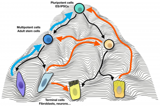 Changing cell fates. Source: MDPI
In vivo reprogramming of cells is a red hot topic in restorative research at the moment. The ability to change the fate of mature cells inside of an organism represents a ‘holy grail’-like goal for regenerative medicine.
Rather than transplanting ‘foreign’/external cells into the body (and having to deal with an immune system response), in vivo cellular reprogramming offers the possibility of simply changing the fate of a cell that is already present inside the body. And there are a lot of research groups around the world now exploring various methods of achieving this goal.
We have discussed the background research associated with in vivo cellular reprogramming for Parkinson’s in a previous SoPD post (Click here to read that post).
In today’s post, we are going to discuss a new research report that was published this month on this topic:
 Title: Glia-to-Neuron Conversion by CRISPR-CasRx Alleviates Symptoms of Neurological Disease in Mice.
Authors: Zhou H, Su J, Hu X, Zhou C, Li H, Chen Z, Xiao Q, Wang B, Wu W, Sun Y, Zhou Y, Tang C, Liu F, Wang L, Feng C, Liu M, Li S, Zhang Y, Xu H, Yao H, Shi L, Yang H.
Journal: Cell. 2020 Apr 1. pii: S0092-8674(20)30286-5. [Epub ahead of print]
PMID: 32272060
In this study, the researchers used a new technology (called CRISPR-CasRx) to reduce levels of a single protein, and the reduction of this protein appears to have aided the conversion of cells in the brains of mice to become dopamine neurons – the population of cells that are severely affected in Parkinson’s.
What is CRISPR-CasRx?
CRISPR-CasRx is a version of CRISPR that targets RNA rather than DNA.
What does any of that actually mean?
Ok, this is going to require some explanation. Let’s start at the beginning.
(AND IF YOU ARE NOT INTERESTED IN THE BIOLOGY LESSON ON CRISPR, THEN JUMP DOWN TO RECAP #2 – I WON’T BE OFFENDED)
In almost every cell of your body, there is a nucleus.
It is the command centre for the cell – issuing orders and receiving information concerning everything going on inside and around the cell. The nucleus is also a storage bank for the genetic blueprint that provides most of the instructions for making a physical copy of you. Those grand plans are kept bundled up in 23 pairs of chromosomes, which are densely coiled strings of a molecule called Deoxyribonucleic acid (or DNA).
 DNA’s place inside the cell. Source: Kids.Britannica
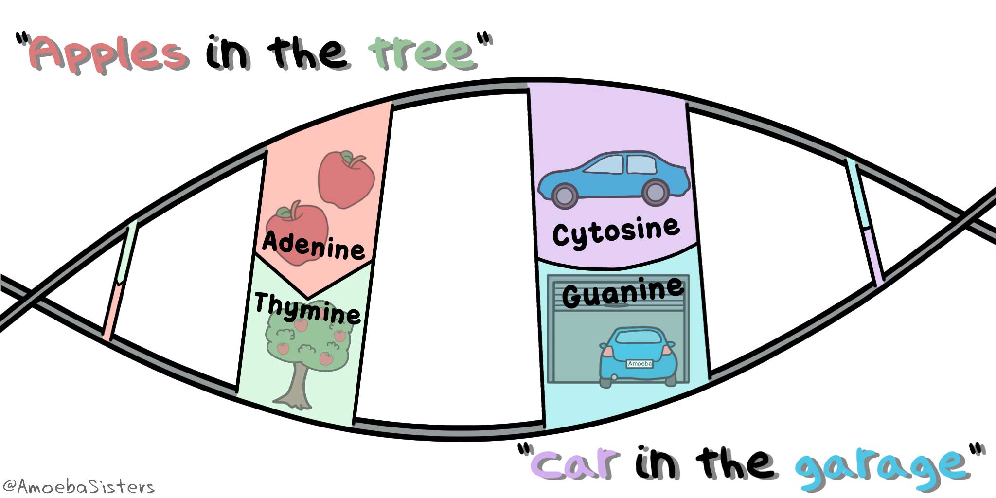
DNA is the ‘double helix’ stuff that biologists get excited about (Click here for the National Institute of Health’s website explanation of DNA). The information about ‘you’ is stored in DNA using a code, made up of four chemical bases: adenine (A), cytosine (C), guanine (G), and thymine (T). These As, Cs, Gs and Ts are paired up and strung together in a huge number of pairings – approximately 3 billion pairs in a single cell in fact.
And when I say ‘pairs’, I mean that these letters pair off — A with T and C with G . An easy way to remember these pairings is Apples are in the Tree and Cars are in Garages:
 Source: Pinterest
These pairings are called base pairs (the A, C, G & T being referred to as bases). Each base is also attached to a sugar molecule and a phosphate molecule. Together, these three components are called a nucleotide.
 The AGTCs of DNA. Source: Knowgenetics
Understand that these simple pairings hold us together. They are utterly critical to the normal biological functions of life – without them, there is no us.
Specific segments of these pairings in the DNA provide the instructions for making a protein. These regions are called ‘protein coding genes’. These segments give rise to a molecule called Ribonucleic acid (or RNA) through a process called transcription. RNA is the intermediate that is used in the production of a protein – a process called translation.
Curiously, large portions of DNA is made up of regions that do not give rise to RNA, while other regions give rise to RNA, but no protein is made using that RNA. These region are called ‘non-coding genes’ (the RNA that is produced has other functions in those situations – Click here to read more about this).
 ‘Protein coding’ or ‘Non-coding’. Source: Nature
Whether an RNA-producing region of DNA is a ‘protein coding’ or ‘non-coding’, these regions are all collectively referred to as genes. They are ‘functional’ segments of DNA. And it’s each of these genes that provide the instructions for keeping you alive.
BUT (and it’s a big BUT), with over 3 billion different base pairs, you will understand that occasionally things may go a little awry. An individual pairing may have an error or a small section of DNA may be deleted, especially during periods when the DNA is being replicated (such as when cells divide). In many cases these errors do not have any effect. And in other cases, they may actually be beneficial (spontaneous errors are one of the foundations in the process of evolution). But these errors can also have profoundly negative consequences, for example they can increase our chances of developing specific medical conditions.
Now, for the longest time…. well, basically all of human history, there has been no way to correct tiny errors (or variations) in our DNA. In fact, it’s only been in the last 60 years of homo sapiens entire existence that we have even become aware of them!
But the point is: Until now your DNA was set in stone – there was no way to correct it.
During the last 20-30 years, however, there have been Herculean efforts to insert functional bits of DNA into cells to replace a gene with an error in it. By inserting the healthy bit of DNA the cell can start to produce the required protein, and in most cases this restores normal functioning to the cell. But this has not altered the underlying fault in the DNA that required the insertion of functional DNA. So what has been required is a means of ‘editing’ DNA itself.
Then in 2013, this happened:
 Title: Multiplex genome engineering using CRISPR/Cas systems.
Authors: Cong L, Ran FA, Cox D, Lin S, Barretto R, Habib N, Hsu PD, Wu X, Jiang W, Marraffini LA, Zhang F.
Journal: Science. 2013 Feb 15;339(6121):819-23.
PMID: 23287718 (This article is OPEN ACCESS if you would like to read it)
Not one research report, but two – published back-to-back:
 Title: RNA-guided human genome engineering via Cas9.
Authors: Mali P, Yang L, Esvelt KM, Aach J, Guell M, DiCarlo JE, Norville JE, Church GM.
Journal: Science. 2013 Feb 15;339(6121):823-6.
PMID: 23287722 (This article is OPEN ACCESS if you would like to read it)
These two research reports introduced the world to CRISPR.
What is CRISPR?
Clustered Regularly Interspaced Short Palindromic Repeats (or CRISPR) are a series of repetitive sequences of DNA that were found in particular bacteria, which form a frontline defensive mechanism against infection from nasty viruses.
During the infection of a bacteria, a phage (a virus that infects bacteria) will inject its DNA into the cell. This DNA will be recognised as foreign DNA by two proteins (called Cas1 and Cas2), and they will chop it up into small pieces which will then be inserted into the bacterial DNA (a region referred to as a “CRISPR locus”). And once it is embedded in the DNA, this CRISPR locus can be passed on to any future cells (if that bacteria decides to divide and multiple).
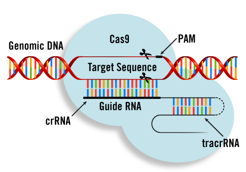
CRISPR regions of the DNA are regularly being transcribed (that is the process of producing RNA). The RNA for each spacer region is called crRNA (CRISPR-RNA). It is basically a small RNA molecule whose sequence matches a region of phage DNA. This crRNA then attaches to another piece of RNA called a tracrRNA.
This crRNA-tracrRNA then joins the Cas9 protein and together they start wandering around the bacteria looking for sections of DNA to check for that particular crRNA.
 How CRISPR works in bacteria. Source: Sciencedirect
Think of CRISPR as a bacterial defensive mechanism against infection from nasty viruses. Like a guard wandering around on patrol duty, looking for any signs of trouble at a prison.
|
# RECAP #1: DNA provides the instructions for making and maintaining an organism. Since the beginning of time this individual blueprint has been largely uncorrectable. New technology (called CRISPR-Cas9) translated from bacteria, now allows us to edit DNA. And this is being applied to all areas of biomedical research. # |
Hang on a second. What exactly is Cas9?
Cas9 (or CRISPR associated protein 9) is the important piece of the puzzle.
It is the protein that does the magic.
It is an endonuclease.
What is an endonuclease?
As I suggested above, replication of DNA is never perfect and there are occasionally errors. Millions of years of evolution has given rise to a very sophisticated, but extremely efficient system of DNA monitoring within cells. Many different proteins are involved in this process, and endonucleases play an important role.
An endonucleases is an enzyme that binds to DNA and cuts it.
Importantly, it does this function in the middle of the chain of DNA, while another class of enzyme (called exonucleases) operate from the end of the chains.
So Cas9 is an protein that connects to DNA and cuts it?
Exactly.
But it does this in a very targeted manner. Cas9 is being guided by pieces of RNA (the crRNA mentioned above) that are produced from the CRISPR regions of bacterial DNA. The crRNA is now often referred to as the ‘guide RNA’.
 Cas9 protein interacting with DNA. Source: Stackexchange
And if a particular Cas9 enzyme joins forces with a “guide RNA” and then finds a matching piece of phage DNA floating around inside the bacteria, then the bacteria will then know that this particular type of phage has infected it and the phage DNA should be disposed of ASAP. I should add that that this is not a conscious process for the bacteria – it is simply an acquired, automatic immune response that helps protect the bacteria.
Interestingly, only 40% of sequenced bacteria and 90% of archaea (single cell organisms that are separate to bacteria) have this CRISPR-Cas9 system. How the rest of the bacterial world are protecting themselves from phage and other nasties is an area of enormous research efforts.
Interesting. But how are researchers using this CRISPR-Cas9 ‘bacterial defense system’?
Well, in the two research reports from 2013 that I mentioned above, the scientists took this ‘bacterial defense system’ and they inserted it into human cells to see if it could work in that setting.
Why?
Because they wanted to know if the CRISPR-Cas9 system could be used to target and edit human DNA.
And did it?
It did.
The researchers took a CRISPR region from two different bacteria Streptococcus thermophilus and Streptococcus pyogenes, and they then designed and inserted spacers/crRNA for a particular human gene into that CRISPR region. Next, they inserted this CRISPR region and the Cas9 protein into the human cells and watched to see what would happen to that particular gene.
The results were very clear – they had demonstrated very accurate editing of DNA.
And it not fake news to suggest that these findings literally changed the world.
 Source: Amazon
It was a really big deal for biology at least. We are still waiting to see the application of the technology in the medical field (but it is coming!).
Since 2013, CRISPR-Cas9 has become a topic that is discussed on a daily basis in molecular biology laboratories all around the world. Everyone in biomedical research is now trying to incorporate this technology into some aspect of their work. And the approach has become much more simplified: now researchers simply insert the Cas9 protein into a cell and specific guideRNAs for the target gene that they want to mutate and then they let the cell do the rest.
 CRISPR experiment for removing (knocking-out) the function of a gene. Source: Genscript
And the CRISPR-Cas9 technology has also been re-engineered in different ways to allow alternative functions to be introduced. For example, the Cas9 protein has been re-engineered so that rather than cutting DNA, a specially designed Cas9 protein now activates that region of DNA where it is directed to be the guideRNA – providing a fantastic means of driving the activity of genes (Click here to read more about this).
Interesting. But is it just DNA that CRISPR works on?
Very interesting question.
And the answer is no. Some phage viruses do not infect bacteria with DNA, but rather RNA. And again, millions of years of evolution appear to have provided bacteria with a method of dealing with these RNA phage as well.
It is called “C2c2”.
Sounds like a Star Wars character, but C2c2 is an RNA-guided RNase.
What is a RNase?
RNases (or ribonucleases) are a broad class of RNA-degrading enzymes. They bind to RNA floating around inside of a cell and break it down.
And the discovery of C2c2 was reported in 2016:
 Title: C2c2 is a single-component programmable RNA-guided RNA-targeting CRISPR effector.
Authors: Abudayyeh OO, Gootenberg JS, Konermann S, Joung J, Slaymaker IM, Cox DB, Shmakov S, Makarova KS, Semenova E, Minakhin L, Severinov K, Regev A, Lander ES, Koonin EV, Zhang F.
Journal: Science. 2016 Aug 5;353(6299):aaf5573.
PMID: 27256883 (This report is OPEN ACCESS if you would like to read it)
The discovery of a RNA-targeting component of the CRISPR system gave biologists a powerful new way of not removing the production of a protein completely, but significantly reducing the level of that protein.
And the RNA-targetting version of CRISPR technology has been further investigated, refined, and improved by the discovery of a more efficient version of the Cas protein named “CasRx“.
Here is the research publication that introduced CasRx:
 Title: Transcriptome Engineering with RNA-Targeting Type VI-D CRISPR Effectors.
Authors: Konermann S, Lotfy P, Brideau NJ, Oki J, Shokhirev MN, Hsu PD.
Journal: Cell. 2018 Apr 19;173(3):665-676.e14.
PMID: 29551272 (This report is OPEN ACCESS if you would like to read it)
In this study, the researchers screened many different versions of the Cas protein (there are quite a few naturally occuring Cas proteins), before identifying Cas13d-NLS from Ruminococcus flavefaciens strain XPD3002 (or “CasRx”) as the most efficient at reducing levels of a targetted RNA in mammalian cells.
And CasRx is also small, which makes it ideal for inserting into a virus that could be used for biological research purposes.
|
# # RECAP #2: CRISPR-Cas9 technology is transforming modern biology, allowing researchers to manipulate DNA and make significant advancements in our understanding of many systems and diseases. CRISPR can also be used to affect RNA and this is now being used to explore the effect of differing levels of genes/proteins. # # |
Ok, so that is what CasRx is. Great. But what does any of this have to do with Parkinson’s?
So this brings us all the way back to the recently published published research report mentioned at the top of this post:
Title: Glia-to-Neuron Conversion by CRISPR-CasRx Alleviates Symptoms of Neurological Disease in Mice.
Authors: Zhou H, Su J, Hu X, Zhou C, Li H, Chen Z, Xiao Q, Wang B, Wu W, Sun Y, Zhou Y, Tang C, Liu F, Wang L, Feng C, Liu M, Li S, Zhang Y, Xu H, Yao H, Shi L, Yang H.
Journal: Cell. 2020 Apr 1. pii: S0092-8674(20)30286-5. [Epub ahead of print]
PMID: 32272060
In this study, the researchers used the CRISPR-CasRx technology to remove a single protein, and the reduction of this protein appears to have aided the conversion of cells in the brains of mice to become dopamine neurons – the population of cells that are severely affected in Parkinson’s.
A “single” protein?
Yep.
And that protein is called polypyrimidine tract-binding protein 1 (Ptbp1).
What is Ptbp1?
Ptbp1 is an RNA-binding protein with various functions related to RNA metabolism and transportation. It plays different roles in many stages of the life-cycle of mRNAs in the nucleus of cells.
And in 2013, researchers discovered something very curious about Ptbp1:
 Title: Direct conversion of fibroblasts to neurons by reprogramming PTB-regulated microRNA circuits.
Authors: Xue Y, Ouyang K, Huang J, Zhou Y, Ouyang H, Li H, Wang G, Wu Q, Wei C, Bi Y, Jiang L, Cai Z, Sun H, Zhang K, Zhang Y, Chen J, Fu XD.
Journal: Cell. 2013 Jan 17;152(1-2):82-96.
PMID: 23313552 (This report is OPEN ACCESS if you would like to read it)
In this study, the researchers reported that reducing levels of Ptbp1 protein was sufficient to cause skins cells to trans-differentiate into functional neurons. That is to say, when they reduced levels of Ptbp1 in skin cells (called fibroblasts), the cells became neurons.
Interestingly, as immature cells naturally develop in the brain and become adult neurons, levels of Ptbp1 drop significantly – suggesting that this protein is some kind of a handbrake on a cell with neuronal ambitions (Click here to read more about this).
The investigators explored the biological mechanisms behind this curious phenomenon, but it was immediately apparent that reducing this protein could have interesting possibilities for regenerative research. And this led to current study where the scientists used the CRISPR-CasRx technology to reduce Ptbp1, which appears to have aided the conversion of cells in the brains of mice to become neurons.
 Source: Pinterest
The investigators used a carefully engineered virus to introduce the CRISPR-CasRx-induced reduction of Ptbp1 into astrocytes.
Astrocytes? Can you remind me what they are?
Astrocytes are ‘helper cells’ in the brain.
They provide nutrients to neurons and make sure the environment surrounding the neurons is balanced and supportive.
 An astrocyte. Source: Wikipedia
The engineered virus was delivered to an area of the brain called the striatum. This is where most of the dopamine in the brain is released, but there are no dopamine neurons in this area. Rather, the dopamine neurons are located in a different region of the brain and they have long branches that then release the dopamine in the striatum.
The goal of delivering the virus to astrocytes in the striatum was to make dopamine neurons in the region where they are required to release dopamine.
The researchers found that 1 month after delivering the CRISPR-CasRx/Ptbp1-reducing virus to normal mice, 50% of the infected astrocytes in the striatum exhibited neuronal features, such as proteins like NeuN. About 8% of these cells showed characteristics of dopamine neurons.
Next, the investigators tested the virus in a mouse model of Parkinson’s (a unilateral 6-OHDA neurotoxin lesion of the right medial forebrain bundle, which causes a complete loss of dopamine neurons on one side of the brain). They found that by 3 months post viral delivery, >30% of the infected astrocytes in the striatum were dopamine-producing neurons (in mice injected with a control virus – no Ptbp1-reducing component – there were no dopamine neurons in the striatum).
And remarkably, these dopamine producing neurons (that used to be astrocytes) were producing enough dopamine in the brain that they almost entirely restored the motor/behavioural features of the Parkinsonian mice.
All together, the researchers concluded that “these results suggested that Ptbp1 knockdown-mediated neuron conversion in the striatum alleviated the motor dysfunctions in the PD mouse model”.
|
# # # RECAP #3: Researchers reported that the reduction of a single protein – called Ptbp1 – in astrocytes (supportive helper cells) in the brains, was enough to convert those cells in to neurons, some of which became dopamine producing neurons. In a mouse model of Parkinson’s, these dopamine neurons (that used to be astrocytes) were plentiful enough to correct the behavioural motor issues that charaterise this model. # # # |
Interesting. So when is the clinical trial starting?
Not for a long time. This is really blue sky stuff that needs to be independently validated and further characterised before it can go anywhere near the clinic. One has to ask, if only 30% of the infected cells become dopamine neurons, what do the rest of those infected cells do? This requires further investigation.
In addition, while it is very interesting that a single protein can have such a major impact on the fate of a cell, it would be interesting to better explore the biology behind this phenomenon and refine it before considering anything more adventurous.
Like I said: Blue sky stuff.
Do we know anything about Ptbp1 in Parkinson’s?
So this is an interesting question.
Because in 2015, researchers reported that Ptbp1 is significantly reduced in individuals with Parkinson’s:
 Title: Network-based metaanalysis identifies HNF4A and PTBP1 as longitudinally dynamic biomarkers for Parkinson’s disease.
Authors: Santiago JA, Potashkin JA.
Journal: Proc Natl Acad Sci U S A. 2015 Feb 17;112(7):2257-62.
PMID: 25646437 (This report is OPEN ACCESS if you would like to read it)
In this study, the researchers were looking for biomarkers in individuals with Parkinson’s. They collected data from four independent studies that had collected RNA from blood taken from people with PD. They found that the highest levels of RNA (compared to control samples) came from a gene called hepatocyte nuclear factor 4 alpha (HNF4A), while the lowest levels of RNA (compared to control samples) came from a gene called…. you guessed it: PTBP1
But when the researchers analysed HNF4A and PTBP1 RNA levels in blood samples collected over time, they found that while HNF4A levels decreased with time, PTBP1 went up.
Now all of this data is collected from blood, which may be different to what is occuring in the brain, but it is still interesting that this gene has already been investigated in the context of Parkinson’s. And the researchers involved in this study have followed up their research and expanded on it.
In a more recent publication (Click here to read that report), they found that levels of PTBP1 were significantly higher in people with Parkinson’s who were cognitively normal compared to individuals with Parkinson’s with mild cognitive impairment.
So what does it all mean?
One of the themes for 2020 here at the SoPD is a focus on regenerative research for Parkinson’s.
I am seeking to explore efforts to restore some of the lost function that the condition has taken away. While there are a lot of clinical trials are focused on agents targeting disease halting approaches, there is a pressing need for more emphasis on regenerative/restorative methods. The current batch of restorative efforts are largely focused on stem cell-based transplantation (see the last SoPD post for more no that).
The research reviewed in this current post is – as I have said – blue sky. It is new and exciting, but the translation of it to humans is still off on the horizon. ‘So why mention it?‘ someone might ask. The answer: to make the community aware that it is being explored and to stimulate some discussion about it. And perhaps even inspire some young researcher who is looking for green pastures to explore, and maybe they have some insight into a missed angle associated with this research.
Beyond that, I just find it really interesting.
Be safe.

All of the material on this website is licensed under a
Creative Commons Attribution 4.0 International License
You can do whatever you like with it!
The banner for today’s post was sourced from interaliamag.

Thanks for sharing this really informational article. Keep sharing your good work.
LikeLike
Well written and incredibly informative. Thank you so very much!
LikeLike
Hi Andrew,
Thanks – glad you liked the post.
Kind regards,
Simon
LikeLike