|
# # # # Recently a lot of media attention has been focused on a new study that reported the replacement of lost dopamine neurons in a mouse model of Parkinson’s, which resulted in the correction the associated behavioural/motor issues. The researchers involved achieved this amazing feat by simply reducing a single protein in a special type of helper cell in the brain, called astrocytes. By lowering the levels of the protein, they were able to transform the astrocytes into dopamine neurons. Intriguingly, the study represented independent replication of a previous study that demonstrated a similar result – transformation of astrocytes inside a mouse brain into dopamine neurons by reducing a single protein. The protein in both studies is called Ptbp1, and in today’s post we will discuss what this protein does, what the new study found, and what the implications of this work could be. # # # # |
 Source: Howtogeek
Earlier this year, I stated in my 2020 wish list for Parkinson’s research (Click here to read that post) that one of the big themes I was hoping to see more of was further research on regenerative approaches for the condition.
We have discussed this a few times, but any “curative” treatment for Parkinson’s will require 3 components:
- A disease halting mechanism – to slow/stop the progression of the disease
- A neuroprotective agent – to protect the remaining cells & provide a nurturing environment for,
- Some form of restorative/regenerative therapy – replacing what has been lost
Now the encouraging news is that if you look at the SoPD “The Road Ahead: 2020” post, you will see that there is a great deal of research being conducted on all three of these components at the clinical stage (in addition to vast amounts of work on the preclinical level).
But it is fair to say that the bulk of the clinical research being conducted on restorative therapy for Parkinson’s is centred around the transplantation of stem cell-derived dopamine neurons to replace the cells that have been lost in Parkinson’s (click here to read a recent SoPD post on this topic).
 Embryonic stem cells in a petridish. Source: Wikipedia
In my wish list for 2020, I was hoping to see regenerative approaches beyond the well trodden path of cell transplantation (growing cells in culture and then injecting them into the brain).
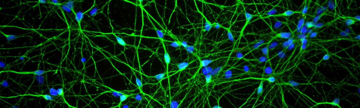 Dopamine neurons (green) in cell culture. Source: Axolbio
Rather, I was hoping to see more research on new regenerative approaches that target/manipulate endogenous pathways in the brain – forcing changes within the central nervous system itself.
I didn’t have high expectations in this department, but I have to admit that now I have been pleasantly surprised by the number of research reports that have been published thus far this year highlighting novel regenerative approaches. We have discussed several of them here on the SoPD already (Click here and here for examples), and today we are going to review another which was recently published in the prestigious scientific journal Nature.
This is what all the news papers have been talking about?
Indeed. There has been a lot of media attention focused on this research report.
So what does the new study report?
This is the study in question:
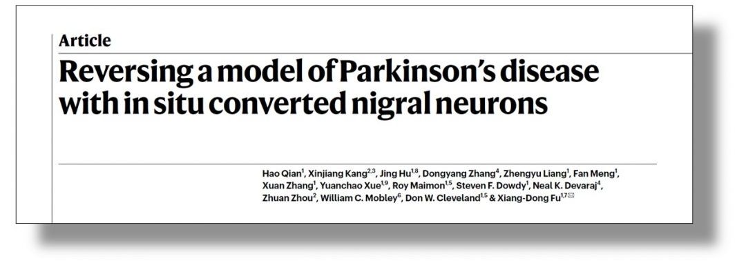 Title: Reversing a model of Parkinson’s disease with in situ converted nigral neurons.
Authors: Qian H, Kang X, Hu J, Zhang D, Liang Z, Meng F, Zhang X, Xue Y, Maimon R, Dowdy SF, Devaraj NK, Zhou Z, Mobley WC, Cleveland DW, Fu XD.
Journal: Nature. 2020 Jun;582(7813):550-556.
PMID: 32581380
In this report, the researchers demonstrated a method of transforming astrocytes into dopamine neurons.
Astrocytes? Can you remind me what they are?
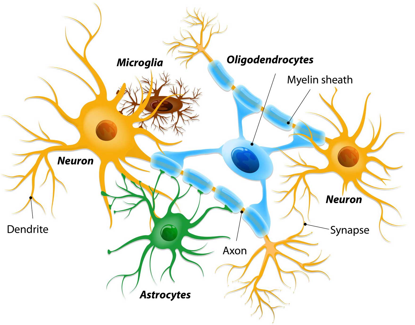
There are different cell types in the brain. Neurons are one of the main types of cells, and they are involved in cognitive activity. But in addition to these there are numerous supporting (or “helper”) cells, which include microglia, oligodendrocytes, or astrocytes.
 Different types of cells in the brain. Source: Dreamstime
Microglia are the resident immune cells in the brain, keeping an eye out for disease causing pathogens and cleaning up when there is damage. Oligodendrocytes insulate neurons and help with rapid transmission of signals across the brain (Click here to read a recent SoPD post on oligos).
Astrocytes are really the key supportive cells for neurons. They provide them with nutrients and make sure the environment surrounding the neurons is balanced and agreeable. They are plentiful and work hard to keep neurons happy and functioning.
 A human astrocyte. Source: Wikipedia
I see. And the researchers turned these astrocytes into dopamine neurons?
Yes, a rather amazing achievement considering they are very different types of cells.
Mmmm. But what exactly is a dopamine neuron?
Dopamine is a chemical in the brain that helps to pass signals between neurons. In this fashion, it is referred to as a neurotransmitter – transmitting a signal from one neuron to the next. There are specific groups of neurons in the brain that produce dopamine, and these are called dopamine neurons (an original name, right?).
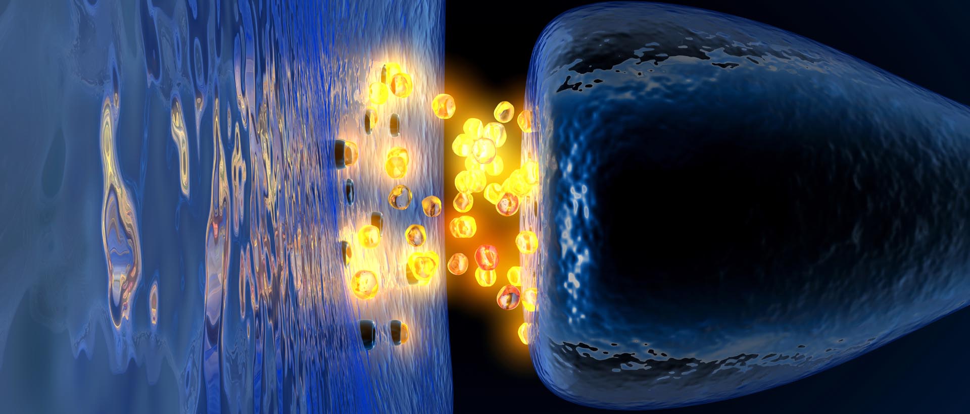 Dopamine being released by one cell and binding to another. Source: Truelibido
These dopamine neurons are critically involved in normal motor function – without dopamine, movement becomes very inhibited.
And this relationship demonstrates itself in Parkinson’s: By the time a person is presenting the motor features characteristic of Parkinson’s (rigidity, slowness of movement, and tremor) and being referred to a neurologist, they have already lost approximately 50% of the dopamine producing neurons in an area of the brain called the midbrain.
 The dark pigmented dopamine neurons in the substantia nigra are reduced in the Parkinsonian brain (right). Source:Memorangapp
Until we have developed methods that can identify Parkinson’s long before these cells are lost and the motor features appear, some form of cell replacement therapy is required to introduce new cells to take up the lost function.
Hence the reason why transforming astrocytes into dopamine neurons is a pretty cool trick.
|
# RECAP #1: Researchers have recently attracted a lot of media attention as they demonstrated a method for changing astrocytes into neurons that produce the chemical dopamine – a neurotransmitter that is severely reduced in the brains of people with Parkinson’s. Astrocytes are helper cells in the brain that support neurons and help to keep them happy. # |
But how did the researchers change the astrocytes into dopamine neurons?
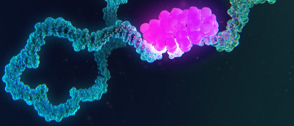
By reducing levels of a protein called polypyrimidine tract-binding protein 1 (Ptbp1).
What is Ptbp1?
Ptbp1 is an RNA-binding protein with various functions related to RNA metabolism and transportation. It plays different roles in many stages of the life-cycle of RNAs in the nucleus of cells. Like other RNA-binding proteins, Ptbp1 helps to stablise RNAs as they are transported out of the nucleus of a cell.
 Source: Merck
But in 2013, researchers discovered something very curious about Ptbp1:
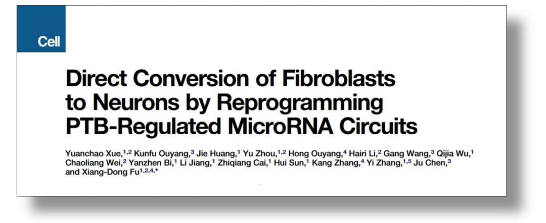 Title: Direct conversion of fibroblasts to neurons by reprogramming PTB-regulated microRNA circuits.
Authors: Xue Y, Ouyang K, Huang J, Zhou Y, Ouyang H, Li H, Wang G, Wu Q, Wei C, Bi Y, Jiang L, Cai Z, Sun H, Zhang K, Zhang Y, Chen J, Fu XD.
Journal: Cell. 2013 Jan 17;152(1-2):82-96.
PMID: 23313552 (This report is OPEN ACCESS if you would like to read it)
In this study, the researchers reported that reducing levels of Ptbp1 protein was sufficient to cause adult mouse skins cells to transform into functional neurons. That is to say, when they reduced levels of Ptbp1 in skin cells (called fibroblasts), the cells became neurons.
Interestingly, as immature cells naturally develop in the brain and become adult neurons, levels of Ptbp1 drop significantly – suggesting that this protein is some kind of a handbrake on a cell with neuronal ambitions (Click here to read more about this).
But when the researchers attempted to repeat this study in adult human fibroblasts, they found that it was a little more complicated. They reported their human skin cell findings in this study:
 Title: Sequential regulatory loops as key gatekeepers for neuronal reprogramming in human cells.
Authors: Xue Y, Qian H, Hu J, Zhou B, Zhou Y, Hu X, Karakhanyan A, Pang Z, Fu XD.
Journal: Nat Neurosci. 2016 Jun;19(6):807-15.
PMID: 27110916 (This report is OPEN ACCESS if you would like to read it)
In this study, the researchers found that reducing Ptbp1 protein in adult human fibroblasts was insufficient to cause them to transform into neurons. They found that they needed to sequential inactivate Ptbp1 and then a paralog of Ptbp1, called nPTB.
What is a paralog?
A paralog is a copy of a gene (created by a duplication event within the genome).
So there is a duplicate copy of Ptbp1 in human DNA?
There are two paralogs Ptbp1 in humans: nPTB (which is mainly present in brain cells) and ROD1 (which is mostly found in blood cells).
The researchers found that in human adult fibroblasts they needed to reduce both Ptbp1 and then nPTB in order to transform the skin cell to a neuron. So in humans there appears to be two ‘gatekeepers’ in this process.
Ok, so getting back to the new study, what did the researchers find when they transformed the astrocytes to neurons?
In their new study, the researchers wanted to see if changing astrocytes to neurons could be used in a neurodegenerative condition like Parkinson’s as a means of introducing new cells and correcting the condition.
Title: Reversing a model of Parkinson’s disease with in situ converted nigral neurons.
Authors: Qian H, Kang X, Hu J, Zhang D, Liang Z, Meng F, Zhang X, Xue Y, Maimon R, Dowdy SF, Devaraj NK, Zhou Z, Mobley WC, Cleveland DW, Fu XD.
Journal: Nature. 2020 Jun;582(7813):550-556.
PMID: 32581380
The investigators firstly examined whether reducing Ptbp1 alone would cause the transformation of astrocytes to neurons in cell culture. Curiously, they found that in both mouse and human cells, only reducing Ptbp1 was required to convert astrocytes to neurons.
Next they wanted to explore if this transformation would work inside the brain, so they injected a carefully designed virus into the brains of mice. The virus was engineered to infect astrocytes and cause them to reduce Ptbp1. To their amazement, just 3 weeks after injecting the mice, the researchers found 20% of infected astrocytes exhibited characteristics of neurons. And by 10 weeks, this percentage had climbed to 80% (important to remember that this is the percentage of infected astrocytes exhibiting neuronal characteristics).
But most interesting of all, when the investigators reduced levels of Ptbp1 in the midbrain, they saw the local infected astrocytes convert to dopamine neurons.
One interesting aspect of these experiments is that the researchers saw a regional effect with the transformation of astrocytes to neurons. By this I mean that when they reduced Ptbp1 levels in the astrocytes in the substantia nigra of the midbrain in mice, they saw the astrocytes transform into dopamine neurons (neurons from this region). But when they performed the same experiment in the cortex (a different region of the brain) the astrocytes in that area transformed into more regionally appropriate neurons (with the same level of conversion efficiency). The researchers concluded that this may relate to the regional specific nature of astrocytes (Click here to read more about this).
Ok, so the scientists could generate dopamine neurons from astrocytes. What did they do next?
Next they looked to see if the ‘new’ dopamine neurons could send out branches (or axons) to the appropriate regions of the brain and make connections with the right targets.
And by 12 weeks post viral infection in the midbrain, the researchers began observing increased numbers of new dopamine fibres in the striatum – this is the region that dopamine neurons in the substantia nigra send their branches to and where they release the bulk of their dopamine.
In the image below you can see a comparison between the dopamine neurons (blue) in the substantia nigraof the human brain with the dopamine neurons in the same region of the mouse brain (blue). Both sets of neurons project their branches to the striatum (it doesn’t really need to be said, but these brains are not drawn to scale).
 Source: Semanticscholar
Following this proof-of-principle in a normal mouse, the researchers next asked if this conversion technique could be used to replenish the stock of dopamine neurons in the midbrain of a model of Parkinson’s and correct the behavioural issues associated with that model.
The investigators used a neurotoxin to degenerate the dopamine neurons on one side of the brain and they infected astrocytes in the midbrain on that side of the brain with the Ptbp1-reducing virus. 10-12 weeks after injecting the virus, the researchers started to see new dopamine neurons appearing in the midbrain and dopamine branches in the striatum of the Ptbp1-reducing virus treated animals (they saw no new dopamine neurons in mice treated with a control virus).
When they counted the number of dopamine neurons in the substantia nigra of the mice, the researchers found that the neurotoxin killed 90% of the original total, but the treatment with the Ptbp1-reducing virus restored this by 1/3. A similar percentage of dopamine branches were observed in the striatum of the mice treated with the Ptbp1-reducing virus. And this resulted in a significant increase in the level of dopamine being released in the striatum (approximately 65% of the intact side of the brain in the mice treated with the Ptbp1-reducing virus – compared to just 25% in the control virus treated mice).
And this increase in dopamine being released was enough to correct the behavioural issues associated with this mouse model of Parkinson’s (at approximately 3 months post treatment).
Finally, to finish off their studies the researchers asked whether they needed to use viruses that cause the continuous reduction of Ptbp1 levels – could the same effect be generated by a short/transcient reduction in Ptbp1 levels?
So they tested this idea by using antisense oligonucleotides.
What are antisense oligonucleotides?
Antisense oligonucleotides are small pieces of DNA or RNA that are designed to target and bind to very specific strands of RNA (in this case Ptbp1 RNA). By doing this, the antisense oligonucleotide blocks the ability of that piece of RNA to be used to make a protein. This reduces the amount of that protein in the cell and the inhibited piece of RNA is eventually disposed of. We have discussed how antisense oligonucleotides are being developed for Parkinson’s in a previous SoPD post (Click here to read that post).
 Source: rdmag
In this current study, the researchers injected Ptbp1-targetting antisense oligonucleotides into the midbrain of a neurotoxin Parkinson’s mouse model, which resulted in a short-term reduction of Ptbp1 levels. Once again, at 12 weeks post treatment, the researchers observed an increase in the number of dopamine neurons in the substantia nigra of the mice injected with Ptbp1-targetting antisense oligonucleotides (vs no increase in the control animals). And this resulted in a correction in the behavioural motor issues associated with the model.
The investigators concluded their study by suggesting that these “antisense oligonucleotide-based experiments illustrate a potentially clinically feasible approach for treatment of patients with Parkinson’s“.
|
# # RECAP #2: By reducing levels of a protein called polypyrimidine tract-binding protein 1 (or Ptbp1) in astrocytes, researchers observed the transformation of these cells in the midbrain into dopamine neurons. They performed this transformation technique in a model of Parkinson’s and they were able to restore the number of dopamine neurons by 1/3, which resulted in an increase in dopamine and the correction of behavioural motor issues associated with the model. # # |
Amazing. Has anyone ever seen these sorts of results before?
In April of this year another (independent) research team published this report:
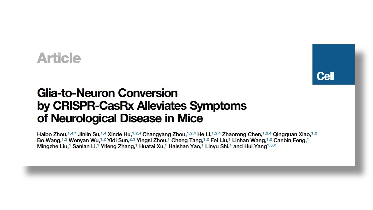 Title: Glia-to-Neuron Conversion by CRISPR-CasRx Alleviates Symptoms of Neurological Disease in Mice.
Authors: Zhou H, Su J, Hu X, Zhou C, Li H, Chen Z, Xiao Q, Wang B, Wu W, Sun Y, Zhou Y, Tang C, Liu F, Wang L, Feng C, Liu M, Li S, Zhang Y, Xu H, Yao H, Shi L, Yang H.
Journal: Cell. 2020 Apr 1. pii: S0092-8674(20)30286-5. [Epub ahead of print]
PMID: 32272060
We have discussed this report in a previous SoPD post (Click here to read that post), but in effect these scientists saw the same result – if you reduce Ptbp1 in astrocytes in the mouse brain, they will transform into dopamine neurons and correct some of the motor impairments in a model of Parkinson’s. There are some subtle differences between the study, but they basically validate each other.
Wow! So where can I sign up for the clinical trial?
It is easy to get excited by all of the media coverage that this new study has received (and it is an impressive piece of work), but it is also important to remember that all of this research has been conducted in mice or cells in culture. Translation to humans (if possible) will be a long process.
 Source: Pinterest
It is encouraging to note that the authors of the new Nature report state that “The University of California, San Diego has filed a patent application on neuronal reprogramming induced by inactivating PTB by any means for treatment of neurological disorders”.
But any potential clinical use of this technology is going to be tricky.
The first clinical applications may come from the field of ophthalmology (the treatment of eye disorders). Before going into the brain, we could achieve more rapid proof of priniciple in eye conditions. In the Zhou et al report (from April), the researchers actually demonstrated conversion of glial cells into retinal ganglion cells in the mouse retina – leading to the correction of symptoms associated with loss of vision – before then attempting the conversion process in the brain.
A similar proof of principle approach has been taken with the clinical development of induced pluripotent stem cells – in Kyoto, before attempting cell transplantation in Parkinson’s, clinical trials were initiated to treat age-related macular degeneration in the eye (Click here to read more about this).
 Source: Youtube
With regards to Parkinson’s, the next step mostly will come in the form of further characterisation in different models of Parkinson’s (will the transformation method have the same efficiency/effect in alpha synuclein models?). Importantly, most of the current data has been conducted in very young mice (2 months old), it will be necessary to further explore the efficancy of the conversion process in older mice. Then there will be feasibility and dosing studies in non-human primates (is it possible to turn astrocytes into neurons – particularly dopamine neurons – in the aged primate brain?).
If this work, it is likely that human clinical trials will take a long time – as is the case with all first-in-human clinical trials (see recent SoPD post on a vaccine for Parkinson’s – a Phase I study that took 4 years!). We are currently awaiting news from various Phase I clinical trials for cell transplantation in Parkinson’s, which also have very long follow up periods.
And I hate to sound like I’m raining on the parade here, but in addition to these clinical process matters, there will be some specific technical challenges with translating to work to humans. Beyond getting the dose of treatment right, delivery will also be an issue that will need attention.
Delivery?
Yes, specifically the targetting of the delivery.
The proposed antisense oligonucleotide approach will basically affect all cell types in the region that this treatment is delivered into, which may not be desired. In addition, I am not currently aware of any clinical grade viral vectors (used in gene therapy) that only target astrocytes (and I am happy to be corrected on this).
But the targetting astrocytes specifically will be required.
Why do you say that?
Just days after the Nature report was published, another study highlighting Ptbp1 was made public. This one was focused on Alzheimer’s:
 Title: Selective Neuronal Vulnerability in Alzheimer’s Disease: A Network-Based Analysis.
Authors: Roussarie JP, Yao V, Rodriguez-Rodriguez P, Oughtred R, Rust J, Plautz Z, Kasturia S, Albornoz C, Wang W, Schmidt EF, Dannenfelser R, Tadych A, Brichta L, Barnea-Cramer A, Heintz N, Hof PR, Heiman M, Dolinski K, Flajolet M, Troyanskaya OG, Greengard P
Journal: Neuron. 2020 Jun 24:S0896-6273(20)30439-6. Online ahead of print.
PMID: 32603655
In this study, the researchers were exploring large data bases of information relating to types of neurons in the brain that are vulnerable in Alzheimer’s. Specifically, they were looking for increases or decreases in levels of proteins that could be making certain neurons more vulnerable to the disease.
And take a wild guess which protein they identified?
I’m going out on a limb here, but… Ptbp1?
Yep.
They found that levels of Ptbp1 were higher in various regions of the Alzheimer’s brain when compared to unaffected control brains.
Interesting. But how is that a problem? Maybe reducing Ptbp1 could help with Alzheimer’s?
The researchers went on to explore what effect reducing Ptbp1 in neurons would have and they observed a significant reduction in levels of Alzheimer’s associated Tau, BUT they also witnessed a large increase in levels of SNCA RNA.
What is SNCA RNA?
SNCA RNA is the RNA that provides the instructions for making the Parkinson’s-associated alpha synuclein (yes, that alpha synuclein, the protein that clumps together and is believed to cause trouble in the PD brain).
So reducing Ptbp1 in neurons may increases the levels of the Parkinson’s-associated protein alpha synuclein – which might not be an ideal outcome.
Thus, delivery to the right cells will be critical. There is a great deal of research still to be done on this new ‘break through’. And while I will be watching intently for any new data, this conversion approach is definitely in my ‘blue sky’ basket.
So what does it all mean?
Two groups of researchers have recently presented data suggesting that reducing levels of a single protein in astrocytes in the mouse brain can result in those cells transforming into neurons. This conversion has led to the correction of mouse models of Parkinson’s, and generated media headlines such as ‘one and done’ suggesting that a curative therapy may be just ‘5 years away’.
One of the really impressive aspects of this research is that two independent research groups have rapidly come to the same result, demonstrating replication of an amazing biological effect in a relatively short space of time. It is mind boggling how fast preclinical developments progressing at the moment (even in the midsts of a pandemic!).
But while independent replication of this approach is very encouraging, I think it is important to be careful about managing expectations here. The new report is a fascinating example of a novel “regenerative approach that targets/manipulates endogenous pathways in the brain” for our 2020 ‘wish list’, but headlines like ‘one and done’ suggest a level of simplicity that is unlikely. Despite all of the media attention, this is still very blue sky research – exciting yes, but a lot of research is required before this conversion technology will get close to the clinic.
Fingers crossed it gets there though.

All of the material on this website is licensed under a
Creative Commons Attribution 4.0 International License
You can do whatever you like with it!
The banner for today’s post was sourced from embl.

This is fabulous research which *restores lost brain function*! It is not palliative–it is genuinely restorative. Yes, it is going to take some time before humans can benefit from it, but this is our light at the end of the tunnel so far as motor (and motivation) issues are concerned.
Having said that, it does not stop the autoimmune process that grinds down dopamine neurons, so it would be setting back a clock that would nonetheless continue to run. It might therefore have to be repeated.
And it would presumably do nothing to stop the spread of aggregated alpha-synuclein through the brain, which results in autonomic issues and dementia.
But…if this successfully makes its way into clinical practice, it will take a huge bite out of PD. It’s the best news we have had for a very long time.
I’m wondering: do astrocytes have the ability to replenish themselves? Or do patients simply already have far more of them than they require?
LikeLike
Hi Lou,
Good to hear from you – thanks for the interesting comment. Yes, it is exciting stuff, and no it would likely do nothing to slow disease progression and inflammation (improve motor features perhaps, but the underlying disease biology would still be unresolved). Hence, one can only refer to this as a symptomatic therapy? That said, as we discussed at the start of the post, this is just one component (#3) of a larger ‘curative therapy’ picture.
As for the astrocyte depletion issue, fear not. While we have a finite number of neurons (and they are not replaced when lost), astrocytes are being replenished throughout life (this is a good review on the topic: https://dev.biologists.org/content/143/7/1075). The number of astrocytes can vary between different regions of the brain (https://link.springer.com/article/10.1007/s11064-020-02959-7). Further research is required, however, to determine how the neurons that were dependent on the now transforming astrocytes handle their absence. Astrocytes are the supporting cells after all.
Kind regards,
Simon
LikeLike
My WordPress account became temporarily inaccessible and so I missed your reply until now. Thank you for that very helpful answer.
That’s an interesting question you ask, regarding whether the therapy is only symptomatic as opposed to curative. I prefer to think in terms of the similar dichotomy: manipulative vs. restorative.
All medical treatments include both a manipulative and a restorative aspect. Sometimes, they lean heavily toward one end or the other of that continuum, but there is generally some of each involved.
So here we are manipulating astrocytes into regressing to a state closer to their stem cell roots, so that from that starting point they can re-manifest as dopamine neurons.
But then after that manipulation, those re-purposed cells implant and extend axons from the substantia nigra into the striatum, in much the same way that they do when a person is in the early stages of development. That part is a natural process; it is just occurring during adulthood instead of during the earliest stages of development. So a developmental process is wonderfully repurposed as a healing process.
I would certainly describe that repurposed process as restorative. The only reason it cannot be described as a cure is because there are other aspects of the disease process that remain unaddressed. But because it includes those restorative elements, I would describe it as a “partial cure.”
Ah, so you’re saying that the repurposed astrocytes might have had specific relationships with other surviving neurons, which would be disrupted by their going off to neuron college, thus leaving behind their responsibilities to those neurons.
So would other astrocytes take over those responsibilities? Well, yes, whenever we mess with something, we risk unforeseen consequences; such is the nature of hubris. Sound like a chance worth taking, though.
LikeLike
Lou T. said: “… there are other aspects of the disease process that remain unaddressed.”
Well, it looks like one of those aspects is now being addressed, via a different ASO [1].
[1] Anti-a-synuclein ASO delivered to monoamine neurons prevents a-synuclein accumulation in a Parkinson’s disease-like mouse model and in monkeys, Diana Alarcon-Aris et al., EBioMedicine 59 (2020) 102944.
https://www.sciencedirect.com/science/article/pii/S2352396420303200
LikeLiked by 1 person
On the alpha-synuclein front, we also have the fact that both curcumin and EGCg have the effect of preventing alpha-synuclein aggregation and also of disentangling alpha-synuclein that has already undergone aggregation. These therapies are available right now. But they are only helpful; not a complete solution to the problem.
LikeLike