In 2018, there is one particular clinical trial that I will be watching, because the drug being tested could have a big impact on certain kinds of Parkinson’s.
The clinical trial is focused on people with cancer and they will be treated with a drug called TVB-2640. TVB-2640 is an inhibitor of an enzyme called fatty acid synthase (or FAS).
In today’s post we will discuss why TVB-2640 might be a useful treatment for certain kinds of Parkinson’s.
Mitochondria and their location in the cell. Source: NCBI
Regular readers of this blog are probably getting sick of the picture above.
I use it regularly on this website, because a.) it nicely displays a basic schematic of a mitochondrion (singular), and where mitochondria (plural) reside inside a cell. And b.) a lot of evidence is pointing towards mitochondrial dysfunction in Parkinson’s.
What are mitochondria?
Mitochondria are the power stations of each cell. They help to keep the lights on. Without them, the party is over and the cell dies.
How do they supply the cell with energy?
They convert nutrients from food into Adenosine Triphosphate (or ATP). ATP is the fuel which cells run on. Given their critical role in energy supply, mitochondria are plentiful (some cells have thousands) and highly organised within the cell, being moved around to wherever they are needed.
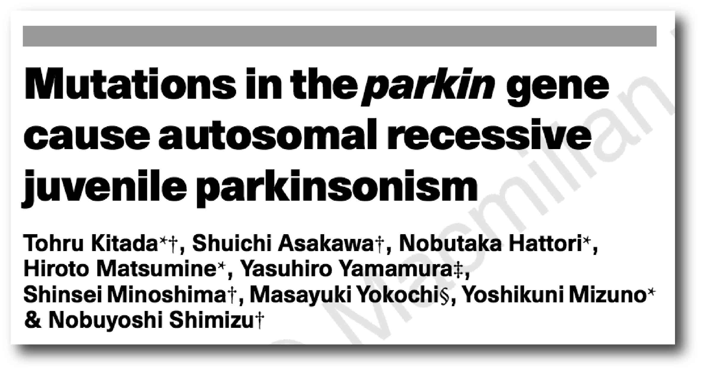
Last week, as everyone was preparing for Christmas celebrations, researchers at the pharmaceutic company Novartis published new research on a gene that is involved with Parkinson’s, called PARKIN (or PARK2).
They used a new gene editing technology – called CRISPR – to conduct a large screening study to identify proteins that are involved with the activation of PARKIN.
In today’s post we will look at what PARKIN does, review the research report, and discuss how these results could be very beneficial for the Parkinson’s community.
As many people within the Parkinson’s community will be aware, 2017 represented the 200th anniversary of the first report of Parkinson’s disease by James Parkinson.
It also the 20th anniversary of the discovery of first genetic mutation (or variant) that increases the risk of developing Parkinson’s. That genetic variation occurs in a region of DNA (a gene) called ‘alpha synuclein’. Yes, that same alpha synuclein that seems to play such a critical role in Parkinson’s (Click here to read more about the 20th anniversary).
In 2018, we will be observing the 20th anniversary of the second genetic variation associated with Parkinson.
That gene is called PARKIN:
Title: Mutations in the parkin gene cause autosomal recessive juvenile parkinsonism. Authors: Kitada T, Asakawa S, Hattori N, Matsumine H, Yamamura Y, Minoshima S, Yokochi M, Mizuno Y, Shimizu N Journal: Nature. 1998 Apr 9; 392(6676):605-8 PMID: 9560156
In 1998, Japanese researchers published this report based on 5 individuals from 4 Japanese families who were affected by juvenile-onset Parkinson’s. In family 1, the affected individual was a female, 43 years old, born of first-cousin parents, and her two younger brothers are healthy. Her condition was diagnosed in her teens and it had then progressed very slowly afterwards. Her response to L-dopa was very positive, but L-dopa-induced dyskinesia were frequent. In family 2-4, affected individuals (born to unrelated parents) exhibited very similar clinical features to the subject in family 1. The age of onset was between 18 to 27 years of age.
Using previous research and various techniques the investigators were able to isolate genetic variations that were shared between the 5 affected individuals. They ultimately narrowed down their search to a section of DNA containing 2,960 base pairs, which encoded a protein of 465 amino acids.
Denali (Koyukon for “the high one”; also known as Mount McKinley) in Alaska is the highestmountain peak in North America, with a summit elevation of 20,310 feet (6,190 m) above sea level. The first verified ascent to Denali’s summit occurred on June 7, 1913, by four climbers Hudson Stuck, Harry Karstens, Walter Harper, and Robert Tatum.
Tatum (left), Karstens (middle), and Harper (right). Source: Gutenberg
Robert Tatum later commented, “The view from the top of Mount McKinley is like looking out the windows of Heaven!”
More recently another adventurous group associated with ‘Denali’ have been trying to scale lofty heights, but of a completely different sort from the mountaineering kind.
There is a protein in most of the cells in your body called “PTEN-induced putative kinase 1″ (or simply PINK1). It plays an important role in keeping your cells healthy.
Genetic variations in the PINK1 gene have been shown to increase ones risk of developing Parkinson’s.
This week researchers have identified a method by which the function of the PINK1 protein can be inhibited and this results in increased vulnerability to Parkinson’s. In this post, we will look at what PINK1 does, how it is inhibited, and what this could mean for the Parkinson’s community.
Mitochondria (green) in health cells (left) and in unhealthy cells (right). The nucleus of the cell is in blue. Source: Salk Institute
I have previously spoken a lot about mitochondria and Parkinson’s on this website.
For the uninitiated, mitochondria are the power house of each cell. They help to keep the lights on. Without them, the party is over and the cell dies.
Mitochondria and their location in the cell. Source: NCBI
You may remember from high school biology class that mitochondria are tiny bean-shaped objects within the cell. They convert nutrients from food into Adenosine Triphosphate (or ATP). ATP is the fuel which cells run on. Given their critical role in energy supply, mitochondria are plentiful (some cells have thousands) and highly organised within the cell, being moved around to wherever they are needed.
Like you and I and all other things in life, however, mitochondria have a use-by date.
As mitochondria get old and worn out (or damaged) with time, the cell will recycle them via a process called mitophagy (a blending of the words mitochondria and autophagy which is the waste disposal system of each cell).
What does this have to do with Parkinson’s disease?
In American slang, to ‘nix‘ something is to ‘put an end to it’.
Curiously, a protein called NIX may be about to help us put an end to Parkinson’s disease, at least in people with specific genetic mutations.
In today’s post we will look at what NIX is, outline a new discovery about it, and discuss what this new information will mean for people living with Parkinson’s disease.
Before we start, I would like the reader to appreciate that I am putting trans-Tasman rivalry side here to acknowledge some really interesting research that is being conducted in Australia at the moment.
And this is really interesting.
I have previously spoken a lot about mitochondria and Parkinson’s on this website. For the uninitiated, mitochondria are the power house of each cell. They help to keep the lights on. Without them, the party is over and the cell dies.
Mitochondria and their location in the cell. Source: NCBI
You may remember from high school biology class that mitochondria are tiny bean-shaped objects within the cell. They convert nutrients from food into Adenosine Triphosphate (or ATP). ATP is the fuel which cells run on. Given their critical role in energy supply, mitochondria are plentiful (some cells have thousands) and highly organised within the cell, being moved around to wherever they are needed.
Like you and I and all other things in life, however, mitochondria have a use-by date.
As mitochondria get old and worn out (or damaged) with time, the cell will recycle them via a process called mitophagy (a blending of the words mitochondria and autophagy – the waste disposal system of each cell).
What does this have to do with Parkinson’s disease?
Well, about 10% of Parkinson’s cases are associated with particular genetic variations that render people vulnerable to developing the condition. Some of these mutations are in sections of DNA (called genes) that provide the instructions for proteins that are involved in the process of mitophagy. Two genes, in particular, are the focus of a lot of Parkinson’s-related research – they are called PARKIN and PINK1.
In addition to looking at current Parkinson’s disease research on this website, I like to look at where technological advances are taking us with regards to future therapies.
In July of this year, I wrote about a new class of engineered viruses that could potentially allow us to treat conditions like Parkinson’s disease using a non-invasive, gene therapy approach (Click here to read that post). At the time I considered this technology way off at some point in the distant future. Blue sky research. “Let’s wait and see” – sort of thing.
So imagine my surprise when an Italian research group last weekend published a new research report in which they used this futurist technology to correct a mouse model of Parkinson’s disease. Suddenly the distant future is feeling not so ‘distant’.
In today’s post we will review and discuss the results, and look at what happens next.
Technological progress – looking inside the brain. Source: Digitial Trends
I have said several times in the past that the pace of Parkinson’s disease research at the moment is overwhelming.
So much is happening so quickly that it is quite simply difficult to keep up. Not just here on the blog, but also with regards to the ever increasing number of research articles in the “need to read” pile on my desk. It’s mad. It’s crazy. Just as I manage to digest something new from one area of research, two or three other publications pop up in different areas.
But it is the shear speed with which things are moving now in the field of Parkinson’s research that is really mind boggling!
In February of this year, researchers published an article outlining how a drug derived from the spiny dogfish could completely suppress the toxic effect of the Parkinson’s associated protein Alpha Synuclein (Click here to read that post).
And then in May (JUST 3 MONTHS LATER!!!), a biotech company called Enterin Inc. announced that they had just enrolled their first patient in the RASMET study: a Phase 1/2a randomised, controlled, multi-center clinical study evaluating a synthetic version of squalamine (called MSI-1436) in people with Parkinson’s disease. The study will enrol 50 patients over a 9-to-12-month period (Click here for the press release).
Yeah, I thought so too, but then this last weekend a group in Italy published new research that completely changed my ideas on the meaning of the word ‘fast’. Regular readers will recall that in July I discussed amazing new technology that may one day allow us to inject a virus into a person’s arm and then that virus will make it’s way up to the brain and only infect the cells that we want to have a treatment delivered to. This represents non-invasive (as no surgery is required), gene therapy (correcting a medical condition with the delivery of DNA rather than medication). This new study used the same virus we discussed in July.
The title of this post probably reads like the mad, drug-fuelled scream of a drunk Saturday night party animal, but the elements of it may be VERY important for a particular kind of Parkinson’s disease.
Mutations in a gene called DJ-1 can cause an early onset form of Parkinson’s disease. The protein of DJ-1 plays an important role in how cells handle oxidative stress – or the increase in damaging free radicals (explained below).
This week researchers announced that they have found an interesting new therapeutic target for people with DJ-1 associated Parkinson’s disease: A chemical called Isocitrate.
In this post, we will discuss what DJ-1 is involved with Parkinson’s disease, how isocitrate helps the situation, and what the results of new research mean for future therapeutic strategies.
In 2017, we are not only observing the 200 year anniversary of the first description of Parkinson’s disease (by one Mr James Parkinson), but also the 20th anniversary of the discovery of the first genetic variation associated with the condition (Click here to read more about that). Our understanding of the genetics of Parkinson’s disease since 1997, has revolutionised the way we look at Parkinson’s disease and opened new doors that have aided us in our understanding.
During the last 20 years, we have identified numerous sections of DNA (these regions are called genes) where small errors in the genetic coding (mutations or variants) can result in an increased risk of developing Parkinson’s disease. As the graph below indicates, mutations in some of these genes are very rare, but infer a very high risk, while others are quite common but have a low risk of Parkinson’s disease.
Some of the genetic mutation need to be provided by both the parents for Parkinson’s to develop (an ‘autosomal recessive‘ mutation – the yellow circles in the graph above); while in other cases the genetic variant needs only to be provided by one of the parents (an ‘autosomal dominant’ mutation – the blue circles). Many of the genetic mutations are very common and simply considered a region of increased risk (green circles).
Importantly, all of these genes provide the instructions for making a protein – which are the functional parts in a cell. And each of these proteins have specific roles in biological processes. These functions tell us a little bit about how Parkinson’s disease may be working. Each of them is a piece of the jigsaw puzzle that we are trying to finish. As you can see in the image below, many of the genes mentioned in the graph above give rise to proteins that are involved in different parts of the process of autophagy – or the waste disposal system of the cell. You may notice that some proteins, like SCNA (otherwise known as alpha synuclein), are involved in multiple steps in this process.
In today’s post we are going to look at new research regarding just one of these genes/proteins. It is called DJ-1, also known as Parkinson disease protein 7 (or PARK7).
In October 2015, researchers from Georgetown University announced the results of a small clinical trial that got the Parkinson’s community very excited. The study involved a cancer drug called Nilotinib, and the results were rather spectacular.
What happened next, however, was a bizarre sequence of disagreements over exactly what should happen next and who should be taking the drug forward. This caused delays to subsequent clinical trials and confusion for the entire Parkinson’s community who were so keenly awaiting fresh news about the drug.
Earlier this year, Georgetown University announced their own follow up phase II clinical trial and this week a second phase II clinical trial funded by a group led by the Michael J Fox foundation was initiated.
In todays post we will look at what Nilotinib is, how it apparently works for Parkinson’s disease, what is planned with the new trial, and how it differs from the ongoing Georgetown Phase II trial.
This week the U.S. Food and Drug Administration (FDA) has given approval for a multi-centre, double-blind, randomised, placebo-controlled Phase IIa clinical trial to be conducted, testing the safety and tolerability of Nilotinib (Tasigna) in Parkinson’s disease.
This is exciting and welcomed news.
What is Nilotinib?
Nilotinib (pronounced ‘nil-ot-in-ib’ and also known by its brand name Tasigna) is a small-molecule tyrosine kinase inhibitor, that has been approved for the treatment of imatinib-resistant chronic myelogenous leukemia (CML).
What does any that mean?
Basically, it is the drug that is used to treat a type of blood cancer (leukemia) when the other drugs have failed. It was approved for treating this cancer by the FDA in 2007.
The title of this post is a play on a Thomas Jefferson quote (“the olive tree is surely the richest gift of heaven“). Jefferson, the third President of the United States (1801 to 1809), was apparently quite the lover of food. During the Revolutionary War, while he was a U.S. envoy to France, Jefferson travelled the country. In Aix-en-Provence, he developed an admiration for olive trees, calling them “the most interesting plant in existence”.
Being huge food lovers ourselves, we here at the SoPD wholeheartedly agree with Jefferson. But we also think that olives are interesting for another reason:
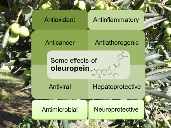
They contain a chemical called Oleuropein.
In today’s post we’ll explore what is known about this chemical and discuss what it could mean for Parkinson’s disease.
The olive, also known by the botanical name ‘Olea europaea,’ is an evergreen tree that is native to the Mediterranean, Asia and Africa, but now found around the world. It has a rich history of economic and symbolic importance within western civilisation. And the fruit of the tree also tastes good, either by themselves or in a salad or pasta dish.
Traditional diets of people living around the Mediterranean sea are very rich in extra-virgin olive oil. Olives are an excellent source of ‘good’ fatty acids (monounsaturated and di-unsaturated), antioxidants and vitamins. Indeed, research has shown that the traditional Mediterranean diet reduces the risk of heart disease (Click here to read more on this).
There are also chemicals within the olive fruit that may have very positive benefits for Parkinson’s disease.
But before you rush out and gorge yourself on olives, we have one small piece of advice:
The chemical is called Oleuropein, and it is usually removed from olives due to its bitterness.
What is Oleuropein?
Oleuropein is a ‘phenylethanoid’ – a type of phenolic compound that is found in the leaf and the fruit of the olive. Phenolic compounds are produced by plants as a protective measure against different kinds of stress.
The main phenolic compounds found in olives are hydroxytyrosol and oleuropein – both of which give extra-virgin olive oil its bitter taste and both have demonstrated neuroprotective effects.
More research has been done on oleuropein so we will focus on it here (for more on hydroxytyrosol – please click here).
Oleuropein has been found to have many interesting properties, such as:
What neuroprotective research has been done on Oleuropein?
Thus far, most of the research addressing this question has been conducted on models of Alzheimer’s disease. The first study
Title: Oleuropein aglycone protects transgenic C. elegans strains expressing Aβ42 by reducing plaque load and motor deficit. Authors: Diomede L, Rigacci S, Romeo M, Stefani M, Salmona M. Journal: PLoS One. 2013;8(3):e58893. PMID:23520540 (This article is OPEN ACCESS if you would like to read it)
The Italian researchers who conducted this study treated a microscopic worm model of Alzheimer’s disease with oleuropein aglycone. We should not that oleuropein aglycone is a hydrolysis product of oleuropein (a hydrolysis product is a chemical compound that is broken apart by the addition of water). The microscopic worm used in the study are called Caenorhabditis elegans:
Caenorhabditis elegans (or simply C. Elegans) are tiny creatures that are widely used in biology because they can be easily genetically manipulated and their nervous system is very simple and well mapped out (they have just 302 neurons and 56 glial cells!). The particular strain of C. elegans used in this first study produced enormous amounts of a protein called Aβ42.
Amyloid beta (or Aβ) is the bad boy/trouble maker of Alzheimer’s disease; considered to be critically involved in the condition. A fragment of this protein (called Aβ42) begins clustering in the brains of people with Alzheimer’s disease. This clustering of Aβ42 goes on to form the plaques that are so characteristic of the Alzheimer’s affected brain.
The Italian researchers conducting this study had previously shown that oleuropein can inhibit the ability of Aβ42 to aggregate in cells growing in culture dishes (Click here to read more about that study), and they wanted to see if oleuropein had the same properties in actual live animals. So they chose the C. Elegans that had been genetically engineered to produce a lot of Aβ42 to test this idea.
In the C. Elegans that produce a lot of Aβ42 gradually become paralysed and their lives are shortened. By treating these worms with oleuropein, however, the Italian researchers found that there was less aggregation of Aβ42 (though the levels of the protein stayed the same), resulting in less plaque formation, and improved mobility (>50% reduction in paralysis) and survival compared to untreated Aβ42 producing C. Elegans.
Encouraged by this result, the researchers next moved on to studies in mice:
Title: The polyphenol oleuropein aglycone protects TgCRND8 mice against Aß plaque pathology. Authors: Grossi C, Rigacci S, Ambrosini S, Ed Dami T, Luccarini I, Traini C, Failli P, Berti A, Casamenti F, Stefani M. Journal: PLoS One. 2013 Aug 8;8(8):e71702. PMID: 23951225 (This article is OPEN ACCESS if you would like to read it)
For this study, the Italian researchers used the genetically engineered TgCRND8 mice. These mice have a mutant form of amyloid precursor protein (which, similar to Aβ42, is associated with Alzheimer’s disease). In the brains of these mice, amyloid clustering begins at 3 months of age, and dense plaques are evident from 5 months of age. The mice also exhibit a clear learning impairment from 3 months of age.
By treating these mice with oleuropein aglycone, the researchers observed a remarkable reduction in plaques in the brain, and those that were present appeared less compact and “fluffy” (their very technical description, not ours). In addition, there was a reduction in the activation of astrocytes and microglia (the helper cells in the brain), indicating a healthier environment.
These same researchers have observed the same results in a rat model of Alzheimer’s disease in a report published the next year (Click here to read more about this).
Interestingly, the oleuropein treated TgCRND8 mice also displayed a major increase in autophagy activity. As we discussed in our previous post (Click here to read that post), autophagy is the rubbish disposal/recycling system of each cell, and increasing the activity of this system can help to keep cells health (particularly if there is a lot of a genetically engineered protein present!).
The Italian researchers repeated this study, and published the results this year, with an interesting twist:
Title: Oleuropein aglycone and polyphenols from olive mill waste water ameliorate cognitive deficits and neuropathology. Authors: Pantano D, Luccarini I, Nardiello P, Servili M, Stefani M, Casamenti F. Journal: Br J Clin Pharmacol. 2017 Jan;83(1):54-62. PMID:27131215
In this study, the researchers tested the same genetically engineered mice, but with two different treatments:
Two much lower doses of oleuropein (4 and 100 times lower)
A mixture of polyphenols from olive mill concentrated waste water
The lowest dose of oleuropein (100 times less oleuropein than the previous study) did not provide any significant improvements for the mice, but the intermediate dose (only 4 times less oleuropein than the previous study) did provide significant benefits. These result indicate that there is a dose-dependent range to the beneficial properties of oleuropein.
The researchers also observed very similar beneficial effects from the mice drinking a mixture of polyphenols from olive mill concentrated waste water. Given these results, the investigators are now seeking to design appropriate conditions to perform a clinical trial to assess better the possible use of oleuropein (or a mix of olive polyphenols) against Alzheimer’s disease.
Ok, but what research has been done with oleuropein and Parkinson’s disease?
Unfortunately, not much.
A research group in Iran has looked at the effect of oleuropein in aged rodents and found an interesting result:
Title: Antioxidant role of oleuropein on midbrain and dopaminergic neurons of substantia nigra in aged rats. Authors: Sarbishegi M, Mehraein F, Soleimani M. Journal: Iran Biomed J. 2014;18(1):16-22. PMID:24375158 (This article is OPEN ACCESS if you would like to read it)
In this study, the investigators took twenty aged rats (18-month-old) and randomly assigned them to two groups: a treatment group (which received a daily dose of 50 mg/kg of oleuropein for 6 months) and a control group (which received just water). Following these treatments, the investigators found an increase in the activity of anti-oxidant agents (such as superoxide dismutase, catalase and glutathione) in the treatment group compared to control group. The treated rats also had significantly more dopamine neurons in the region of the brain affected by Parkinson’s disease (the substantia nigra). The investigators concluded that oleuropein consumption in a daily diet may be useful in reducing oxidative stress damage by increasing the antioxidant activity in the brain.
This first study was followed more recently by a report from a group in Quebec (Canada) who investigated oleuropein use in a cell culture model of Parkinson’s disease:
Title: Oleuropein Prevents Neuronal Death, Mitigates Mitochondrial Superoxide Production and Modulates Autophagy in a Dopaminergic Cellular Model. Authors: Achour I, Arel-Dubeau AM, Renaud J, Legrand M, Attard E, Germain M, Martinoli MG. Journal: Int J Mol Sci. 2016 Aug 9;17(8). PMID:27517912 (This article is OPEN ACCESS if you would like to read it)
The researcher conducting this study wanted to determine if oleuropein could prevent neuronal degeneration in a cellular model of Parkinson’s disease. They exposed cells to the neurotoxin 6-hydroxydopamine (6-OHDA) and then investigated mitochondrial oxidative stress and autophagy.
What is mitochondrial oxidative stress?
Mitochondria are the power house of each cell. They keep the lights on. Without them, the lights go out and the cell dies.
Mitochondria and their location in the cell. Source: NCBI
Oxidative stress results from too much oxidation. Oxidation is the loss of electrons from a molecule, which in turn destabilises the molecule. Think of iron rusting. Rust is the oxidation of iron – in the presence of oxygen and water, iron molecules will lose electrons over time. Given enough time, this results in the complete break down of objects made of iron.
The exact same thing happens in biology. Molecules in your body go through a similar process of oxidation – losing electrons and becoming unstable. This chemical reaction leads to the production of what we call free radicals, which can then go on to damage cells. A free radical is an unstable molecule – unstable because they are missing electrons.
How free radicals and antioxidants work. Source: h2miraclewater
In an unstable format, free radicals bounce all over the place, reacting quickly with other molecules, trying to capture the much needed electron to re-gain stability. Free radicals will literally attack the nearest stable molecule, to steal an electron. This leads to the “attacked” molecule becoming a free radical itself, and thus a chain reaction is started. Inside a living cell this can cause terrible damage, ultimately killing the cell.
Now if this oxidative process starts in the mitochondria, it can be very bad for a cell.
And what is autophagy?
Yes, the researchers also looked at autophagy levels in their cells. Autophagy is an absolutely essential function in a cell. Without autophagy, old proteins and mitochondria will pile up making the cell sick and eventually it dies. Through the process of autophagy, the cell can break down the old protein, clearing the way for fresh new proteins to do their job.
Think of autophagy as the waste disposal/recycling process of the cell.
Waste material inside a cell is collected in membranes that form sacs (called vesicles). These vesicles then bind to another sac (called a lysosome) which contains enzymes that will breakdown and degrade the waste material. The degraded waste material can then be recycled or disposed of by spitting it out of the cell.
Ok, so what did the researchers find?
Well, by pretreating the their cells with oleuropein 3 hours before exposing them to the neurotoxin, the investigators found a significant neuroprotective effect. There was a significant reduction in mitochondrial production of free radicals, and the investigators found an important role for oleuropein in the regulation of autophagy.
And the good news is that other research groups have observed similar beneficial effects of oleuropein in cell culture models of Parkinson’s disease (Click here to read more about that).
The bad news is: that is all the published research on oleuropein and Parkinson’s disease we could find (and we would be happy to be corrected on this if people are aware of other reports!).
So what does Oleuropein do in the brain?
This is a good question, but with so little research done in this area, it is hard to answer.
We know that oleuropein is well absorbed by the human body and that it is relatively stable (Click here to read more on this). In addition, it can cross the blood-brain-barrier – in rodents at least (Click here to read more on that).
Obviously (based on the research we described above), we know that oleuropein has anti-oxidant promoting activities. In addition, it appears to be doing something with regards to autophagy. And it may be regulating autophagy by acting as an inhibitor of mammalian target of rapamycin (mTOR) activation.
What is mTOR?
mTOR is a protein that binds with other proteins to form the nexus of a signalling pathway which integrates both intracellular and extracellular signals (such asnutrients, growth factors, and cellular energy status) and then serves as one of the central instructors of how the cell should respond.
For example, insulin can signal to mTOR the status of glucose levels in the body. mTOR also deals with infectious or cellular stress-causing agents, thus it could be involved in a cells response to conditions like Parkinson’s disease.
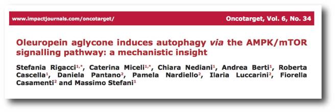
One important property of mTOR is its ability to block autophagy (the recycling process of the cell that we discussed above). Recently, the Italian researchers whose work we reviewed above, found that oleuropein can activate autophagy by blocking the mTOR pathway:
Title: Oleuropein aglycone induces autophagy via the AMPK/mTOR signalling pathway: a mechanistic insight. Authors: Rigacci S, Miceli C, Nediani C, Berti A, Cascella R, Pantano D, Nardiello P, Luccarini I, Casamenti F, Stefani M. Journal: Oncotarget. 2015 Nov 3;6(34):35344-57. PMID:26474288 (This article is OPEN ACCESS if you would like to read it)
The researchers conducting this study found that treatment with oleuropein caused an increase in autophagy in both cell culture and in a mouse model of Alzheimer’s disease, and they demonstrated that it achieved this by blocking the mTOR pathway.
Has anyone ever looked at oleuropein in the clinic?
No, not to our knowledge (and we are happy to be corrected on this).
It is a widely available supplement that a lot of people use to help lower bad cholesterol and blood pressure, so yes it can be considered safe. But any decision to experiment with oleuropein should only be made in consultation with your regular medically trained physician.
Why? Because there are always caveats.
Importantly, individuals with low blood pressure and diabetes may suffer even lower blood pressure and blood glucose levels as a result of consumption of oleuropein. Oleuropein may also interact with other pharmaceutical drugs that are designed to lower blood pressure or regulate diabetes. Such interactions could be dangerous.
And this is a particularly important factor for Parkinson’s disease as up to 30% of people with Parkinson’s may be glucose intolerant (Click here to see our post on Parkinson’s & diabetes).
Those who experience symptoms such as headache, nausea, flu-like symptoms, fainting, dizziness, and other life threatening symptoms should medical attention immediately.
What does it all mean?
We are grateful to regular reader (Don) who brought oleuropein to our attention. It is a very interesting chemical and we are definitely intrigued by it. We would certainly like to see more research on oleuropein in models of Parkinson’s disease.
Attentive readers will have noticed that most of the research discussed above have been conducted in the last 5-10 years. This suggests that oleuropein research is still in its infancy, particularly with regards to research on neurological conditions. And we hope that by reporting on it here, we will be bringing it to the attention of researchers.
Oleuropein is extracted from all parts of the olive tree (the leaves, bark, root, and fruit). It forms part of the defence system of the olive tree against stress or infection. Perhaps we could apply some of its interesting properties to Parkinson’s disease.
EDITORIAL NOTE: Under absolutely no circumstances should anyone reading the material on this website consider it medical advice. The information provided here is for educational purposes only. Before considering or attempting any change in your treatment regime, PLEASE consult with your doctor or neurologist. While some of the drugs and supplements discussed on this website are clinically available, they may have serious side effects. We urge caution and professional consultation before altering any treatment regime. SoPD can not be held responsible for any actions taken based on the information provided here.
The banner for this post was sourced from jrbenjamin
“Here they make all sorts of cutlery-ware, but especially that of edged-tools, knives, razors, axes, &. and nails; and here the only mill of the sort, which was in use in England for some time was set up, for turning their grindstones, though now ’tis grown more common”
Sheffield has a long history of metal work, thanks largely to its geology: The city is surrounded by fast-flowing rivers and hills containing many of the essential raw materials such as coal and iron ore.
And given this fortunate circumstance and an industrious culture, the city of Sheffield particularly prospered during the industrial revolution of the mid-late 1800s (as is evident from the population growth during that period).
The population of Sheffield over time. Source: Wikipedia
But traditional manufacturing in Sheffield (along with many other areas in the UK) declined during the 20th century and the city has been forced to re-invent itself in the early 21st century. And this time, rather than taking advantage of their physical assets, the city is focusing on its mental resources.
Great. Interesting stuff. Really. But what does this have to do with flies, fish and Parkinson’s disease???
Indeed. Let’s get down to business.
The Sheffield Institute for Translational Neuroscience (SITraN) was officially opened in 2010 by Her Majesty The Queen. It is the first European Institute purpose-built and dedicated to basic and clinical research into Motor Neuron Disease as well as related neurodegenerative disorders such as Parkinson’s and Alzheimer’s disease.
Since its opening, the institute has published some pretty impressive research, particularly in the field of Parkinson’s disease.
We have previously discussed “Pink” flies and their critical role in Parkinson’s research (Click here to read that post).
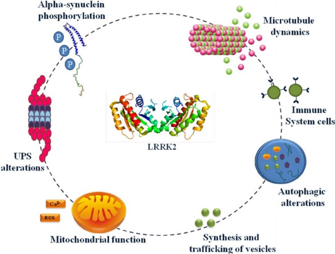
Today we are going to talk about Lrrk2 flies.
What is Lrrk2?
This is Sergey Brin.
He’s a dude.
One of the founders of the search engine company “Google”. Having changed the world, he is now turning his attention to other projects.
One of those other projects is close to our hearts: Parkinson’s disease.
In 1996, Sergey’s mother started experiencing numbness in her hands. Initially it was believed to be RSI (Repetitive strain injury). But then her left leg started to drag. In 1999, following a series of tests, Sergey’s mother was diagnosed with Parkinson’s disease. It was not the first time the family had been affected by the condition: Sergey’s late aunt had also had Parkinson’s disease.
Both Sergey and his mother have had their DNA scanned for mutations that increase the risk of Parkinson’s disease. And they discovered that they were both carrying a mutation on the 12th chromosome, in a gene called PARK8 – one of the Parkinson’s disease associated genes. Autosomal dominant mutations (meaning if you have just one copy of the mutated gene) in the PARK8 gene dramatically increase one’s risk of developing Parkinson’s disease.
PARK8 provides the instructions for making an enzyme called Leucine-rich repeat kinase 2 (or Lrrk2).
Also known as ‘Dardarin‘ (from the Basque word “dardara” which means trembling), Lrrk2 has many functions within a cell – from helping to move things around inside the cell to helping to keep the power on (involved with mitochondrial function).
NOTE: Curiously, mutations in the PARK8 gene are also associated with Crohn’s disease (Click here and here for more on this) – though the mutation is in a different location for PD.
Now, not everyone with this particular mutation will go on to develop Parkinson’s disease, and Sergey has decided that his chances are 50:50. But he does not appear to be taking any chances though. Being one of the founders of a large company like Google, has left Sergey with considerable resources at his disposal. And he has chosen to focus some of those resources on Lrrk2 research (call it an insurance policy). He has done this via considerable donations to groups like the Michael J Fox foundation.
Actor Michael J Fox was diagnosed at age 30. Source: MJFox foundation
So just as Pink flies derive their name from mutations in the Parkinson’s associated Pink1 gene, Lrrk2 flies have mutations in the Lrrk2 gene.
So what have the researchers at Sheffield done with the Lrrk2 flies?
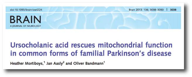
In 2013, the Sheffield researchers published an interesting research report:
Title: Ursocholanic acid rescues mitochondrial function in common forms of familial Parkinson’s disease Authors: Mortiboys H, Aasly J, Bandmann O. Journal: Brain. 2013 Oct;136(Pt 10):3038-50. PMID:24000005
In this study, the investigators took 2000 drugs (including 1040 licensed drugs and 580 naturally occurring compounds) and conducted a massive screen to identify drugs that could rescue mitochondrial dysfunction in PARK2 (Parkin) mutant cells.
Mitochondria are the power house of each cell. They keep the lights on. Without them, the lights go out and the cell dies.
Mitochondria and their location in the cell. Source: NCBI
In certain genetic forms of Parkinson’s disease (such as those associated with mutations in the PARK2 gene), the mitochondria in cells becomes dysfunctional and may not be disposed of properly (Click here to read our previous post related to this).
In their huge screen of 2000 drugs, the researchers in Sheffield identified 15 drugs that could rescue the mitochondria dysfunction in the PARK2 skins cells. Of those 15 compounds, two were chosen for further functional studies. They were:
Ursocholanic acid
Dehydro(11,12)ursolic acid lactone
Neither ursocholanic acid nor dehydro(11,12)ursolic acid lactone are FDA-licensed drugs. We have little if any information regarding their use in humans. Given this situation, the researchers turned their attention to the chemically related bile acid ‘ursodeoxycholic acid’, which has been in clinical use for more than 30 years.
What is Ursodeoxycholic Acid?
Ursodeoxycholic Acid (or UDCA) is a drug that is used to to improve bile flow and reduce gallstone formation. In the USA it is also known as ‘ursodiol’.
Bile is a fluid that is made and released by your liver, and it stored in the gallbladder. Its function is to help us with digestion. UDCA occurs naturally in bile – it is basically a bile acid and can therefore be useful in dissolving gallstones. UDCA has been licensed for the treatment of patients since 1980. UDCA also reduces cholesterol absorption.
So what did the Sheffield researchers find with UDCA?
The researchers tested UDCA on mitochondrial function in PARK2 skin cells, and they found that the drug rescued the cells. They then tested UDCA on skin cells from people with Parkinson’s disease who had mutations in the PARK8 (Lrrk2) gene (G2019S).
The researchers had previously found impaired mitochondrial function and morphology in skin cells taken from people with PARK8 associated Parkinson’s disease (Click here to read more about this), and other groups had reported similar findings (Click here for more on this).
And when they treated the Lrrk2 cells with UDCA, guess what happened?
UDCA was able to rescue the mitochondrial effect in those cells as well!
Obviously these results excited the Sheffield scientists and they set up a collaboration with researchers at York University and from Norway, to look at the potential of UDCA in rescuing the fate of Lrrk2 flies. The results of that study were published two years ago:
Title: UDCA exerts beneficial effect on mitochondrial dysfunction in Lrrk2 (G2019S) carriers and in vivo. Authors: Mortiboys H, Furmston R, Bronstad G, Aasly J, Elliott C, Bandmann O. Journal: Neurology. 2015 Sep 8;85(10):846-52. PMID:26253449 (This article is OPEN ACCESS if you would like to read it).
The researchers tested UDCA on flies (or drosophila) with specific Lrrk2 mutations (G2019S) display a progressive loss of photoreceptor cell function in their eyes. The mitochondria in the photoreceptor are swollen and disorganised. When the investigators treated the flies with UDCA, they found approximately 70% rescue of the photoreceptor cells function.
The researchers in Sheffield concluded that UDCA has a marked rescue effect on cells from a Parkinson’s disease-associated gene mutation model, and they proposed that “mitochondrial rescue agents may be a promising novel strategy for disease-modifying therapy in Lrrk2-related PD, either given alone or in combination with Lrrk2 kinase inhibitors” (for more information about the Lrrk2 inhibitors they refer, click here).
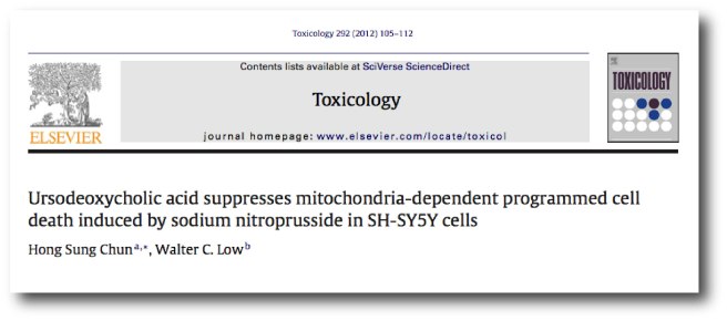
And the good news regarding this line of research: other research groups have also observed similar beneficial effects with UDCA in models of Parkinson’s disease:
Title: Ursodeoxycholic acid suppresses mitochondria-dependent programmed cell death induced by sodium nitroprusside in SH-SY5Y cells. Authors: Chun HS, Low WC. Journal: Toxicology. 2012 Feb 26;292(2-3):105-12. PMID:22178905
This research group also demonstrated that UDCA could reduce cell death in a cellular model of Parkinson’s disease.
And this study was followed by another one from a different research group, which involved testing UDCA in animals:
Title: Ursodeoxycholic Acid Ameliorates Apoptotic Cascade in the Rotenone Model of Parkinson’s Disease: Modulation of Mitochondrial Perturbations. Authors: Abdelkader NF, Safar MM, Salem HA. Title: Mol Neurobiol. 2016 Mar;53(2):810-7. PMID:25502462
These researchers found UDCA rescued a rodent model of Parkinson’s disease (involving the neurotoxin rotenone). UDCA not only improved mitochondrial performance in the rats, but also demonstrated anti-inflammatory and anti-cell death properties.
Given all this research, the Sheffield researchers are now keen to test UDCA in clinical trials for Parkinson’s disease.
Has anyone tested UDCA in the clinic for Parkinson’s disease?
Not that we are aware of, but two groups are interested in attempting it.
Firstly, the University of Minnesota – Clinical and Translational Science Institute has registered a trial (Click here to read more about this). This trial will not, however, be testing efficacy of the drug on Parkinson’s symptoms. It will focus on measuring UDCA levels in individuals after four weeks of repeated high doses of oral UDCA (50mg/kg/day), and determining the bioenergetic profile and ATPase activity in those participants. Basically, they want to see if UDCA is safe and active in people with Parkinson’s disease.
The CurePD trust (in the UK) is also currently seeking to run a clinical trial for UDCA (Click here for more on this). The group are currently organising the funding for that trial.
EDITOR’S NOTE HERE: Before we move on, the team at the SoPD would like to say that while UDCA is a clinically available drug, it is still experimental for Parkinson’s disease. There is no indication yet that it has beneficial effects in people with Parkinson’s disease. In addition, UDCA is also is known to have side effects, which include flu symptoms, nausea, diarrhea, and back pain. And individuals have been known to have allergic reactions to UDCA treatment (Click here and here for more on the side effects of UDCA). Thus we must impress caution on anyone planning to experiment with this drug. Before attempting any kind of change in a current treatment regime, PLEASE discuss your plans with a medically qualified physician who is familiar with your case history.
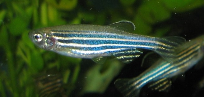
Ok, so that was the flies research, what about the fish? And the… uh, tigar?
Yes. The fish are called Zebrafish (or Danio rerio).
They are a tropical freshwater fish that is widely used in biological research.
Biology researchers love these little guys because their genome has been fully sequenced and they has well characterised and testable behaviours. In addition, their development is very rapid (3 months), and its embryos are large and transparent.
And the researchers at Sheffield are using these fish to study Parkinson’s disease.
How did they do that?
Title: TigarB causes mitochondrial dysfunction and neuronal loss in Pink1 deficiency Authors: Flinn LJ, Keatinge M, Bretaud S, Mortiboys H, Matsui H, De Felice E, Woodroof HI, Brown L, McTighe A, Soellner R, Allen CE, Heath PR, Milo M, Muqit MM, Reichert AS, Köster RW, Ingham PW, Bandmann O. Journal: Ann Neurol. 2013 Dec;74(6):837-47.
PMID: 24027110 (This article is OPEN ACCESS if you would like to read it)
Firstly, the group at Sheffield generated zebrafish that had a mutation in the Parkinson’s associated gene ‘PARK6’. This gene provides the plans for the production of a protein called Pink1 (we have previously discussed Pink1 – click here to read more on this).
In normal healthy cells, the Pink1 protein is absorbed by mitochondria and eventually degraded as it is not used. In unhealthy cells, however, this process becomes inhibited and Pink1 starts to accumulate on the outer surface of the mitochondria. Sitting on the surface, it starts grabbing another Parkinson’s associated protein called Parkin. This pairing is a signal to the cell that this particular mitochondria is not healthy and needs to be removed.
Pink1 and Parkin in normal (right) and unhealthy (left) situations. Source: Hindawi
The process by whichmitochondria are removed is called mitophagy. Mitophagy is part of the autophagy process, which is an absolutely essential function in a cell. Without autophagy, old proteins and mitochondria will pile up making the cell sick and eventually it dies. Through the process of autophagy, the cell can break down the old protein, clearing the way for fresh new proteins to do their job.
Think of autophagy as the waste disposal/recycling process of the cell.
Waste material inside a cell is collected in membranes that form sacs (called vesicles). These vesicles then bind to another sac (called a lysosome) which contains enzymes that will breakdown and degrade the waste material. The degraded waste material can then be recycled or disposed of by spitting it out of the cell.
In the case of a PARK6 mutations, Pink1 protein can not function properly with Parkin and the autophagy process breaks down. As a result, the old or unhealthy mitochondria start to pile up in the cell, resulting in the cell getting sick and dying.
Now back to the Zebrafish.
When the Sheffield researchers mutated PARK6 in the zebrafish, they noticed that the fish had a very early and persistent loss of dopamine neurons in their brains. These fish also had enlarged, unhealthy mitochondria and reduced mitochondrial activity.
Given this result, the investigators next wanted to identify which genes have increased or decreased levels of activity as a result of this genetic manipulation. They identified 108 genes that were higher in the PARK6 mutant, and 146 genes had lower activity.
One gene in particular had activity levels 12 times higher in the PARK6 mutant fish than the normal zebrafish.
The name of that gene? TP53-Induced Glycolysis And Apoptosis Regulator (or Tigar).
What is Tigar?
Tigar is a gene that provides the instructions for making a protein that is activated by p53 (also known as TP53).
What does that mean?
p53 is a protein that has three major functions: controlling cell division, DNA repair, and apoptosis (or cell death). p53 performs these functions as a transcriptional activator (that is a protein that binds to DNA and helps produce RNA (the process of transcription) – see our previous post explaining this).
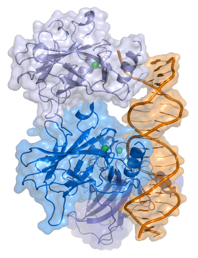
p53 protein structure, bound to DNA (in gold). Source: Wikipedia
In regulating the cell division, p53 prevents cells from dividing too much and in this role it is known as a tumour suppression – it suppresses the emergence of cancerous tumours. Genetic mutations in the p53 gene result in run away cell division, and (surprise!) as many as 50% of all human tumours contain mutations in the p53 gene.
In DNA repair, p53 is sometimes called “the guardian of the genome” as it prevents mutations and helps to conserve stability in the genome. This function also serves to prevent the development of cancer, by helping to repair potentially cancer causing mutations….and in this role it is known as a tumour suppression. Obviously, if there is a mutation in the p53 gene, less DNA repair will occur – increasing the risk of cancer occurring.
And finally, in cell death, p53 plays a critical role in telling a cell when to die. And (continuing with the cancer theme), if there is a mutation in the p53 gene, fewer cells will be told to die – increasing the risk of cancer occurring. And in this role p53 is known as a tumour suppression.
In normal cells, the levels of p53 protein are usually low. When a cell suffers DNA damage and stress, there is often an increase in the amount of p53 protein. If this increases past a particular threshold, then the cell will be instructed to die.
If you haven’t guessed yet, p53 is a major player inside most cell, and it controls the activity of a lot of genes.
And one of those genes is Tigar.
But what does Tigar actually do?
So we have explained the “TP53-Induced” part of the “TP53-Induced Glycolysis And Apoptosis Regulator” name, let’s now focus on the “Glycolysis And Apoptosis Regulator”
Tigar is an interesting protein because it is an enzyme that primarily functions as a regulator of the breaking down of glucose (“Glycolysis” involves the conversion of glucose into a chemical called pyruvate). In addition to this role, however, Tigar acts in preventing cell death (or apoptosis).
Increased levels of Tigar protects cells from oxidative-stress induced apoptosis, by decreasing the levels of free radicals. In this way, it promotes anti-oxidant activities.
But hang on a second, anti-oxidant activity should be good for the cell right? Why are the dopamine cells are dying if Tigar levels are increasing in the PARK6 mutants?
Fantastic question!
The answer: TIGAR is also a negative regulator of a process called mitophagy. As we discussed above, mitophagy is the process of removing mitochondria by autophagy. Increases in the levels of TIGAR blocks mitophagy in a cell, and results in an increased number of swollen and unhealthy mitochondria in those cells (Click here to read more about this). These swollen mitochondria are comparable to the enlarged mitochondria identified the PARK6 zebrafish by the Sheffield researchers.
And the researchers believe that this may be the cause of the cell death in the PARK6 zebrafish – the double impact of PARK6 and Tigar induced problems with mitophagy.
NOTE: Problems with mitophagy is believed to be an important mechanism in the development of early-onset Parkinson’s disease (Click here for a recent review on this)
Ok, and what did the Sheffield researchers do next?
Given that there was such a huge increase in Tigar levels in the PARK6 zebrafish, the investigators decided to reduce Tigar levels in the PARK6 zebrafish to see what impact this would have on the fish (and their mitochondria).
Remarkably, reductions of Tigar levels resulted in complete rescue of the dopamine neurons in the PARK6 fish. It also increased mitochondrial activity in those cells, and reduced the activation of the microglia cells, which can also play a role in the removal of sick cells in the brain.
The researchers concluded that the results demonstrate that TIGAR is “a promising novel target for disease‐modifying therapy in Pink1‐related Parkinson’s disease”.
And what are the researchers planning to do next with Tigar?
Prof Oliver Bandmann, the senior scientist who ran the study, has said that they “need to finish studying TIGAR levels in the brains of people with Parkinson’s and want to better understand how this protein is involved in maintaining the cell batteries – called ‘mitochondria'” (Source).
Our guess is that the group will also be conducting studies looking at Tigar reduction in rodent models of Parkinson’s disease to determine if this is a viable target in mammals. If Tigar reduction in rodents is found to be effective, the researchers will probably turn their attention to drug screening studies to identify currently available drugs that can reduce the activity of Tigar. Such a drug would provide us with yet another potential treatment for Parkinson’s disease.
We’ll be keeping an eye out for these pieces of research.
This is all very interesting. What does the future hold for Parkinson’s research in Sheffield?
The goal of the company – the first of its kind – is to combine world-leading research from the University with funding and expertise from the charity to help develop revolutionary drugs for Parkinson’s disease.
What is virtual about it? The biotech won’t be building its own labs, employing a team of specialist laboratory scientists, or buying any high-tech equipment (which would all be incredibly expensive). Rather they will form partnerships with groups that do specific tasks the best.
Here is a video of Dr Author Roach (director of Research at Parkinson’s UK) explaining the idea behind this endeavour:
By seeking a collaboration with Sheffield in the creation of a spin-out biotech company, Parkinson’s UK is not only acknowledging Sheffield’s track record, but also making an investment in their future research. While we cannot be entirely sure of what the long-term future holds for Parkinson’s research in Sheffield, we do know that Keapstone will be an important aspect of it in the immediate future.
Could this be a model for the future of Parkinson’s disease research? Only time will tell. We will have a closer look at Keapstone Therapeutics in an upcoming post.
Click here to learn more about the virtual biotech project.
So what does it all mean?
In 2017, we here at the SoPD have decided to begin highlighting some of the Parkinson’s disease research centres as an addition feature on the blog. We have not been approached by the research group in Sheffield or the University itself, and our selection of this city as our first case study was based purely on the fact that we really like what is happening there with regards to Parkinson’s research!
The research group in Sheffield has undertaken multiple lines of research which could potentially providing us with several novel treatment options for Parkinson’s disease. These lines of research have focused not only on clinically available drugs, but also identifying novel targets. We like what they are doing and will keep a close eye on progress there.
And over the next year we will select additional centres of Parkinson’s research based on the same criteria (us liking what they are doing). Our next case study will be the Van Andel Research Institute in Grand Rapids, Michigan (we would hate to be accused of having a UK bias).
EDITORIAL NOTE: Under absolutely no circumstances should anyone reading the material on this website consider it medical advice. The information provided here is for educational purposes only. Before considering or attempting any change in your treatment regime, PLEASE consult with your doctor or neurologist. While some of the drugs discussed on this website are clinically available, they may have serious side effects. We urge caution and professional consultation before altering any treatment regime. SoPD can not be held responsible for any actions taken based on the information provided here.


