HIV and Parkinson’s disease

hiv-aids-definition2


I was recently made aware of an interesting fact:

Approximately 5% of people with Human immunodeficiency virus (HIV) infections develop Parkinson’s disease-like features.

Why is this?

In today’s post we will try to understand what is going on, and what it may mean for Parkinson’s disease.


hiv-budding-colo2r

HIV (in green) budding (being released) from a blood cell (lymphocyte). Source: Wikipedia

Ok, let’s start at the beginning:

What is HIV?

Human immunodeficiency virus (or HIV) – as the name suggests – is the virus.

It causes the infection which gives rise to Acquired Immune Deficiency Syndrome (or AIDS). AIDS is a progressive failure of the immune system – the body loses its ability to fight infections. Without treatment, average survival period after infection with HIV is between 9 – 12 years.

HIV can be spread by the transfer of bodily fluids, such as blood and semen. The World Health Organisation (WHO) has estimated that approximately 36.9 million people worldwide were living with HIV/AIDS at the end of 2014 (that is equivalent to the entire population of Canada!).

hi-virion-structure_en-svg

The structure of the HIV virus. Source: Wikipedia

Does HIV affect the brain?

Yes.

At postmortem examinations, less than 10% of the brains from HIV infected individuals are histologically normal (Source).

HIV is a member of the lentivirus family of viruses, which readily infect immune cells (such as blood cells). HIV can also infect other types of cells though, including those in the brain. HIV will usually enter the central nervous system within the first month following infection. It enters the brain via infected blood cells which come into contact with brain ‘immune system/helper’ cells such as microglia and macrophages at the blood-brain-barrier.

f1-large

How HIV enters the brain. Source: Disease Models and Mechanisms

HIV can also infect astrocytes (albeit at a lower frequency than microglia and macrophages), by direct cell-cell contact with infected T cells (blood cells) at the blood-brain-barrier (No. 1 in the image above). After infecting astrocytes, there is dysfunction in the astrocyte and it will no longer be so supportive to the local neurons (No. 2 in the image above). Once inside the brain, HIV-infected macrophages will allow for infection of other macrophages and microglia (No. 3 in the image above), and all together these HIV-infected astrocytes and microglia will cause damage to neurons by releasing viral proteins (two in particular, called Tat and gp120) and additional nasty chemicals which are bad for the neurons (No. 4 in the image above). Finally, as the disease progresses, the protective layer of the blood-brain-barrier becomes compromised and HIV-infected T cells eventually enter the brain and they cause damage to neurons by releasing pro-inflammatory chemicals (making the environment harsh for neurons).

There is remarkably little evidence of HIV actually infecting neurons (Click here for a review on this), so any cell loss in the brain that is associated with HIV does not result from neurons themselves being infected. This may be due to the fact that neurons do not have the HIV receptors (such as CD4) on their cell membrane. Similarly, oligodendrocytes (a supporting cell) does not appear to be easily infected by HIV. The bulk of the infected cells in the brain appear to be of the microglial, macrophage and astrocytes. And without these supporting cells doing their jobs in a normal fashion, it is easy to see how neurons can start dying off.

The severity, characteristics and distribution of HIV-induced injury in the brain varies greatly between affected individuals. It is most likely associated with the viral load (or the number of viral particles) in the brain, which can vary from a few thousand to more than a million copies per mL.

Do HIV-infected people show any signs of the virus entering the brain?

For the majority of people infected with HIV, this entry of the virus into the nervous system is neurologically asymptomatic (meaning they will not notice it), except for the occasional mild headache (for more on this read this review). As a result of the HIV virus entering the brain, however, many infected individuals will suffer from a specific set of neurological disorders, collectively called the AIDS dementia complex (ADC) (also known as HIV-associated cognitive/motor complex, or simply HIV dementia).

So how does HIV infection result in Parkinson’s disease-like features?

As we have suggested in the introduction to this post, on rare occasions (approximately 5% of cases), HIV-infected patients may present an illness virtually identical to Parkinson’s disease. More commonly, people with HIV will exhibit an increased sensitivity to dopamine receptor-blocking agents, such as drugs with a low potential for inducing Parkinsonism, (for example prochlorperazine and metoclopropamide).

The exact mechanism by which HIV infection results in Parkinson’s disease-like features is the subject of debate, but what is clear is that the basal ganglia (a structure involved in Parkinson’s disease) faces the brunt of the HIV infection in the brain. HIV-infected microglia and macrophage are most prominent in the basal ganglia when compared to other brain regions (Click here and here for more on this), and the basal ganglia is where the chemical dopamine from the midbrain is being released.

In addition, there are other changes in the brains of HIV infected people which may aid in the appearance of Parkinsonian features:


viraltitle

Title: Increased frequency of alpha-synuclein in the substantia nigra in human immunodeficiency virusinfection.
Authors: Khanlou N, Moore DJ, Chana G, Cherner M, Lazzaretto D, Dawes S, Grant I, Masliah E, Everall IP; HNRC Group.
Journal: J Neurovirol. 2009 Apr;15(2):131-8.
PMID: 19115126       (This article is OPEN ACCESS if you would like to read it)

The researchers in this study used staining techniques to look at the amount of alpha synuclein – the Parkinson’s associated protein – in slices of brain tissue taken from postmortem autopsies of 73 HIV+ individuals aged between 50 and 76 years of age.

The presence of alpha synuclein in the substantia nigra (an area of the brain affected by Parkinson’s disease) was a lot higher in the HIV+ brains when compared with healthy control samples (16% of the HIV+ brains had high levels of alpha synclein vs 0% for the healthy brains).

Interestingly, nearly all of the brains analysed (35 out of 36 HIV+ brains) had high levels of the Alzheimer’s disease associated protein, beta amyloid (which again raises the question of whether beta amyloid could be playing a defensive role in infections – see our previous post on this). Also interesting, was that there was no correlation between these proteins being present and the age of the person at death – that is to say, older brains did not have more of these proteins when compared with younger brains.

There are also additional ways in which HIV could be causing Parkinson’s-like features, such as:

  • HIV has been shown to affect the protein levels of Parkinson’s disease associated proteins, such as DJ1 and Lrrk2 (Click here and here to read more on this).
  • HIV can, in some cases, increase the level of Dopamine transporter, which would reduce the levels of free floating dopamine in the brain (Click here to read more about this).

How is HIV treated?

aidspills

Treating HIV. Source: NPR

There is currently no cure for HIV infection.

There are, however, treatments which help to slow the virus down. These are called Anti-retroviral drugs (HIV is a retrovirus). There are different kinds of anti-retroviral drugs, which act at different stages of the HIV life cycle. Combinations of several anti-retroviral drugs (generally three or four) is known as ‘Highly Active Anti-Retroviral Therapy'(or HAART).

hiv-drug-classes-svg

Mechanism by which four classes of anti-retroviral drugs work against HIV. Source: Wikipedia

As the schematic image above highlights, there are many ways to slow down the HIV virus. For example, you can prevent it from attaching to a cell and fusing with the cell membrane (fusion inhibitors). By treating HIV infected people with multiple medications attacking different parts of the HIV life cycle, the virus has been slowed down.

Does HAART treatments for HIV help with these Parkinson’s-like features?

In some cases, the answer appears to be yes.

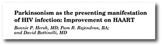
There are numerous case studies in the literature which demonstrate the alleviation of HIV-associated Parkinsonian symptoms with HAART, such as this report:
hersh

Title: Parkinsonism as the presenting manifestation of HIV infection: improvement on HAART.
Authors: Hersh BP, Rajendran PR, Battinelli D.
Journal: Neurology. 2001 Jan 23;56(2):278-9.
PMID: 11160977

In this study the researchers described the case of a 37 year old man who developed Parkinson’s like features in the setting of an HIV infection, which were resolved after 1 year of HAART.

Over a period of 4 months, the man developed co-ordination issue, clumsiness and an irregular tremor in his right hand (there was, however, no resting tremor). He noted a generalised slowness and exhibited a tendency towards decreased right arm swinging. He also developed dystonia in the right hand/arm. Following L-dopa treatment (25/100; one tablet 3x per day) there was improvement in balance & co-ordination, speech, facial expression, and the tremor (L-dopa does appear to improve most cases of HIV-associated Parkinson’s-like features).

Six months after first displaying these Parkinsonian features (and two month after initiating L-dopa treatment), the subject was placed on HAART treatment. Four months later, he discontinued L-dopa treatment and 12 months after starting the HAART regime his Parkinsonian features were largely resolved.

More case studies of HAART alleviating HIV-associated Parkinsonisms can be found by clicking here and here.

What does this mean for Parkinson’s disease?

This post was written for the research community rather than people with Parkinson’s disease. I thought the fact that some people with HIV can start to have Parkinson’s like features was an interesting curiosity and wanted to share/spread the information.

Having said that, this post raises some really interesting questions, such as if a virus like HIV can have this effect on the brain, could other viruses be having similar effects? Could some cases of Parkinson’s disease simply be the result of a viral infection? Either multiple hits from a particular virus or different viruses each taking a varying toll over the course of a life time.

This idea would explain many of the curious features of Parkinson’s disease, such as:

  • the asymmetry of the symptoms (people with Parkinson’s usually have the disease starting on one side of the body.
  • the fact that some cells in the brain are more vulnerable to the disease than others (perhaps they are more receptive to a particular virus).
  • the protein clusterings in the cells (Lewy bodies may be defensive efforts against viral infections).

As we have previous mentioned, theories of viral causes for Parkinson’s have been circulating ever since the 1918 flu pandemic (Click here to read our previous post on this topic). About the same time as the influenza virus was causing havoc around the world, another condition began to appear called ‘encephalitis lethargica‘. This disease left many of the victims in a statue-like condition, both motionless and speechless – similar to Parkinson’s disease. Initially, it was assumed that the influenza virus was the causal factor, but more recent research has left us not so sure anymore.

The point is, however, perhaps it is time for us to re-examine the possibility of a viral agent being involved in the development of Parkinson’s disease.

There is new technology that allows us to determine the viral history of each individual from a simple blood test (Click here for more on this), so it would be interesting to compare blood samples from people with Parkinson’s disease with healthy controls to determine any differences.

In addition to the overall question of a viral role in Parkinson’s disease, there also remains the question of why only a small fraction of people with HIV are affected by Parkinsonisms. It could be interesting to genetically screen those people with HIV that exhibit Parkinsonisms and compare them with people with HIV that do not. Do those affected individuals have recognised Parkinson’s related genetic mutations? Or do they have novel genetic variations that could tell us more about Parkinson’s disease?

Food for thought. Would be happy to hear others thoughts.


The banner for today’s post was sourced from AidsServices

George H and Vascular Parkinsonism

george-hw-bush

During Super Bowl 51, ex-president George HW Bush was visibly wheel chair bound. He has in fact been using motorised scooters and wheelchairs since 2012.

His doctors have indicated that he suffers from Vascular Parkinsonism.

In today’s post we will discuss what Vascular Parkinsonism is and how it differs from Parkinson’s disease.


gettyimages-173500627

During a visit to the White house. Source: Heavy

An important concept to understand about the subject matter here:

Parkinsonism is a syndrome, while Parkinson’s is a disease.

A syndrome is a collection of symptoms that characterise a particular condition, while a disease is a pathophysiological response to internal or external factors. The term ‘Parkinsonism’ is an umbrella term that encompasses many conditions which share some of the symptoms of Parkinson’s disease.

There are many different types of Parkinsonism, such as:

  • Idiopathic Parkinson’s disease (the most common type of parkinsonism)
  • Progressive Supranuclear Palsy (PSP)
  • Corticobasal Degeneration (CBD)
  • Multiple System Atrophy (MSA)
  • Essential tremor
  • Vascular Parkinsonism
  • Drug-induced Parkinsonism
  • Dementia with Lewy bodies
  • Inherited Parkinson’s disease
  • Juvenile Parkinson’s disease

All of these conditions fall under the syndrome title of ‘Parkinsonism’, but are all considered distinct/separate diseases in themselves.

So what is Vascular Parkinsonism?

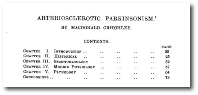
Vascular Parkinsonism was first described in 1929 by Dr Macdonald Critchley (King’s College Hospital, London).

409633

Macdonald Critchley. Source: Npgprints

vascularpd

Title: Arteriosclerotic Parkinsonism.
Author: Critchley, M.
Journal: Brain (1929) 52, 23–83
PMID: N/A                                (this article is accessible by clicking here)

It is estimated that approximately 3% to 6% of all cases of Parkinsonism may have a vascular cause. Vascular (or Arteriosclerotic) Parkinsonism is results from a series of small strokes in the basal ganglia area of the brain and can lead to the appearance of symptoms that look like Parkinson’s disease: slow movements, tremors, difficulty walking, and rigidity.

Walking problems are particularly prominent with Vascular Parkinsonism, as the lower half of the body is usually more affected than the upper half. Another sign of Vascular Parkinsonism can be a poor or no response to L-dopa treatment, as production of dopamine is not the problem. Using brain scanning techniques we can see that some people with Vascular Parkinsonism will have a normal Dopamine transporter (DAT) scan – which demonstrates appropriate levels of dopamine being released and reabsorbed in the striatum (the red-white areas in the image below).

f1-large

DAT-scan and MR images of 62-y-old male  with Vascular Parkinsonism (A) and 62-y-old male with Parkinson’s disease (B). Source: JNM

The brain scans above are from a person with Vascular Parkinsonism (A) and another person with Parkinson’s disease (B). Firstly, note the reduced levels of red-white areas in the image (B) – this reduction is due to less dopamine is being released and reabsorbed in the striatum in Parkinson’s disease (as there are less dopamine fibres present). Compare that with the relatively normal levels of red-white areas in the image (A), indicating normal levels of dopamine turnover (suggesting dopamine fibres are still present). Next, look at the black and white image in panel (A) and you will see a red arrow pointing at damaged areas (darkened regions) of the striatum – indicative of mini strokes. A dopamine receptor scan may show a reduction in the levels of dopamine receptors as a result of the strokes, meaning that the released dopamine will not be having much effect.

Do we know what can cause the strokes associated with Vascular Parkinsonism?

The symptoms of Vascular Parkinsonism tend to appear suddenly and generally do not progress, unlike Parkinson’s disease. We don’t know for sure what causes the mini strokes associated with Vascular Parkinsonism, and it probably varies from person to person, In general, however, doctors believe that high blood pressure and diabetes are the most likely causal factors (heart disease may also play a role).

What does it all mean?

Some people of Parkinson’s disease may actually have Vascular Parkinsonism, which can result from mini strokes in the basal ganglia region of the brain. They will usually be unresponsive to L-dopa and have more motor issues with their lower half of the body.

While Ex-President George HW Bush’s situation is extremely unfortunate, it reminds us that not all forms of Parkinsonism are Parkinson’s disease – an important factor to keep in mind when considering treatment regimes. We have posted this information here to make the Parkinson’s community more aware of this form of Parkinsonism. Later in the year we will discuss another form of Parkinsonism.


The banner for today’s post was sourced from Ew

PARIS is always a good idea

bzn8qhy

Audrey Hepburn was taking about the city when she uttered the words that title this post, but today we will be talking about the protein that bears the same name: PARIS.

Last week new research was published which demonstrated that in the absence of Parkin and Pink1 protein, the protein PARIS builds up and becomes toxic for cells.

Today’s post will review that research and we’ll discuss what it all means for Parkinson’s disease.


paris

No label required. A magnificent city. Source: HathawaysofHaworth

Today’s post has nothing to do with the city of Paris, but it is always nice to have photos of this European capital gracing the page.

We have recently discussed the Parkinson’s associated proteins Pink1 and Parkin (click here for that post). Today we will be revisiting these proteins as we discuss another protein that they interact with: PARIS (specifically PARIS1).

What is PARIS?

PARIS (aka TBC1D2 or TBC1 Domain Family Member 2) is a GTPase-activating protein.

What does that mean?

Getting a signal from outside of a cell into the interior is a complicated affair. There are numerous ways to do it, but one of the most common involves ‘G-proteins‘. These are involved with transmitting a signal from the outside of a cell into the interior, and when inside the cell G-proteins act as molecular switches.

G-proteins are located inside the cell membrane and are activated by G-protein-coupled receptors. When a signaling molecule binds to the G-protein-coupled receptor on the outside of the cell membrane, the portion of the receptor inside the cell activates the G-protein which then starts of a chain of events resulting in the signal being passed on.

11_07bgprotcoupledrec-l

Source: Bio1151

The role of GTPase-Activating Proteins in this process is to turn the G protein’s activity off. In step 4 of the image above, a GTPase-Activating Protein (which is not shown) binds to the G-protein and terminate the activity of the signalling event – returning it to an inactive state.

Thus, GTPase-Activating Proteins – like PARIS – are important regulators of signalling inside the cell.

What do we know about PARIS1 in Parkinson’s disease?

So a few years ago, a group of researchers led by Prof Ted Dawson at John Hopkins School of Medicine published this study:

cell

Title: PARIS (ZNF746) repression of PGC-1α contributes to neurodegeneration in Parkinson’s disease.
Authors: Shin JH, Ko HS, Kang H, Lee Y, Lee YI, Pletinkova O, Troconso JC, Dawson VL, Dawson TM.
Journal: Cell. 2011 Mar 4;144(5):689-702.
PMID: 21376232        (This article is OPEN ACCESS if you would like to read it)

In this study, the researchers noticed that the protein PARIS was accumulating in cells that did not have the Parkinson’s associated protein, Parkin. In those cells, the Parkin gene was mutated so that the Parkin protein was not produced properly. The researchers discovered that Parkin was important for labelling old PARIS protein for disposal. Thus in the absence of Parkin, PARIS protein would not be disposed of and simply piled up.

This build up of PARIS resulted in the loss of dopamine neurons in mice that did not produce Parkin. When the researchers re-introduced normal Parkin protein, the researchers were able to rescue the cell loss. Interestingly, the researchers also found that over production of PARIS in normal mice resulted in cell loss which could be rescued by a similar over production of Parkin.

When they looked in postmortem human brains, the researchers found that levels of PARIS protein were more than two times higher in regions affected by Parkinson’s disease (the striatum and the substantia nigra) of people with sporadic Parkinson’s disease when compared to healthy controls. Interestingly, this increase was only seen with PARIS protein, and not PARIS RNA (where the scientists saw no different with control samples), suggesting a build up of PARIS protein in the Parkinsonian brain.

The investigators concluded that this meant PARIS was could be playing a role in the cell loss associated with Parkinson’s disease.

They followed up this research a few years later with this publication:

parkins

Title: Parkin loss leads to PARIS-dependent declines in mitochondrial mass and respiration.
Authors: Stevens DA, Lee Y, Kang HC, Lee BD, Lee YI, Bower A, Jiang H, Kang SU, Andrabi SA, Dawson VL, Shin JH, Dawson TM.
Journal: Proc Natl Acad Sci U S A. 2015 Sep 15;112(37):11696-701.
PMID: 26324925     (This article is OPEN ACCESS if you would like to read it)

In this study, the same researchers found that when they remove the Parkin protein from the brains of adult mice there would be a decrease in the size and number of mitochondria. We have previous discussed mitochondria – the power stations of the cell – and their loss is bad news for a cell (click here to read more on mitochondria).

The researchers next demonstrated that this loss of mitochondria could reversed by removing PARIS protein from the Parkin mutant mice, and this prevented the loss of dopamine neurons. They also showed that the loss of mitochondria (and loss of dopamine neurons) could be caused by over production of PARIS in normal mice.

These results pointed towards an important role for both Parkin and PARIS in the maintenance of healthy mitochondria.

So what new research has been published about PARIS1?

This study was published last week:

dawson

Title: PINK1 Primes Parkin-Mediated Ubiquitination of PARIS in Dopaminergic Neuronal Survival.
Authors: Lee Y, Stevens DA, Kang SU, Jiang H, Lee YI, Ko HS, Scarffe LA, Umanah GE, Kang H, Ham S, Kam TI, Allen K, Brahmachari S, Kim JW, Neifert S, Yun SP, Fiesel FC, Springer W, Dawson VL, Shin JH, Dawson TM.
Journal: Cell Rep. 2017 Jan 24;18(4):918-932.
PMID: 28122242       (This article is OPEN ACCESS if you would like to read it)

In their study the researchers found that Parkin is not the only Parkinson’s associated protein in the PARIS story.

We have previously talked about the protein Pink1 (click here to read more on) – and yes, you would be forgiven if you start to think that all Parkinson’s related proteins start with the latter ‘P’. Pink1 grabs Parkin and causes it to bind to dysfunctional mitochondria. Parkin then signals to the rest of the cell for that particular mitochondria to be disposed of. In this study, the researchers found that Pink1 also grabs PARIS and signals for Parkin to dispose of it. In the absence of Pink1, normal Parkin protein does not label old PARIS protein for disposal and PARIS starts to pile up.

The researchers then began manipulating the levels of Pink in the brains of mice and they observed PARIS-dependent cell loss – that is to say, in the absence of Pink1, cells died only when PARIS was present.

These findings suggest that therapies targeting PARIS could be used in people with Parkinson’s disease who are carrying either a Parkin or a Pink1 mutation (both very common in early onset Parkinson’s disease).

What does it all mean?

People with early onset Parkinson’s disease quite often have a genetic mutation in one of a small number of genes – Pink1 and Parkin being prominent amongst these genes. The researchers who conducted the study that we have reviewed today have identified a common mechanism by which both of these proteins could be acting in their roles in Parkinson’s disease: a protein called PARIS.

Currently there is no treatment (that we are aware of) that targets the PARIS protein – nothing in the clinic nor being experimentally tested. Obviously, however, PARIS represents a VERY interesting protein for further investigations. The Dawson lab has several patents on PARIS (Click here and here for more on these), so evidently people will be working on drug candidates that inhibit PARIS.

There is a naturally occurring inhibitor, a micro RNA cluster miR-17-92 (also known as oncomir-1), which reduces the production of PARIS protein by blocking PARIS RNA (Click here for more on this). Using this micro RNA to target PARIS will be very difficult (both activating/delivering the micro RNA and unknown off target effects).

We are assuming that Prof Dawson and colleagues are rapidly screening compounds to determine which can block or inhibit PARIS activity and we will eagerly wait to see the results of that work.

Watch this space.


The banner for today’s post was sourced from Wallpapercave


EDITORIAL NOTE: Yay, 100 posts!

Something ‘new and fresh’ from Korea

42734181_l-1024x682

The word ‘Kainos‘ comes from ancient Greek, meaning ‘new’ or ‘fresh’.

A company in South Korea has chosen to use this word as their name.

Why?

In today’s post we will discuss a clinical trial that started this week that is taking a ‘new and fresh’ approach to treating Parkinson’s disease.


south_korea

Enchanting country. Source: Eoasia

South Korea is an amazing place, with a long and proud history of innovation and technological development. This week a biotech company there called Kainos Medicine has added itself to that history by initiating a clinical trial that takes a new approach to treating Parkinson’s disease.

As Kainos Medicine points out on their website, the current treatment options for Parkinson’s disease function by alleviating symptoms, for example L-dopa simply replaces the lost dopamine rather than treating the underlying disease. Kainos’s new experimental treatment, called KM-819, is trying to help in a different way: it is trying to slow down the cell death that is associated with Parkinson’s.

How does it do that?

KM-819 is an inhibitor of Fas Associated Factor 1 (or FAF1).

And what is FAF1?

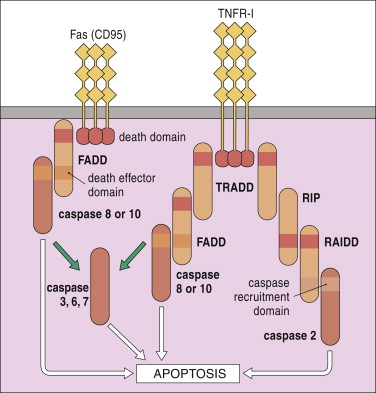
Fas Associated Factor 1 is a protein that interacts with and enhances the activity of a protein on the surface of cells with the ominous name: Fas Cell Surface Death Receptor…and yes, the use of the word ‘death’ in that name should give you some indication as to the function of this protein. When Fas Cell Surface Death Receptor gets activated on any given cell, things have definitely taken a turn for the worse for that particular cell.

Fas Cell Surface Death Receptor (also called CD95) is an initiator of apoptosis.

3-s2-0-b9780323069472100203-f20-05-9780323069472

FasSource: Sciencedirect

What is apoptosis?

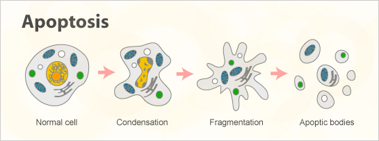
Apoptosis (from Ancient Greek for “falling off”) is the process of programmed cell death – a cell initiates a sequence of events that result in the cell shutting down and dying.

apoptosis_b

The process of apoptosis. Source: Abnova

Apoptosis is a very clean and organise process of a cell being removed from the body, with it eventually being broken down into small units (called apoptotic bodies) which are consumed by other cells.

Sounds interesting, but what research has been done on FAF1 and Parkinson’s disease?

Back in 2008, this research report was published:

faf1

Title: Fas-associated factor 1 and Parkinson’s disease.
Authors: Betarbet R, Anderson LR, Gearing M, Hodges TR, Fritz JJ, Lah JJ, Levey AI.
Journal: Neurobiol Dis. 2008 Sep;31(3):309-15.
PMID: 18573343   (This article is OPEN ACCESS if you would like to read it)

The researcher who conducted this study noticed that the FAF1 gene was located in the ‘PARK 10’ region of chromosome 1. PARK regions are areas of our DNA where mutations (or disruptions to the sequence of DNA) can result in increased vulnerability to Parkinson’s disease (there are currently at least 20 PARK regions). PARK 10 is a region of DNA in which mutations have been associated with late-onset Parkinson’s disease. The scientists thought this was interesting and investigated FAF1 in the context of Parkinson’s disease.

When they looked at postmortem brains, the researchers found that FAF1 levels were significantly increased in brains from people with Parkinson’s disease when compared to brains from healthy control cases. In addition, increased levels of FAF1 exaggerated the cell death observed in different cell culture models of Parkinson’s disease, suggesting an important role for FAF1 in sporadic Parkinson’s disease.

NOTE: More recently, a closer analysis of the PARK10 region resulted in a shrinking of the area which resulted in FAF1 falling outside the PARK10 domain (click here and here to see that research). We are currently not sure if genetic variations in the FAF1 gene infer vulnerability to PD.

This initial work led others to researching FAF1 in the context of Parkinson’s disease and in 2013 this research report was published:

faf2

Title: Accumulation of the parkin substrate, FAF1, plays a key role in the dopaminergic neurodegeneration.
Authors: Sul JW, Park MY, Shin J, Kim YR, Yoo SE, Kong YY, Kwon KS, Lee YH, Kim E.
Journal: Hum Mol Genet. 2013 Apr 15;22(8):1558-73.
PMID: 23307929

These researchers found that Parkinson’s associated protein, Parkin (which we have briefly discussed in a previous post) labels FAF1 for disposal. And they found in the absence of Parkin there was a build up of FAF1, making the cells more vulnerable to apoptosis. They followed this finding up by demonstrating that FAF1-mediated cell death was rescued by re-introducing the normal parkin protein. Interestingly, there was no rescue when the mutant parkin protein was re-introduced. These results suggest that normal Parkin acts as an inhibitor FAF1.

To further investigate this finding, the researchers next modelled Parkinson’s disease in genetically engineered mice which had the FAF1 gene removed. They found that the behaviour motor problems and loss of dopamine cells in the brain was significantly reduced in the FAF1 mutant mice, indicating that the FAF1 pathway could be a worthy target for future Parkinson’s disease treatment.

And this and other research has led those same researchers to the clinical trial started in Korea by Kainos Medicine.

So what is the clinical trial all about?

The company will be conducting a phase 1 dose-escalation clinical trial in South Korea, which will evaluate the safety, tolerability, and biochemical properties of their drug KM-819 in 48 healthy adults (click here to read more about the trial).

This is the very first step in the clinical trial process.

The study is split in two parts: Part A is a single dose of KM-819 or a placebo given in ascending doses to participants. And Part B is the same except that multiple ascending doses of the compound will be given to the participants.

The trial will last around six weeks, and – according to the press release – the first subject has just been dosed.

What does it all mean?

Parkinson’s disease is a neurodegenerative condition, which means that certain cells in the brain are dying. Medication that could block that cell death from occurring represents an interesting way of treating the disease and this is what Kainos are attempting to do.

Blocking or slowing cell death is a tricky business, however, because in other parts of the body, cell death is a very necessary biological process. In some areas of our body, cells are born, conduct a particular function and die off relatively quickly. By slowing that cell death in the brain which may be a good thing, we may be causing issues elsewhere in the body, which would be bad.

In addition there has recently been concerns raised about the clinical use of apoptosis inhibitors, such as this study:

liver

Title: Caspase Inhibition Prevents Tumor Necrosis Factor-α-Induced Apoptosis and Promotes Necrotic CellDeath in Mouse Hepatocytes in Vivo and in Vitro.
Authors: Ni HM, McGill MR, Chao X, Woolbright BL, Jaeschke H, Ding WX.
Journal: Am J Pathol. 2016 Oct;186(10):2623-36.
PMID: 27616656

The researchers who conducted this study found that using apoptosis inhibitors on a mouse model of liver disease did stop apoptosis from occurring, but this didn’t save the cells which eventually died via another cell death mechanism called necrosis (from the Greek meaning “death, the act of killing” – lots of Greek in this post!). In necrosis, rather than breaking down in a systematic and organised fashion (apoptosis), a cell will simply rupture and fall apart. Very messy.

Thus there is the possibility with the Kainos drug, KM-819, will protect cells in the Parkinsonian brain from dying via apoptosis, but as the disease continues to progress those cells may become more ill and eventually disappear as a result of necrosis. That said, if the drug can slow down Parkinson’s disease, it would still represent a major step forward in our treatment of the condition!

The connection with Parkin is also very interesting.

It would be wise for future phase 2 and 3 trials – which will test efficacy – to include (or specifically recruit) people with Parkinson’s disease who have mutations in the Parkin gene. This is a very small proportion of the overall Parkinson’s community (approx. 20% of people with early onset PD have a Parkin mutation – click here to read more on this), but if the drug is going to be effective, these would be the best people to initially test it in.

This will be a very interesting set of clinical trials to watch. The phase 1 safety trial will be very quick (6 weeks), and hopefully Kainos Medicine will be able to progress rapidly to a phase 2 efficacy trial. Fingers crossed for positive results.


The banner for today’s post was sourced from Koreabizwire

A smartphone application for Parkinson’s disease

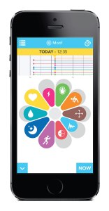
umotif

Here at the SoPD, we like our gadgets and new technology.

And we believe that there is enormous potential for people with Parkinson’s disease to benefit hugely even from some of the small technological advances that seem to be occurring on a day basis.

Today’s post will review a recent study that looked at tested the benefits of a smartphone application for people with Parkinson’s disease.


selfcare-infographic-english

A schematic illustrating the limited monitoring of Parkinson’s. Source: Riggare

On her great blog, Swedish engineer and ‘proud mother’ Sara Riggare posted the image above to illustrate the ridiculous current situation regarding the standard monitoring of Parkinson’s disease.

As the schematic perfectly illustrates, in 2014 Sara spent 8,765 hours conducting ‘self care’. That is, she was applying her own knowledge and experience to managing her Parkinson’s disease. For just one hour in that year was her Parkinson’s actually being monitored by a medical clinician (8,766 being the number of hours in a year).

This is actually a very serious problem – for not only the Parkinson’s community – but anyone suffering from a long-term medical condition. How are they to gage their current situation on a day to day basis when they have such infrequent visits to their medical specialist?

And this is where technology can help.

But, before we begin:

FULL DISCLOSURE NO.1: the author of this blog is an author in the study that will be discussed (#ThisIsNotShamelessSelfPromotion).

FULL DISCLOSURE NO.2: We here at the SoPD are in no way benefitting from mentioning the study here. The company behind the product, umotif, has not asked us nor been contacted by us regarding this post (in fact, they are completely unaware that we are posting this). We are writing this post simply because we thought that it would be of interest to the wider Parkinson’s community. And yes, as other technology comes to along, we will bring it to you attention by posting about it here.

With all of that out of the way, the study is AMAZING!

smart

Title: Using a smartphone based self-management platform to support medication adherence and clinical consultation in Parkinson’s disease: Results from the SMART-PD Randomised Controlled Trial v4.
Authors: Lakshminarayana R, Wang D, Burn D, Barker RA, Chaudhuri KR, Galtrey C, van Guzman N, Hellman B, Pal S, Stamford J, Steiger M, Stott SRW, Teo J, Barker RA, Wang E, Bloem BR, van der Eijk M, Rochester L, & Williams A
Journal: NPJ Parkinson’s disease (2017), 3, 2.
PMID: N/A          (This article is OPEN ACCESS if you would like to read it)

The company behind the application approached various clinical research groups around the UK and proposed to run a study of their new product. The software had many features (including information about medication, a reminder alarm for when medication should be taken, tests/games, and links to other resources online).

41531_2016_3_fig4_html

A programmable reminder system for medication. Source: Nature

The primary focus of the software, however, was a flower-petal shaped motif that the participants could manipulate to indicate how they were feeling.

umotif-app-screenshot-2

The umotif flower motif. Source: SalusDigital

Participants could drag each coloured petal in or out to indicate how they were feeling at a particular moment in time. The smaller the petal, the more lower the score. And each petal represented different aspects of daily life, for example the moon and stars (dark blue) petal allowed an indication of how one slept.

Each time the participant indicated their current status on the flower motif, the information was recorded and could be tracked over days, weeks, and months. This level of information allowed people to begin to see patterns in their own behaviour over time, with some people getting poorer sleep during the middle of the month for example. And different variables could be compared (such as sleep score with exercise score), providing users with a more dynamic idea of their situation over time.

41531_2016_3_fig5_html-1

Comparing scores between measures over time. Source: Nature

A total of 215 people with Parkinson’s disease were randomly assigned to either receive the application (106 subjects) or not (acting as a control subject; 109 subjects). Both groups were contacted by the investigators fours times during the 16 week trial and feedback was provided in addition to any changes in their treatment regimes.

72% of participants application group continued to use and engage with the application throughout the 16-week trial. By the end of the study, the application group demonstrated significantly improved adherence to their treatment regime when compared to the control group. Curiously, the application also significantly improved patients’ perception of quality of follow up consultations, demonstrating unexpected benefits.

And at the end of the study all of the control group participants in the study were allowed to begin using the application, while the application group continued to use it.

One interesting aspect of the study was the lack of interaction with technology by the target population. 180 people who were initially invited to take part in the study could not because they did not have smartphones (with iPhone/iPad or Android operating systems). So obviously there are opportunities for alternative approaches to this kind of tracking (other than a smartphone).

What does it all mean?

This smartphone application is a user friendly approach to tracking someone’s Parkinson’s over time, getting around the ‘lack of monitoring’ issue that concerns many in the community.

Umotif and Parkinson’s UK have kindly made this video about the study results so we might just sit back and let them explain what it all means and point out all of the benefits.

We are working on additional posts about wearable tech for Parkinson’s disease, which will be coming soon.

So stay tuned.


The banner for today’s post was sourced from ParkinsonsMovement

An interesting commentary on the interpretation of the Nilotinib trial results

DSK_4634s

“The devil is in the detail”

A frequently used quote and sage words when analysing scientific data, especially clinical trial data.

Nilotinib is a cancer drug from Novartis that has the Parkinson’s community very excited. In October 2015, researchers at Georgetown University announced that a phase 1 open-label clinical study involving 12 people with Parkinson’s had demonstrated some pretty impressive results (click here to read more about this). The results of that first clinical trial have been published (click here to read more on this), but follow up studies have been hampered by study design issues (click here for more on this).

Today a letter to the editor of the Journal of Parkinson’s disease (published in this months issue) was brought to our attention (click here to read the letter). It queries one important aspect of the results from that first Nilotinib clinical trial for Parkinson’s disease.

In the letter, Prof Michael Schwarzschild of Massachusetts General Hospital (Boston) notes that 8 of the 11 subjects in the study had their monoamine oxidase-B (MAO-B) inhibitor treatment withdrawn less than a month after starting the trial. The change of treatment regime was made due to “increased psychosis in the first 2–4 weeks after Nilotinib administration”.


For reasons which we will outline below, a small change like this in a clinical trial could have major implications for the end results.

What are MAO-B inhibitors?

After the chemical dopamine is used by a neuron, it is reabsorbed by the dopamine cell and broken down for disposal. MAO-B is the enzyme that breaks down dopamine.

maoi-inhibitor
Selegiline is an example of a MAO-B inhibitor. Source: KnowMental

As the schematic above illustrates, dopamine is released by dopamine neurons and then binds to a receptor on a neighbouring cell. After this process has occurred, the dopamine detaches and it is reabsorbed by the dopamine neuron via a particular pathway called the dopamine transporter. Back inside the dopamine cell, dopamine is quickly broken down by the enzyme MAO-B into 3,4-Dihydroxyphenylacetic acid (or DOPAC).

Now, by blocking MAO-B, more dopamine is left hanging around inside the cell where it can be recycled and used again. Thus, this blockade increases the level of dopamine in the brain, which helps with alleviating the motor features of Parkinson’s disease. This simple concept has lead to the development of MAO-B inhibitors which are used in the treatment of the condition.

Why is this important to the Nilotinib results?

Dopamine is broken down by MAO-B into DOPAC. DOPAC can be further broken down into Homovanillic acid (HVA), and both DOPAC and HVA are often used in research studies to indicate levels of dopamine activity. Higher levels of both (in theory) should indicate higher levels of dopamine. It is a means of inferring greater dopamine production.

In the published results of the Nilotinib clinical trial, the researchers used increased HVA levels as an indication of greater dopamine production as a result of taking Nilotinib. But Prof Schwarzschild is correct in providing a cautionary warning of over-interpreting this result. You see, by discontinuing the treatment of MAO-B inhibitors shortly after starting the study, one would expect to see a rise in HVA levels regardless of any effect Nilotinib may be having. Without the MAO-B inhibitors, more dopamine will be broken down thus resulting in increased levels of HVA (compared to the baseline measurements at the start of the study).

And this issue is particularly important since HVA measurements taken at the start of the study (before the MAO-B inhibitors were removed) were compared with HVA measurement taken at the end of the study.

Another commentary discussing the Nilotinib results published in July of last year (in the same journal) actually questioned the value of measuring HVA levels, saying that prior studies have suggested that HVA levels can vary greatly between subjects at similar disease stages, and in general do not correlate well with disease progression.

Whether the removal of MAO-B inhibitors alters the overall interpretation of the first clinical study results is a subject for debate. Something interesting did appear to be happening in the participants involved in the first trial (whether this could have been a placebo effect could also be debated). Obviously, as Prof Schwarzschild’s letter indicates, what we really require now is a carefully designed, placebo-controlled, randomised clinical trial to determine if the initial results can be replicated.

And we are still awaiting news regarding a start date for that delayed trial.

Improving the SoPD blog – any thoughts?

improve-yourself1

It has been a week since we posted our discussion regarding where we think things are going in 2017 in the world of Parkinson’s disease research. Today’s short post is a follow up piece on how we can improve the SoPD blog.

Specifically, we would like to ask for your thoughts as to what particular improvements you would like to this on this blog.


cwk7va-xgaa-vip

PPI in action. Source: Parkinson’s UK

Patient and public involvement (PPI)

PPI is a big deal in the world of Parkinson’s research.

It involves researchers and people affected by Parkinson’s disease (both sufferers and carers/family/friends) work in partnership to plan, design, implement, manage, evaluate and disseminate research. It is a win-win situation for everyone involved as it seeks to achieve a more patient-centric approach to the research.

Parkinson’s UK provides lots of very useful information on PPI (Click here to read more).

Here at SoPD, we see great value in PPI and we would like to embrace it by asking for your feedback on what we are doing here.

It is very easy in science to get very exciting about the details and fail to see the big picture (a ‘not seeing the forest for the trees’ scenario). This situation can make us blind to what the reader of this blog may be looking for. Similarly, we have certain ideas about how this blog is developing and where it could be going which may not be the best way to serve the Parkinson’s community.

So in this post, we will review where things at the SoPD currently stand, and then what future plans are being developed, before we then invite your feedback.

sotu201211

The State of the Union address. Source: Tngop

So lets begin with where we are at present.

The state of the blog:

The blog has been running since the 9th Sept, 2015. We currently have 90 posts dealing with all manner of Parkinson’s disease research-related content. If you are interested in a particular topic, you can use our site map page to search for key words across all of those post.

In addition, we have a menu bar of key topics related to Parkinson’s disease, such as dopamine and tremor. We also have a page of lectures that we would like to expand in the new year.

The post are usually focused around a particular topic, recent research publication, or clinical trial. We try to provide an easy to understand background on the topic before delving into what new discovery or result has been announced. At the end of each post, we try to sum up what it all means for the Parkinson’s community.

For shorter and more regular updates, we also have a twitter account that you can follow.

Future directions:

In this new year, we are planning to:

  • add more pages to our menu bar dealing with key aspects of Parkinson’s disease (such as “what is a Lewy body?”)
  • encourage great involvement and participation in Parkinson’s research
  • put some videos on the site which will explain some of the more commonly asked questions regarding Parkinson’s disease.

There are some other ideas, but these are the ones we are prepared to put on paper and be held to.

And this brings us to your feedback.

maxresdefault

Source: Youtube

What improvements can we make?

We are seeking feedback here – either in the comments section below or by contacting us directly by email – regarding what features or changes you would like to see on this blog.

Specifically:

  • what could we improve or do better that we currently do on the blog?
  • what new features could we add?
  • are there alternative ways of bringing the same information to you that would be better/easier for you to consume?

Any and all thoughts would be greatly appreciated. Please help us to improve the service we are providing.

We look forward to hearing from you.

The team at SoPD


The banner for today’s post was sourced from OnthecontraryKelly

The road ahead – Parkinson’s research in 2017

road

With the end of the 2016, we thought it would be useful and interesting to provide an overview of where we believe things are going with Parkinson’s disease research in the new year. This post can be a primer for anyone curious about the various research activities, and food for thought for people who may have some fresh ideas and want to get involved with the dialogue.

Never before has so much been happening, and never before has there been greater potential for real change to occur. It is a very exciting time to be involved in this field, and it really does feel like we are on the cusp of some major discoveries.

In today’s post we will outline what to expect from Parkinson’s research in 2017.


bright-future-ahead-1024x772

Things to look forward to. Source: Dreamstime

Before we start: something important to understand –

The goal of most of the research being conducted on Parkinson’s disease is ultimately focused on finding a cure.

But the word ‘cure’, in essence, has two meanings:

  1. The end of a medical condition, and
  2. The substance or procedure that ends the medical condition

These are two very different things.

And in a condition like Parkinson’s disease, where the affected population of people are all at different stages of the disease – spanning from those who are not yet aware of their condition (pre-diagnosis) to those at more advanced stages of the disease – any discussion of a ‘cure for Parkinson’s disease’ must be temporal in its scope.

In addition to this temporal consideration, everyone is different.

A ‘cure’ for one person may not have an impact on another person – particularly when genetics is included in the equation. Currently there is a clinical trial which is only being tested on people with Parkinson’s disease who have a particular genetic mutation (Click here to read more about this).

With all of that said, there are 4 key areas of ongoing/future research:

  • Defining and understanding the biology of the condition
  • Early detection
  • Slowing/halting the disease
  • Replacing what has been lost

EDITOR’S NOTE HERE: While we appreciate that this list does not take into account important research dealing with the improving the day-to-day living and quality of life of those affected by Parkinson’s disease (such as prevention of falling, etc), we are primarily focusing here on finding a ‘cure’.

Let’s now have a look at and discuss each of these key areas of research:

Defining and understanding the biology

maxresdefault

Complicated stuff. Source: Youtube

The first key area of research feeds into all of the others.

It is only through a more thorough understanding of the mechanisms underlying Parkinson’s disease that we will be able to provide early detection, disease halting therapies, and cell replacement options. A better conception of the disease process would open doors in all of the other areas of research.

Given the slow pace of progress thus far, you will understand that this area of research is not easy. And it is made difficult by many issues. For example, it may be that we are blindly dealing with multiple diseases that have different causes and underlying mechanisms, but display the same kinds of symptoms (rigidity, slowness of movement and a resting tremor). Multiple diseases collectively called ‘Parkinson’s’. By not being able to differentiate between the different diseases, we have enormous confounding variables to deal with in the interpretation of any research results. And this idea is not as far fetched as it may sound. One of the most common observations within a group of people with Parkinson’s disease is the variety of disease features the group presents. Some people are more tremor dominate, while others have severe rigidity. Who is to say that these are not manifestations of different diseases that share a common title (if only for ease of management).

This complication raises the possibility that rather than being a disease, ‘Parkinson’s’ may actually be a syndrome (or a group of symptoms which consistently occur together).

Recently there have been efforts to deal with this issue within the Parkinson’s research community. We have previously written about the improved diagnostic criteria for Parkinson’s disease (click here to read that post). In addition, as we mentioned above, some new clinical trials are focusing on people with very specific types of Parkinson’s disease in which the subjects have a particular genetic mutation (Click here to read more about this). Better stratification of the disease/s will help us to better understand it. And with the signing into law of the 21 century Cures Act by President Obama, the Parkinson’s research community will have powerful new data collection tools to use for this purpose – in addition to more funding for research at the National Institute of Health (Click here to red more on this).

More knowledge of the basic biology of Parkinson’s disease is critical to the road forward. Whether the Parkinson’s disease-associated proteins, like alpha synuclein, are actually involved with the cell death associated with the condition is a question that needs to be resolved. If they are simply the bio-product of an alternative (unseen) disease process is important to know.

It is impossible to know what the new year will bring for new discoveries in the basic biology of Parkinson’s disease. Compared to 20 years ago, however, when the new discoveries were few and far between, 2017 will bring with it major new discoveries every month and we’ll do our best to report them here.

Early detection for Parkinson’s disease

test

Consider the impact of a pregnancy test on a person’s life. Source: Wikipedia

Ethically, the ‘early detection’ area of research can be a bit of a mine field, and for good reasons. You see, if we suddenly had a test that could accurately determine who is going to get Parkinson’s disease, we would need to very carefully consider the consequences of using it before people rush to start using it in the clinic.

Firstly there are currently no disease halting treatments, so early knowledge of future potential events may not be useful information. Second, there is the psychological aspect – such information (in light of having no treatment) may have a dramatic impact on a person’s mental wellbeing. And thirdly, such information would have huge implications for one’s general life (for example, individuals are legally bound to tell their banks and insurance companies about such information). So you see, it is a very tricky field to tackle.

Having said all of that, there are some very positive aspects to early detection of Parkinson’s disease. Early indicators (or biomarkers) may tell us something new about the disease, opening novel avenues for research and therapeutic treatments. In addition, early detection would allow for better tracking of the disease course, which would enhance our ideas about how the condition starts and changes over  time.

There are numerous tests being developed – from blood tests (click here and here for posts we wrote about this topic) to saliva tests (again, click here for our post on this topic). There are even a simple urine test (click here for our post on this) and breath analysis test (click here for more on this) being developed. And there are ever increasing brain imaging procedures which may result in early detection methods (Click here for more on this).

How does the Parkinson’s research community study early detection of Parkinson’s disease though? Well, we already know that people with rapid eye movement (REM) sleep disorder problems are more likely to develop Parkinson’s disease. Up to 45 percent of people suffering REM sleep behaviour disorders will go on to develop Parkinson’s disease. So an easy starting point for early detection research is to follow these people over time. In addition, there are genetic mutations which can pre-dispose individuals to early onset Parkinson’s disease, and again these individuals can be followed to determine common ‘biomarkers’ (aspects of life that are shared between affected individuals).  Epidemiological studies (like the Honolulu Heart study – click here for more on this) have opened our eyes to keep features and aspects of Parkinson’s disease that could help with early detection as well.

Slowing/halting Parkinson’s disease

stop

Source: MyThaiLanguage

One of the most significant findings in Parkinson’s disease research over the last few years has been the discovery that transplanted dopamine cells can develop Lewy bodies over time. It is very important for everyone to understand this concept: healthy embryonic cells were placed into the Parkinsonian brain and over the space of one or two decades some of those cells began to display the key pathological feature of Parkinson’s disease: dense, circular clusters of protein called Lewy bodies.

The implications of this finding are profound: Healthy cells (from another organism) developed the features of Parkinson’s disease. And this is (presumably) regardless of the genetic mutations of the host. It suggests that the disease spreads by being passed from cell to cell. There is a very good open-access article about this in the journal Nature (click here to read that article).

Slowing down the progression of Parkinson’s disease is where most of the new clinical trials are focused. There are numerous trials are focused on removing free-floating alpha synuclein (the main protein associated with Parkinson’s disease). This is being done with both vaccines and small molecules (such as antibodies). Beyond possibly slowing the disease, whether these clinical trials are successful or not, they will most definitely provide an important piece of the puzzle that is missing: is alpha synuclein involved with the spread of the disease? If the trials are successful, this would indicate ‘Yes’ and by blocking alpha synuclein we can slow/halt the spread of the disease. If the trials fail, this would suggest that alpha synuclein is not responsible, and indicate that we need to focus our research attention elsewhere.

2017 will be very big year for Parkinson’s disease as some of these clinical trials will be providing our first glimpse at resolving this major question.

Replacing what is lost

fig1-liste

Cell transplantation for Parkinson’s disease. Source: AtlasofScience

So if we discover a means of stopping the disease with a vaccine or a drug, this will be fantastic for people who would be destined to develop the condition… but what about those still living with the disease. Halting the condition will simply leave them where ever they are on the course of the disease – a rather unappealing situation if one is in the latter stages of the condition.

Cell transplantation is one means of replacing some of the cells that have been lost in this disease. Most of the research is focused on the dopamine neurons whose loss is associated with the appearance of the movement features of Parkinson’s disease.

Unfortunately, this area of research is more ‘blue sky’ in terms of its clinical application. It will be some time before cell transplantation has a major impact on Parkinson’s disease. And while many research groups have plans to take this approach to the clinic, there are currently just two ongoing clinical trials for cell transplantation in Parkinson’s disease:

The former is behind schedule due to the technical matters (primarily the source of the tissue being transplanted) and the latter is controversial to say the least (click here and here to read more). In the new year we will be watching to see what happens with a major research consortium called G-Force (strange name we agree). They are planning to take dopamine neurons derived from embryonic stem cells to clinical trials in 2018. Embryonic stem cells represents a major source of cells for transplantation as they can be expanded in a petri dish (millions of cells from just one cell). If they can be pushed in the right direction and they develop into dopamine neurons, they would allow people to start having some of the cells that they have lost to Parkinson’s disease to be replaced.


Above we have discussed the key areas of Parkinson’s disease (dealing with ‘finding a cure’) for 2017. We would love to hear your thoughts on them. If not, here on the SoPD, then somewhere else. Please get involved with the discussion in which ever forum you choose. Speak up and add your personal account of things to the discussion.

It is only through the sharing of ideas, information, and experiences that we are going to figure out this debilitating condition.


And now we are going to change focus and discuss what we are expecting/hoping for in the new year (particularly from the clinical side of things):

Bright future ahead

beach-at-sunset-pics

Looking ahead to better times. Source: Journey with Parkinson’s (Great blog!)

So looking ahead, what is happening:

Recently some major players have come together to focus on Parkinson’s disease:

  1. Bayer and healthcare investment firm Versant Ventures joined forces to invest $225 million in stem-cell therapy company BlueRock Therapeutics. This venture will be focused on induced pluripotent stem cell (iPSC)-derived therapeutics for cardiovascular disease and neurodegenerative disorders, particularly Parkinson’s disease (co-founders Lorenz Studer and Viviane Tabar are world renowned experts in the field of cell transplantation for Parkinson’s disease). Importantly, BlueRock has acquired rights to a key iPSC intellectual property from iPS Academia Japan, and with 4 years of funding they will be looking to make things happen (Click here to read more on this).
  2. Evotec and Celgene are also jumping into the IPS cell field, but they are collaborating to screen for novel drug targets. (Click here to read more on this).
  3. For a long time we have been hearing that the major tech company Apple is working on software and devices for Parkinson’s disease. They already provide ResearchKit and CareKit software/apps. Hopefully in the new year we will hear something about their current projects under development (Click here to read more on this).
  4. In February of 2016, seven of the world’s largest pharmaceutical companies signed up to Critical Path for Parkinson’s set up by Parkinson’s UK. It will be interesting in the new year to see what begins to develop from this initiative.
  5. Parkinson’s UK has also set up the Virtual Biotech, which is looking at providing faster means for new drugs to be brought to market. Hopefully this will take off in 2017.

In addition, there are many clinical trials starting and also announcing results. Here are the top 20 that we are keeping an eye on:

  1. Herantis Pharma, a Finnish pharmaceutical company, will begin recruiting 18 people with Parkinson’s disease for their Conserved Dopamine Neurotrophic Factor (or CDNF) clinical trial. CDNF is very similar to GDNF which we have previously discussed on this site (Click here for that post). Herantis will be collaborating with another company, Renishaw, to deliver the CDNF into the brain (Click here to read more on this trial).
  2. The results of the double-blind, placebo controlled clinical study of the diabetes drug Exenatide will be announced in 2017. We have previously discussed this therapy (click here and here for more on this), and we eagerly await the results of this study.
  3. AAV2-hAADC, which is a gene therapy treatment – a virus that works by allowing cells in the body other than neurons to process levodopa. The results of the phase one trial were successful (click here to read about those results) and the company (Voyager Therapeutics) behind the product are now preparing for phase 2 trials (Click here for more on this).
  4. Donepezil (Aricept®) is an Alzheimer’s therapy that is being tested on dementia and mild cognitive impairment in Parkinson’s disease (Click here for more on this trial).
  5. Oxford Biomedica is attempting to proceed with their product, OXB-102, which is a gene therapy treatment – a virus that modifies neurons so that they produce dopamine. Phase 1 successful, but did not show great efficacy. Phase 2 is underway but not recruiting (click here for more on this trial).
  6. Biotie is proceeding with their product, SYN120, which is new class of combination drug (dual antagonist of the serotonergic 5-HT6 and 5-HT2A receptors) which is being tested as a treatment of cognition and psychosis in Parkinson’s disease (Click here for more on this).
  7. Acorda Therapeutics is continuing to take the new inhalable form of L-dopa, called CVT-301 to the clinic. Phase 1 trials were successful (Click here and here to read more) and phase 2 trials are being planned.
  8. Related to caffeine, Istradefylline, is an A2A receptor antagonist, already approved in Japan, that is designed to reduce “off” time and suppress dyskinesias. Phase 1 testing was successful (Click here for more on this) and phase 2 trials are being planned.
  9. Another product from Biotie, Tozadenant, is an A2A receptor antagonist designed to reduce “off” time and suppress dyskinesias.
  10. UniQure was developing AAV2-GDNF – A gene therapy treatment – a virus designed to deliver GDNF (a naturally occurring protein that may protect dopamine neurons) in the brain (Click here for more on this trial). The company has recently announced cost cutting, however, and removed AAV2-GDNF from it’s list of products under development, so we are unsure about the status of this product.
  11. AstraZeneca are taking their myeloperoxidase (MPO) inhibitor, AZD3241, through phase 2 trials at the moment (Click here for more on this trial). Oxidative stress/damage and the formation of excessive levels of reactive oxygen species plays a key role in the neurodegeneration associated with Parkinson’s disease. MPO is a key enzyme involved in the production of reactive oxygen species. By blocking it, AstraZeneca hopes to slow/halt the progression of Parkinson’s disease.
  12. Genervon Biopharmaceuticals will be hopefully be announcing more results from their phase 2 clinical trial of GM 608 (Click here for more on the trial). GM 608 has been shown to protect neurons against inflammatory factors floating around in the brain. Initial results looked very interesting, though the study was very small (Click here for more on those results).
  13. Neurimmune (in partnership with Biogen) is proceeding with their product, BIIB054, which is an immunotherapy – an antibody that clears free floating alpha-synuclein in an attempt to halt the spread of the disease (Click here for more on this trial).
  14. Neuropore is continuing to move forward with their product, NPT200-11, which is a drug designed to stabilize alpha-synuclein, preventing it from misfolding and aggregating. Phase 1 trial was successful (Click here and here to read more on this). Phase 2 trials are being planned.
  15. Prothena are very pleased with their product, PRX002 (an immunotherapy – an antibody that clears free floating alpha-synuclein in an attempt to halt the spread of the disease (similar to BIIB054 described above)). Phase 1 trials were successful (Click here for more on this).
  16. Edison Pharma is currently conducting a phase  2 trial of Vatiquinone on Visual and Neurological Function in Patients with Parkinson’s Disease (Click here for more on this trial). Vatiquinone modulates oxidative stress by acting on the mitochondria on cells.
  17. Isradipine (Prescal®) – a calcium channel blocker that is approved for treatment of high blood pressure – is being tested in Parkinson’s disease by the Michael J Fox Foundation (Click here for more on this).
  18. Inosine – which is a nutritional supplement that converts to urate, a natural antioxidant found in the body – is going to be tested in a phase 3 clinical trial (Click here for more on that trial).
  19. In 2015, Vernalis has licensed its adenosine receptor antagonist programme (including lead compound V81444) to an unnamed biotech company. We are hoping to see the results of the phase 1 trial that was conducted on V81444 for Parkinson’s disease sometime in the new year (Click here to read more about that trial).
  20. And finally, we are hoping to see progress with Nilotinib (Tasigna®) – A cancer drug that has demonstrated great success in a small phase 1 trial of Parkinson’s disease. Unfortunately there have been delays to the phase 2 trial due to disagreements as to how it should be run (Click here to read more). We have been following this story (Click here and here and here to read more), and are very disappointed with the slow progress of what could potentially be a ‘game-changer’ for the Parkinson’s community. Hopefully the new year will bring some progress.

Please note that this is not an exhaustive list – we have missed many other compounds being tested for Parkinson’s disease. For example there are always alternative versions of products currently on the market being tested in the clinic (eg. new L-dopa products). We have simply listed some of the novel approaches here that we are particularly interested in.

See the Michael J Fox Foundation Pipeline page for more information regarding clinical trials for Parkinson’s disease.


EDITORIAL NOTE HERE: All of the team at the SoPD wants to wish everyone a very enjoyable festive season where ever you are. And all the very best for the new year!

Happy New Year everyone,

The team at SoPD


The banner for today’s post was sourced from Weknowyourdreams

Mmmm, Chocolate…

chocolate_png38

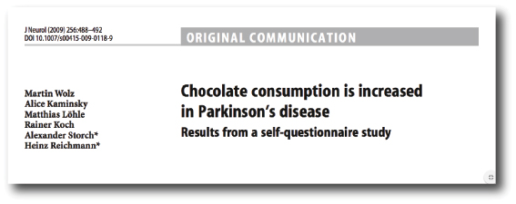
INTERESTING RESEARCH FINDING 1: People with Parkinson’s disease eat more chocolate than people without Parkinson’s.

INTERESTING RESEARCH FINDING 2: This difference is specific to chocolate. There is no difference in the consumption of other forms of sugary treats between the two populations.

Today’s post deals with a topic very dear to me and it’s relationship with Parkinson’s disease.


year-supply-of-chocolate1

Mmmm, chocolate. Source: Saucefinefoods

Everyone likes chocolate, especially at this time of the year. Curious thing is though, people with Parkinson’s seem to like chocolate even more than non-Parkinsonian people.

Why is that? We’re not sure. But some interesting research has been conducted on chocolate and Parkinson’s disease, which we shall review in this post.

But first:

What is chocolate?

Silly question. Fascinating answer.

The word “chocolate” comes from the word xocolātl. This word is from the Aztec language (Nahuatl), and is a combination of the words xococ (meaning ‘sour or bitter’), and ātl (‘water or drink’). This is because for the vast majority of it’s existence, chocolate has been consumed as a drink.

maya-lord-chocolate

“Don’t touch my jar of xocolātl!” Source: Ancient-origins

Fermented beverages made from chocolate date back to 1900 BC in Mesoamerica. The Aztecs believed that cacao seeds were a divine gift from the god of wisdom, ‘Quetzalcoatl’. In fact, the seeds were so precious to the Aztecs that they once had so much value that they were used as a form of currency.

Chocolate was introduced to Europe in the 16th century by the Spanish who added sugar or honey to counteract the natural bitterness. Since then, it has had a rather successful rise in popularity.

child_crown_chocolate_86562_1920x1080

Chocolate: it’s popular stuff. Source: WallPapersCraft

How is chocolate actually made?

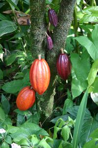
Chocolate comes from the beans of the Theobroma cacao tree – an evergreen native to the tropical regions of Central and South America. The beans are produced in pods, like these:

800px-cocoa_pods

Pods of the of Theobroma cacao tree. Source: Wikipedia

cocoa_beans_in_cocoa_pod_at_el_trapiche_costa_rica

Seeds inside the pod. Source: Wikipedia

Cacao beans are harvested from the pods, and then allowed to ferment over a period two weeks. Two things happen during this process: 1. outside of the pod, the beans are exposed to the warm heat which kills the germinating seed, and 2. natural yeasts – which like the heat – grow and help to develop complex flavours.

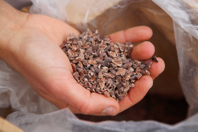
After this fermentation period, the beans are then sun-dried, which helps to preserve them for shipping. Next, the beans are roasted, which helps to further develop complex flavours and to remove unpleasant acidic compounds developed in the fermentation process. This is followed by mechanically separating the valuable nibs (interior of the bean) from the useless shells.

o-cacao-nibs-facebook

Cacao nibs. Source: HuffingtonPost

The nibs must then be refined, which involves extensive grinding. The raw cocoa liquor is then “conched“. This is the stage where the general characteristic tastes, smells and textures of chocolate are developed. Conching is a lengthy process which drives off the rest of the acidic flavoring compounds. The process also helps coat the ground up cocoa particles with fat to reduce the viscosity of the molten chocolate.

fb40xbignxwy87n-medium

Conching. Source: Instructables

Finally, the conched product is tempered which gives chocolate it’s gloss and affects how it melts in your hands/mouth. Tempering is necessary because cocoa butter can crystallise into at least six different forms (form V being the most desired). Each of these forms have different stability and properties. Form V has the most popular texture and ‘feeling in the mouth’, in addition it does not melt too quickly in the hand.

Why is chocolate soooo good?

No one is entirely sure. Chocolate contains many different chemicals which help in making chocolate tasty and (dare we say it) addictive. Among the most important ingredients are stimulants like phenylethylamine and caffeine (all in very small quantities), which can give you a positive boost.

Importantly, chocolate also contains a feel-good chemical called anandamine, which functions in the brain by binding to cannabinoid receptors (these are the same receptors that chemicals in marijuana bind to). Its name actually comes from ananda, the Sanskrit word for “bliss”. Normally anandamide is broken down quite quickly after it is produced, but some researchers believe that the anandamide in chocolate makes the natural anandamide in our brain persist for longer, giving us a longer-lasting “chocolate high”.

And what is the difference between dark and white chocolate?

Ok, now don’t be upset, but technically speaking white chocolate is not really a chocolate.

It is made without any cocoa powder or solids, containing just cocoa butter mixed with milk and sugar. Without the cocoa powder, chocolate has no colour thus it’s white. In addition, white chocolate doesn’t have many of the ‘happy’ ingredients likes caffeine.

Not actually being a chocolate makes white chocolate very useful, however, in research (as you shall see below). Given that it is missing many of the key components of normal chocolate (eg. cocoa, caffeine, etc), white chocolate can be used as a control substance in studies looking at the effect of chocolate on various conditions.

Are there health benefits of eating chocolate?

Mum always told me that chocolate was bad for me, but recent scientific research has altered this perception. Studies of the Kuna Indians of the San Blas islands of Panama, who consume large amounts of a natural cocoa beverage, have found lower blood pressures, better renal function and decreased cardiovascular mortality relative to mainland Panamanian control populations (Click here to read more on this).

hot-chocolate-and-blood-pressure

Kuna indians. Source: Superfoodsrx

The prevalence of hypertension in Kuna indians who have migrated to urban areas on mainland Panama is significantly higher (10.7% of the population compared with just 2.2% of those still on the islands). This is believed to be partly due to the reduction in cacao intake – Kuna indians on the islands eat 10 times more than their mainland equivalents.

Interesting. But what does all of this have to do with Parkinson’s disease?

Right. Down to business. In 2009, this research report was published:

choco-1

Title:  Chocolate consumption is increased in Parkinson’s disease. Results from a self-questionnaire study.
Authors: Wolz M, Kaminsky A, Löhle M, Koch R, Storch A, Reichmann H.
Journal: J Neurol. 2009 Mar;256(3):488-92. doi: 10.1007/s00415-009-0118-9.
PMID: 19277767

These researchers conducted a survey of 274 people with Parkinson’s disease and 234 age-matched controls. They found that people with Parkinson’s disease ate approx. 100g of chocolate per week (on average) compared to just 57.3g for the control subjects.

Using measures of mood (such as the Beck’s Depression Inventory survey), the researchers found that this increased consumption of chocolate was independent of feelings of depression. This interesting observations lead the researchers to conduct this clinical study:

choco-2

Title:  Comparison of chocolate to cacao-free white chocolate in Parkinson’s disease: a single-dose, investigator-blinded, placebo-controlled, crossover trial.
Authors: Wolz M, Schleiffer C, Klingelhöfer L, Schneider C, Proft F, Schwanebeck U, Reichmann H, Riederer P, Storch A.
Journal: J Neurol. 2012 Nov;259(11):2447-51. doi: 10.1007/s00415-012-6527-1.
PMID: 22584952

The researchers in this study (the same people who published the survey study above) tested the effects of 200g of (80% cacao) chocolate on Parkinsonian motor scores (as measured by UPDRS). They assessed 26 people with moderate non-fluctuating Parkinson’s disease at both 1 and 3 hours after eating the chocolate. The researchers used white chocolate as the control treatment in the study, and they (the assessors) were blind to which treatment each subject received.

At 1 hour after consumption, the researchers noted a mild decrease in both treatment groups (most statistically in the dark chocolate group) when compared to the measures taken at baseline (that is before the actual study started). Similar results were observed in the measures taken at 3 hours post consumption. The researchers also took blood samples and found no differences in β-phenylethylamine blood levels (we’ll come back to this shortly).

Altogether, while there was an improvement in motor performance after eating chocolate, the results indicated no difference between dark chocolate and white cacao-free chocolate on Parkinson’s motor function.

What is β-phenylethylamine?

β-phenylethylamine is a naturally occurring chemical in the body, which is produced in chocolate during the thermal processing of cocoa (click here for more on this). Functionally, β-phenethylamine is similar to amphetamine in its action, as it leads to the release of dopamine. Interestingly, people with Parkinson’s disease have almost 50% less β-phenethylamine in the fluid surrounding their brains (Click here to read more on this). Thus, in addition to any stimulant effect of caffeine, increasing β-phenethylamine levels by eating chocolate may be causing an increase in dopamine levels in the brains of people with Parkinson’s disease – resulting in better motor scores.

But the researchers in the clinical study of chocolate reviewed above did not register any change in blood levels of β-phenethylamine. Again, perhaps longer term usage is required in order to detect a significant rise.

What does it all mean?

Here at the SoPD, we feel that the effect of chocolate on Parkinson’s disease have not been fully explored. More research is required. And we are not just saying this because everyone likes chocolate.

Firstly, it would be interesting to replicate what has already been done, particularly the survey of chocolate consumption to determine if people with Parkinson’s disease really do eat more chocolate! This is the most interesting observation reported thus far and needs to be replicated. It would be interesting to determine if the difference pre-dates diagnosis – that is to say, do people who develop Parkinson’s disease eat more chocolate when they are younger (before they are diagnosed)? Could chocolate be actually having a negative effect on the development of the disease?

Second, if a longer term analysis of chocolate and Parkinson’s disease indicates an effect, it would be interesting to further investigate individual ingredients. If we are investigating the ingredients of coffee to assess beneficial components for Parkinson’s disease (click here for more on this), the same analysis of chocolate should be conducted.

I’m going to go off now and contemplate some of this with a piece of dark chocolate…


The banner for today’s post was sourced from pngimg.

Blood transfusions and Parkinson’s disease

blood_646x300

Donating blood helps to save lives. And an awful lot of blood is needed on a daily basis: In the England alone, over 6,000 blood donations are required every day to treat patients.

There has been concerns over the years about what can be transmitted via blood donation (from donor to recipient). The good news is that we now know that Parkinson’s disease is not.

Today’s post looks at recent research investigating this issue and discusses the implications of the findings.


transfusion-superjumbo

Blood transfusions save lifes. Source: New York Times

The average adult human carries approx. 10 pints (about 6 litres) of blood in his body. So much blood, that we actually have an excess – we can survive with a little less. And this allows us to donate blood to blood banks on a regular basis (approx. every 8 weeks). Roughly 1 pint can be given during each blood donation and our bodies will have no trouble replacing it all.

These donations can be used in blood transfusions, replacing blood that has been lost via accident or during surgical procedures. It may surprise you that blood transfusion (from human to human) has been practised for some time. The very first blood transfusion was performed by an obstetrician named Dr. James Blundell in the late 1820’s.

dr

Dr. James Blundell. Source: Wikpedia

The exact date of that first procedure is the subject of debate, but Blundell wrote up his experience in the journal Lancet in 1829:

lancet

Blundell’s article in the journal Lancet. Source: Wikipedia

Since that time, blood transfusions have gradually become an everyday occurrence at hospitals all over the world. And as we suggested above a lot of blood is used on a daily basis, keeping people alive. Determining whether each donation of blood is safe to use is obviously a critical step in this process, and all donated blood is tested for HIV, hepatitis B and C, syphilis and other infectious diseases before it is released to hospitals. But for a long time there has been a lingering concern that not everything is being detected and filtered out.

In fact there has been a serious concern that some neurodegenerative conditions like Alzheimer’s and Parkinson’s disease may be transmissible. If these diseases are being caused by ‘prion-like behaviour’ from the particular proteins involved with these conditions (eg. beta amyloid and alpha synuclein, respectively), then there is a very real possibility that such rogue proteins could be transferred via blood transfusions.

This was a concern (note the past tense) until July of this year when this research report was published (with a rather mis-leading title):

transmissiontitle

Title: Transmission of neurodegenerative disorders through blood transfusion. A cohort study
Authors: Edgren G, Hjalgrim H, Rostgaard K, Lambert P, Wikman A, Norda R, Titlestad KE, Erikstrup C, Ullum H, Melbye M, Busch MP, Nyrén O.
Journal: Ann Intern Med. 2016 Sep 6;165(5):316-24.
PMID: 27368068

The researchers in this study took all of the data from the enormous nationwide registers of blood transfusions in Sweden and Denmark – collectively almost 1.5 million people have received transfusions in these two countries between 1968 and 2012 – and compared the medical records of the recipients to those of the donors (you have to love the Scandinavians for the medical databases!). Approximately 3% of the recipients received a blood transfusion from a donor who was diagnosed with one of the neurodegenerative diseases included in this study (Alzheimer’s, Parkinson’s and Motor neurone disease (or Amyotrophic lateral sclerosis – ALS). There was absolutely no evidence of transmission of any of these diseases.

For the statistic lovers amongst you, the hazard ratio for dementia in recipients of blood from donors with dementia versus recipients of blood from healthy donors was 1.04 (95% CI, 0.99 to 1.09). Estimates for individual diseases, Alzheimer disease and Parkinson disease were 0.99 (CI, 0.85 to 1.15) and 0.94 (CI, 0.78 to 1.14), respectively.

These statistics mean that if Parkinson’s disease is being transmitted via a blood transfusion, it is an extremely rare event.

So what does this mean for our understanding of Parkinson’s disease?

Well, we already know that you can’t catch Parkinson’s disease from your spouse (Click here to read more on this) and there is a lot of other evidence to suggest that Parkinson’s disease is not contagious (Click here to read more on this). So this is one less thing for carers, family members and friends to worry about.

But if Parkinson’s disease is not caused by some contagious agent, this knowledge has major implications for our understanding of the disease. Previous lab-based research has pointed toward a ‘Prion’-like nature to alpha synuclein (the protein most associated with Parkinson’s disease. Prions being small infectious agents made up entirely of protein material, that can lead to disease that is similar to viral infection. And researchers actually found that if you inject specific types of alpha synuclein into the muscles of mice, those animals would start to develop cell loss in the brain (Click here to read more about this).

If Parkinson’s disease is a ‘prion’ condition, then we have to ask one important question: why isn’t it being transmitted via blood transfusion? Alpha synuclein is certainly found in the blood of people with Parkinson’s disease.

It could be that an infectious agent initiated the condition many years ago and it has very slowly been developing (similar to chronic infections resulting from Hepatitis – click here to read more on this).

Research like we have reviewed today may result in a serious re-think of our theory of Parkinson’s disease.


The banner for today’s post was sourced from CampusCluj