
|
Theories of viral agents as possible causal (or influencing) factors in Parkinson’s have long existed. This week a research team from Colorado in the USA published a new report demonstrating that mice infected with a mosquito-borne alphavirus (called Western equine encephalitis virus) develop Parkinson’s-like features. These features include the loss of dopamine neurons, increased neuroinflammation, locomotor issues, and the wide spread presence of aggregated protein (all classical hallmarks of the Parkinsonian brain). In today’s post, we will look at what mosquito-borne alphaviruses are, what this new study found, and how the results could help us to better understand some cases of Parkinson’s.
|
 Electron micro photograph of Influenza viruses. Source: Neuro-hemin
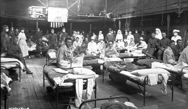
Between January 1918 and December 1920, there were two terrible outbreaks of an influenza virus.
The event became known as the 1918 flu pandemic.
Approximately 500 million people across the globe were infected by the H1N1 influenza virus during this period, and there were approximately 50 to 100 million associated death.
Now, to put that into perspective for you, that was basically 3-5% of the world’s population at that time.
1918 Spanish flu. Source: Chronicle
At the time, much of the world was blind to these events. Given that this pandemic occurred during World War 1, censors limited the media coverage of the pandemic in many countries in order to try and maintain some sort of morale (very thoughtful of them).
The Spanish media, however, were not censored and this is why the 1918 pandemic is often referred to as the ‘Spanish flu’.
But at the same time that H1N1 influenza virus was causing havoc, a Romanian born neurologist named Constantin von Economo noticed something interesting.
What did he notice?
Constantin reported a number of unusual symptoms which were referred to as encephalitis lethargica.
This condition left victims in a statue-like condition, speechless and motionless.

Constantin von Economo. Source: Wikipedia
By 1926, encephalitis lethargica had spread around the world, with nearly five million people being affected. Many of those who survived never returned to their pre-existing state of health. They were left frozen in an immobile state.
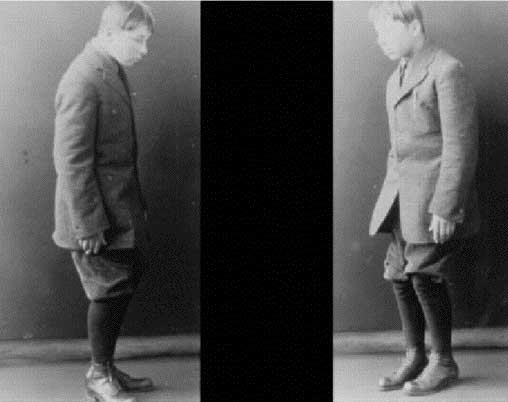
An individual with encephalitis lethargica. Source: Baillement
Historically, it was believed that encephalitis lethargica was caused by the influenza virus from the 1918 Spanish influenza pandemic. This was largely due to the temporal association (things happening at approximately the same time) and the finding of influenza antigens in some of the suffers of encephalitis lethargica (Click here to read more about this).
And then there were also the observations of Dr Oliver Sacks:

Dr Oliver Sacks. Source: Pensologosou
During the late 1960s, while employed as a neurologist at Beth Abraham Hospital’s chronic-care facility in New York, Dr Sacks began working with a group of survivors of encephalitis lethargica, who had been left immobile by the condition. He treated these individuals with L-dopa (the standard treatment for Parkinson’s now, but it was still very experimental at the time) and he observed them become miraculously reanimated. The sufferers went from being completely motionless to suddenly active and mobile. Unfortunately the beneficial effects were very short lived.
You may be familiar with Dr Sacks’ book about his experience of treating these patients. It is called ‘Awakenings’ and it was turned into a film starring actors Robin Williams and Robert De Niro.

Robert De Niro & Robin Williams (as Sacks) in Awakenings. Source: Pinterest
More recent, postmortem analysis of the brains of encephalitis lethargica patients found an absence of influenza RNA – click here for more on this), which has led many researchers to reject the association between influenza and encephalitis lethargica. But there has been evidence to suggest that encephalitis lethargica may have been caused by a enterovirus (Click here to read more about this).
Interesting, but what does this have to do with Parkinson’s?
Well, this week, this research report was published:
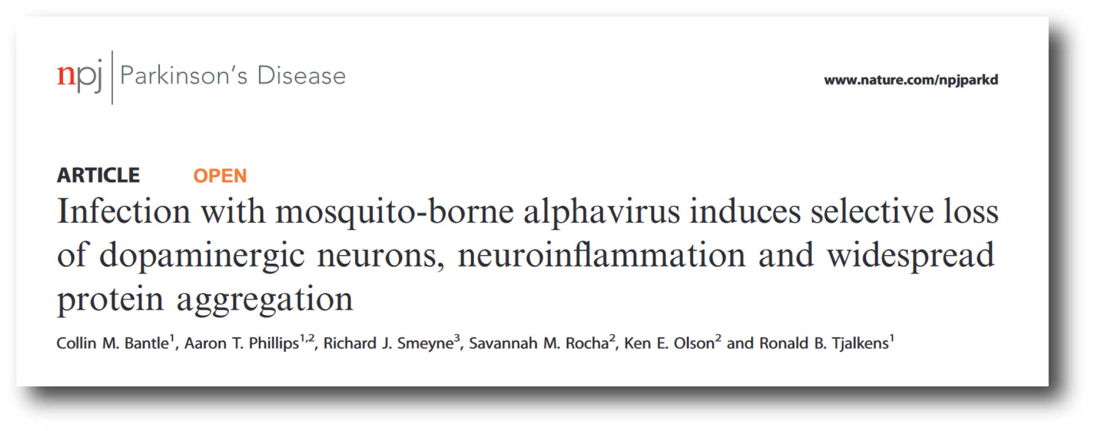 Title: Infection with mosquito-borne alphavirus induces selective loss of dopaminergic neurons, neuroinflammation and widespread protein aggregation
Authors: Bantle CM, Phillips AT, Smeyne RJ, Rocha SM, Olson KE & Tjalkens RB
Journal: npj Parkinson’s Disease, 5, 20 (2019)
PMID: 31531390 (This report is OPEN ACCESS if you would like to read it)
In this study, the researchers from Colorado State University were interested in how a virus that is carried by mosquitos could be involved with Parkinson’s.
I’m sorry, did you say ‘mosquitos’?
Yes.
Mosquitos involved with Parkinson’s?!?
Yes, go with me on this one, there is a bit of backstory to it:
In 1977, a patient was admitted to hospital in Colorado with confirmed western equine encephalitis. Encephalitis is a condition in which the brain becomes inflammed and swollen, usually via an infection. Western equine encephalitis results from an infection of an alpha virus, which is carried by (can you guess?) mosquitos.
What differentiated this individual from others, however, was that they developed Parkinsonian features.
What?!?
And that individual was not the only case. During that period 6 of 25 patients admitted for Western equine encephalitis also developed a progressive form of Parkinsonism (Click here to read more about this).
The features of this Parkinsonism included resting tremor and ‘cogwheel’ rigidity – which persist long after the actual infect was resolved. This may be due to the fact that alphavirus RNA can persist in the rodent brain (at least) for up to 16 months after the actual infection. The persistent presence could potentially cause a sustained inflammatory state.
I didn’t think that mosquitos were so dangerous
Oh, be under no illusions: We are at war with mosquitos!
These seamingly innocent, tiny winged creatures kill more humans each year than any other animal – almost a million deaths per year are attributed to mosquitos. They kill more humans than humans do (580,000 per year) and those “dangerous” snakes and lions are positively minor leagues by comparison (60,000 and 100, respectively).
 Source: Gatesnotes
To be fair, it’s not the mosquito doing the killing. Malaria is the main reason for this top of the tables ranking.
Remind me, what is malaria?
Malaria is a disease caused by a parasite, which is transmitted to humans through the bites of infected female Anopheles mosquitoes.
Interesting fact: Only female mosquitoes feed on blood. Male mosquitoes prefer to feed on plant nectar, and thus do not transmit the disease.
The life cycle of malaria is rather simple: When infected mosquitos inject the infective form of the parasite (called the sporozoite) into the blood stream of an animal, it will make it’s way to the liver, where it will use liver cells to reproduce in a cycle that will ultimately kill the organism is left untreated.
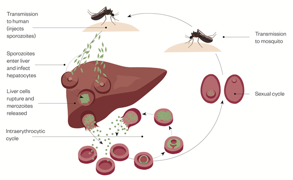 Source: Pharmaceutical-journal
People who have malaria usually feel very sick 10-15 days after being biten, and they will experience a high fever and shaking chills. Individuals with severe untreated malaria infections will often develop anaemia, respiratory distress, and ultimately multi-organ failure.
Remarkable progress has been made in dealing with marlaria (thanks to organisations like the Bill & Melinda Gates Foundation). In fact, a new report published in The New England Journal of Medicine found that the malaria death rate in sub-Saharan Africa has declined by a 57% since 2000 (new report to read more about this).
But we are not here to talk about malaria today. Rather we are going to focus on another mosquito-borne pathogen: the western equine encephalitis virus.
Ok, before I go buy some mosquito repellant, tell me what the researchers behind this new study found? And how does it relate to Parkinson’s?
They infected normal, healthy mice with genetically engineered western equine encephalitis viruses, which were designed to label any cell it infected with a biological marker. The route of infection was made via the nose (a common pathway of normal viral infections), and they idenified a particularly potent virus, which resulted in the loss of dopamine neurons within 4 days of infection.
All of the mice infected with this virus displayed motor deficits by 24 h post infection, and none were able to survive more than 96 hours.
When the researchers looked at the brains of the mice for the genetically engineered biological marker that highlighted infected cells, they found that the substantia nigra (a region of the brain that houses the dopamine neurons which are severely affected by Parkinson’s) was particularly affected.
They also noted the activation of glial cells in the brain. Glial cells (astrocytes and microglia) are the helper cells, which become activated when something is wrong.
 The activation of microglia. Source: Diacomp
When the investigators used immunotherapy (using polyclonal antibodies to the WEEV E1-glycoprotein – to inhibit the extent of infection – given 12 hours+ after virus exposure), they found that the western equine encephalitis virus-infected mice survived and the virus was cleared from the body by 8 weeks post infection. But, the mice did display a selective loss of dopamine neurons in the substantia nigra, in addition to motor problems and a persistent activation of glial cells.
Oh, and the researchers saw alpha synuclein protein aggregation – one of the classic hallmarks of Parkinson’s – which has been reported before in a viral infection model of Parkinson’s (Click here to read more about this).
Wow! Was the alpha synuclein aggregation in dopamine neurons?
No, it was primarily in the activated glial cells.
Has alpha synuclein ever been associated with viral infections before?
Yes, it has actually.
In addition to the studies mentioned above, alpha synuclein has also been shown to have anti-viral properties:

Title: Alpha-Synuclein Expression Restricts RNA Viral Infections in the Brain.
Authors: Beatman EL, Massey A, Shives KD, Burrack KS, Chamanian M, Morrison TE, Beckham JD.
Journal: J Virol. 2015 Dec 30;90(6):2767-82. doi: 10.1128/JVI.02949-15.
PMID: 26719256 (This article is OPEN ACCESS if you would like to read it)
David Beckham (not the football player) and his research colleagues introduced West nile virus to brain cells grown in cell culture and they observed an increase in alpha synuclein production. They also found that the brains of people with West nile infections had increased levels of alpha synuclein.
The researchers then injected West Nile virus into both normal mice and genetically engineered mice (which produced no alpha synuclein) and they found that the genetically engineered mice which produced no alpha synuclein died quicker than the normal mice. They reported that there was an almost 10x increase in viral production in the genetically engineered mice. This suggested to them that alpha synuclein may be playing a role in protecting cells from viral infections.
So what does it all mean?
A recent report from researchers in Colorado has highlighted the potential role of viruses in the course of Parkinson’s. They infected mice and observed a rapid onset of Parkinson’s-like features and the accumulation of alpha synuclein – some of which matches the symptoms observed in humans infected by the same virus.
Whether viruses are a causal agent or simply an influencing factor in the course of Parkinson’s deserves more investigation, particularly as more research hints at the microorganisms of the gut being involved in the bigger picture, and of Parkinson’s-associated protein having anti-viral properties (click here for another example).
You can expect more on this idea in future SoPD posts.
For now, invest in mosquito repellant!
 Source: ABC
(Just kidding – we need to see independent replication of this new research before we wipe out mosquitos for good).
The banner for today’s post was sourced from the Times

Simon,
Thank you for discussing this truly fascinating topic. First I would like to ask you a question regarding virus – alpha-synucleins connection: are the alpha-synucleins that appear in response to the virus infection misfolded? Is it possible that certain genetic defects in the infected cells are changing the way alpha-SN are synthesized in the cells resulting in misfolding? Or is it possible perhaps that the virus itself is capable of changing ASN structure? Is misfolded ASN effective in slowing down virus infection? This is something that could easily be researched in vitro. Was this research ever done? Answering these questions could explain how viral infection turns into a chronic disease as well accumulation of misfolded asn in PD. This certainly would be a major development, right?
Second, could you please shed some light on why I see such lack of interest in viral etiology hypothesis of Parkinson disease? Specifically I am talking about work by Dr. Dourmashkin et al you mentioned on several occasions in this forum. Although many find enterovirus theory quite interesting I couldn’t find anyone who would be interested in repeating that research or do it in a blind experiment manner. I even suggested that financial support would be provided for working on this issue, still there were no takers. There were some questions raised about some methods used by the authors but they are certainly reputable and experienced group of scientists. So why is it that nobody wants to continue this research? If true this theory would explain why pd is not found in other species, give some insight into gut PD connection, pd vaccine development, diagnostic tests development, etc. The pay back potential is huge so why nobody is working on this? I would appreciate it if you could please explain me what I am missing.
Thank you,
Felix
LikeLike
It seems unsurprising that mosquito-born viral diseases can have neurological effects – think of Zika and Guillane-Barre syndrome.
I suspect that there is not a single cause for Parkinson’s, but that it is related to a negative feedback mechanism between inflammation, disruption of autophagy and disruption of energy supplies to the brain. Almost all substances being researched wrt Parkinson’s fall into 3 categories, though some fall into multiple catagories:
Energy / Mitochondrial disfunction – think Exanatide, B vitamins – also used in energy drinks. The outcome of the trial to enhance metabolism using easily available supplements mentioned in the Aug SOP summary will be particularly important.
Junk clean up / autophagy – think ambroxol, trehalose, exercise, fasting. Interestingly Insulin also has a role both in energy supply and autophagy https://www.frontiersin.org/articles/10.3389/fnins.2019.00491/full
Inflammation – think of the recent SOP post on Resolvin. Anti-malarial drugs such as chloroquine and doxycyclin are also anti-inflammatory – and have been identified as having an effect on PD.
Whatever caused the original problem is to some extent irrelevant. Finding the perfect combination of drugs to combat all three aspects may take time, but if all three aspects are partly addressed this may at least slow progression. So the sooner a person knows they are on a slippery slope, the sooner they can look at the best combination of lifestyle / supplements / prescribed drugs to combat these 3 factors.
LikeLike
Linda,
I cannot agree with you. PD is a progressive disease and the symptoms change and get worse and worse over time. While it is true that many illnesses are treated in spite of the fact that their etiology is unknown it is my firm believe that we have to learn the cause of PD in order to have representative animal model and to be able to come up with the disease modifying treatment or preventive vaccine. I am sure that what we call PD in our ‘macro’ world has more than one ‘micro’ world causes. However I found it very interesting that ALL 14 samples of brain tissues from DIFFERENT people who had PD investigated by Dr Dourmashkin ( https://f1000research.com/articles/7-302/v2 ) all had similar virus like particles in different stages of virus development that stained with the same AB. Only one of 7 samples in control group had some evidence of virus activity and microscopically it appeared to be a different virus. To date there is NOTHING I repeat NOTHING that slows the disease down and C/L treatment is ineffective for many as well as causing numerous bad side effects. The animal models currently in use are simply not representative of PD and only lead to waste of time and money. I might be too harsh but I have some serious doubts that there is genuine desire to eradicate this illness. I am waiting for the result reporting for the next stage of the ASN AB clinical trial ( prothena, etc.- Simon has extensive writings on these) but I would not be a bit surprised if it shows no significant disease modification benefit. In fact if the misfolded ASN has antiviral properties ( see the question I asked in my post above) we might see the disease progressing faster in spite of significant ASN reduction. I hope Simon can find some time to answer my questions and explain me why nobody is looking at the virus theory closely. By the way for those who doubt that PD can be caused by an infectious agent here is an interesting fact of PD trivia: 4 other members of MJ Fox crew contracted Parkinson at the same time as he did ( https://www.intmath.com/blog/environment/michael-j-fox-and-the-parkinsons-cluster-782 ) . What are the odds?
Thank you,
Felix
LikeLike
‘Fascinating, well-written. Keep up the good work!
LikeLike
THANK YOU SIMON
LikeLike