|
# # # # Our ability to grow cells in culture (in petri dishes and flasks in laboratories) has been critical to our efforts to learn more about the biology of Parkinson’s and to screen for novel potential therapies. Recently, researchers have employed more sophisticated methods of characterising cells in culture, to achieve greater insights. These effort have led to some interesting work from investigators at Google Research and They used a powerful new technique called “Cell Painting”. In today’s post, we will outline what Cell Painting is, discuss what the new research demonstrates, and explore what their findings could mean for Parkinson’s research. # # # # |
 John Maynard Keynes. Source: NYTimes
I recently read an interesting story about the economist John Maynard Keynes.
In 1918 Keynes was working as a Treasury adviser when a friend – the art critic Roger Fry – told him about a sale of impressionist works that was about to occur in Paris. The collection was from the artist Edgar Degas, who had died in late 1917.
 Edgar Degas. Source: Wikimedia
With the great war still raging in northern France, intrepid Keynes somehow managed to convince not only the UK Government to give him money, but also for the director of The National Gallery, Sir Charles Holmes, to join him on his mad dash to Paris. They boarded a boat to Boulogne and then travelled by train to Paris, carrying a suitcase containing £20,000 in French banknotes (understand that this is equivalent to £1.1 million in todays money).
As the auction started, Paris was rocked by the sound of German artillery. Many of the hopeful bidders at the auction fled, but Keynes and Holmes stood their ground and secured some incredible bargains (not only for the British Government, but also for themselves – such as a still life with seven apples, by Paul Cézanne that Keynes purchased for himself).
 Cézanne’s seven apples. Source: Wikimedia
Upon arrival back in England, Keynes could not carry all of his newly acquired luggage, so he left his Cézanne under a hedge on the side of the road. Upon arrival at their lodgings for the night, he instructed their host that ‘if you’d like to go down to the road, there’s a Cezanne just behind the gate‘.
It was a very Keynesian enterprise as that Cézanne painting alone is probably worth well over £30 million if it went to auction today.
Interesting, but what does this have to do with Parkinson’s?
Nothing, but today’s post is about a different kind of painting, so I thought I’d start off with this little anecdote.
What does painting have to do with Parkinson’s?
In 2016, this report was published:
 Title: Cell Painting, a high-content image-based assay for morphological profiling using multiplexed fluorescent dyes.
Authors: Bray MA, Singh S, Han H, Davis CT, Borgeson B, Hartland C, Kost-Alimova M, Gustafsdottir SM, Gibson CC, Carpenter AE.
Journal: Nat Protoc. 2016 Sep;11(9):1757-74.
PMID: 27560178 (This report is OPEN ACCESS if you would like to read it)
In this study, the researchers were interested in improving phenotypic analysis of cells in culture.
What does phenotypic mean?
An important distinction in biology is the difference between phenotype and genotype.
A genotype refers to aspects of the genetic characteristics of an organism (for example, a mutation in a particular gene). A phenotype deals with the physical characteristics of an organism, which could result from a specific genotype or from environmental factors.
Can you give an example?
Sure. Let’s use eye colour.
 Source: Michiganvca
The visible eye colour of your eyes is your phenotype. It determined by your genotype, but the actual observable trait (the colour of your eyes) is a phenotype.
Overall physical appearance is another good example. My phenotype is ridiculously tall and freakishly good looking – the genotype of which is complicated.
Got it. So what did the phenotypic analysis of cells in culture involve?
In their report, they presented a novel method of morphological profiling cells in culture. It provided a quantitative means of evaluating the cells that could potentially be used to better characterise the effects of drugs or genetic manipulations on the cells being analysed.
They called this new method “Cell Painting“.
It involved using six fluorescent dyes that could be imaged across five different channels, to reveal eight broadly relevant cellular components (also know as organelles).
Organelles?
Organelles are subcellular structures that have one or more specific task to perform inside the cell, such as mitochondira (the energy generating power stations of cells) or lysosomes (part of the waste disposal system of cells). Think of organelles in the cell in the same manner as organs your the body, each with a specific task.
Using certain dyes, the researchers can label the different organelles inside cells. These dyes can be applied in unison, allowing for different organelles to be analysed at the same time. In the image below you can see 5 different dyes being used on two different cell cultures (top row vs bottom row) to label different types of organelles:
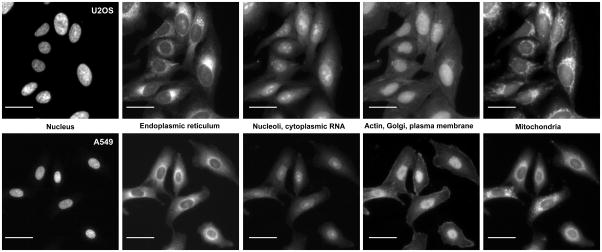 Source: PMC
The researchers imaged different types of cells grown in culture using a high-throughput microscope system, providing them with thousands of images to anlyse. Next, they used automated image analysis software to identify individual cells and then measure ~1,500 different morphological features (such as size, shape, texture, intensity, etc.). As the data built up (resulting from 1500 features assessed across thousands of images), the investigators accumulated a very rich profile which allowed them to detect very subtle phenotypes (visible traits).
Interesting. But what can this technology be used for?
This is potentially a very powerful method. Cell Painting can firstly be used to identify certain subtle phenotypes associated with a particular genotype (genetic manipulation). It can then be used to screen for drugs that revert those subtle phenotypes (or “signatures”) back to normal.
Understand that for hundreds of years, biologists have been using microscopies to study how cells function, but we have now reached a point where we can do this analysis in exquisite detail – with powerful implications.
Think about this from the stand point of drug discovery. Rather than simply determining if a treatment protects cells from a toxic chemical or disease associated protein, cell painting can provide a much richer representation of what the treatment might be doing.
|
# RECAP #1: Phenotypic analysis (of cells in culture) is the study of visible traits resulting from genetic or environmental influences. It provides researchers with a useful method of screening for and investigating novel therapies. Cell painting is a new high through-put, automated phenotypic analysis technique that assesses ~1500 morphological measures in each cell being assessed. # |
Has cell painting been used much in research so far?
A year after cell painting was first presented, this report was published:
 Title: Systematic morphological profiling of human gene and allele function via Cell Painting.
Authors: Rohban MH, Singh S, Wu X, Berthet JB, Bray MA, Shrestha Y, Varelas X, Boehm JS, Carpenter AE.
Journal: Elife. 2017 Mar 18;6:e24060.
PMID: 28315521 (This report is OPEN ACCESS if you would like to read it)
In this study, the researchers selected a series of well-known genes and analysed different genetic variants within those genes in cells grown in culture (using cell painting).
The investigators grew human bone osteosarcoma cells (U-2 OS cells) in cell culture plates that had 384 wells per plate – meaning they could conduct 384 experiments per plate. Using these culture conditions, they evaluated 323 different genetic manipulations (with known phenotypes) across 220 genes. And each experiment was repeated five times.
The researchers found that 50% of the 220 tested genes displayed detectable morphological signatures that could be measured and quantified. These morphological features could also be grouped into biologically meaningful gene clusters consistent with what is already known in the published literature.
In addition to these findings, however, the analysis also revealed previously unknown biological interactions. Specifically, the researchers found an unexpected relationship between two major cellular signaling pathways (Hippo and NF-κB) – pathways that play critical regulatory roles in how cancer starts and develops. And they confirmed this interaction with additional genetic analysis.
Use of cell painting is also now giving rise to vast databases of images and data that can be used by researchers to improve their computational algorithms, resulting in better analyses. For example, this report presents one such database:
 Title: A dataset of images and morphological profiles of 30 000 small-molecule treatments using the Cell Painting assay.
Authors: Bray MA, Gustafsdottir SM, Rohban MH, Singh S, Ljosa V, Sokolnicki KL, Bittker JA, Bodycombe NE, Dancík V, Hasaka TP, Hon CS, Kemp MM, Li K, Walpita D, Wawer MJ, Golub TR, Schreiber SL, Clemons PA, Shamji AF, Carpenter AE.
Journal: Gigascience. 2017 Dec 1;6(12):1-5.
PMID: 28327978 (This report is OPEN ACCESS if you would like to read it)
In this report, the researchers used the same U2OS bone cancer cells that were used in the previous report. They grew them in 384-well cell culture plates, and then treated the cells with one of 30,616 molecules (and they repeated this experiment 4 times!).
This HUGE load of work resulted in a database of data contains 919, 265 different images of five-channel fields of view (that’s approximately 5 million image files in 16-bit TIFF format in a database that is OPENLY available at “The Cell Image Library” repository).
This database “serves as a useful resource for the wider scientific community applying morphological (image-based) profiling“.
|
# RECAP #2: Cell painting can be used to identify new biological interactions and conduct large drug screening studies. Vast databases of images and data are now being generated, which are helping to improve the computational algorithms used to analyse the data. # |
Has this cell painting stuff ever been used in Parkinson’s research?
Funny you ask that. Now we get to the interesting part of this post.
Before we go any further, however, I should say that I am about to be break one of the unwritten rules of science communication (…again).
Until a research report has been through the peer-review process you probably should not be discussing the results in the public domain. But in this particular case, the research is really interesting and relevant to what we discuss on this website. In addition, it has been made available on the OPEN ACCESS preprint depository website called BioRxiv.
![]() Source: BioRxiv
Source: BioRxiv
I should also add that this is not the first time we have discussed manuscripts on BioRxiv (Click here, here and here to read other SoPD posts on Biorxiv manuscripts).
We are regular rule breakers here at the SoPD.
Ok, that said, let’s move on.
Here is the manuscript in question:
 Title: Deep learning and automated Cell Painting reveal Parkinson’s disease-specific signatures in primary patient fibroblasts
Authors: Schiff L, Migliori B, Chen Y, Carter D, Bonilla C, Hall J, Fan M, Tam E, Ahadi S, Fischbacher B, Geraschenko A, Hunter CJ, Venugopalan S, DesMarteau S, Narayanaswamy A, Jacob S, Armstrong Z, Ferrarotto P, Williams B, Buckley-Herd G, Hazard J, Goldberg J, Coram M, Otto R, Baltz EA, Andres-Martin L, Pritchard O, Duren-Lubanski A, Reggio K, NYSCF Global Stem Cell Array Team, Bauer L, Aiyar RS, Schwarzbach E, Paull D, Noggle SA, Monsma Jr. FJ, Berndl M, Yang SJ, Johannesson B.
Database: BioRxiv
DOI: bioRxiv 2020.11.13.380576
In this study, the researchers have applied the cell painting platform to skin cells (called fibroblast cells) collected from people with Parkinson’s.
And we’re not just talking about 3 or 4 people with Parkinson’s.
Oh no, these investigators decided to go ‘all in’ and used cells collected from 91 people.
The cell analysed were collected from:
- 45 healthy controls
- 32 cases of sporadic/idiopathic Parkinson’s,
- 8 cases of GBA-associated Parkinson’s
- 6 cases of LRRK2-associated Parkinson’s
And five of the participants had donated a second skin biopsy 3 to 6 years later. The cell samples came from repository.
 Source: bioRxiv
As a first test of the cell painting method, the researchers wanted to determine if they could identify cells from individual case within the 96 evaluated cases, using just the parameters being measured by the system.
 Source: bioRxiv
Remarkably, their algorithm identified the correct patient case 76% of the time (compared with the expected 1% of the time). This is rather remarkable. That physical characteristics of our individual cells can be used to identify us is pretty breathtaking stuff. And these cell morphology characteristics could also be determined across multiple experiments, using different culture conditions.
Next the researchers shifted their attention to determining if morphological profiles of cells could be used to separate “Parkinson’s cells” (sporadic and LRRK2-associated cases) from the cells collected from unaffected controls.
Impressively, the investigators found that their algorithm was correct 80% of the time in determining PD from non-PD. And when the researchers removed one of the five different channels used in the analysis, it did not significantly affect the result, demonstrating how robust the system is.
 Source: bioRxiv
The investigators used two divergent analytical approaches, and both succeeded in separating “Parkinson’s cells” from unaffected controls cells, providing a within-study validation of the results.
Collectively all of this work resulted in a 48 terabytes database that is OPENLY available for researchers to access (Click here to access that database).
The research was a collaborative effort by Google Research and
Google Research is obviously part of the internet firm Google. As I understand thing, at Google engineering teams and the research teams work very closely together, with the research teams doing more experimental stuff while the engineering teams focus on the product side of things. The goal at Google Research is to tackle “challenges that define the technology of today and tomorrow” and they do this by reimagining “technology across all areas of computer science research“.

But it is interesting to read the discussion section of the manuscript: “To overcome the poor clinical trial outcomes for Parkinson’s disease, it is clear that more physiologically relevant disease models and drug screening approaches that are less reliant on preconceived hypotheses are required“.
And then “Our ability to identify Parkinson’s-specific disease signatures using standard cell labeling and deep learning–based image analysis highlights the generalizable potential of this platform to identify complex disease phenotypes in a broad variety of cell types. This represents a powerful, unbiased approach that may facilitate the discovery of novel precision drug candidates undetectable with traditional target-and hypothesis-driven methods”.
Hopefully, while this manuscript is going through the peer-review process, the researchers at Google Research and will be turning their attention to screening drugs (with the potential of determining not only novel Parkinson’s treatments, but also individual – more personalised – results as well). And this follow up work will be centred around more disease-relevant cell types (such as dopamine neurons), rather than simply skin cells – but this will involve an order of magnitude increase in the amount of work required in terms of preparing the cells.
Take home message: Powerful stuff. Huge potential. Raising the bar significantly for everyone else in the field.
So what does it all mean?
Keynesian economics suggests that economic output is strongly influenced by aggregate demand. Starting this post with a reference to the great economist seems rather apt as there is growing ‘aggregate’ demand for finding novel therapies for Parkinson’s, which is resulting in significant research output.
I know nothing about the valuation of art, but I do appreciate the effort that goes into some of the great classics and the amazing stories associated with them (Picasso’s Guernica being the best example). And the same priniciple applies to the research that we have reviewed in today’s post – particularly in terms of the amount of effort!
The study outlined in the bioRxiv manuscript is HUGE! Both in the scale of the work involved and with regards to the data generated (48 terabytes?!? Are you kidding me???). But also in terms of potential for Parkinson’s research. If other research groups are able to build identical platforms, and they too can tease out similar differences between individual cell samples, then a more personalised approach for Parkinson’s feels more achievable.
By using specific dyes to “paint the cells” with as many fluorescent markers as possible without compromising the ability to build quantitative profiles via a high-throughput platform, researchers have been able to construct a powerful new tool for Parkinson’s research. They are able to differentiate between cells from people with Parkinson’s and unaffected controls, AND identify specific characteristics of cells unique to particular individuals.
I don’t often give opinions here, but I was pretty impressed by this study and will be looking forward to this platform being applied to various aspects of Parkinson’s research going forward (particularly drug screens on dopamine neurons from patient-derived cells).
Watch this space.
 All of the material on this website is licensed under a
Creative Commons Attribution 4.0 International License
You can do whatever you like with it!
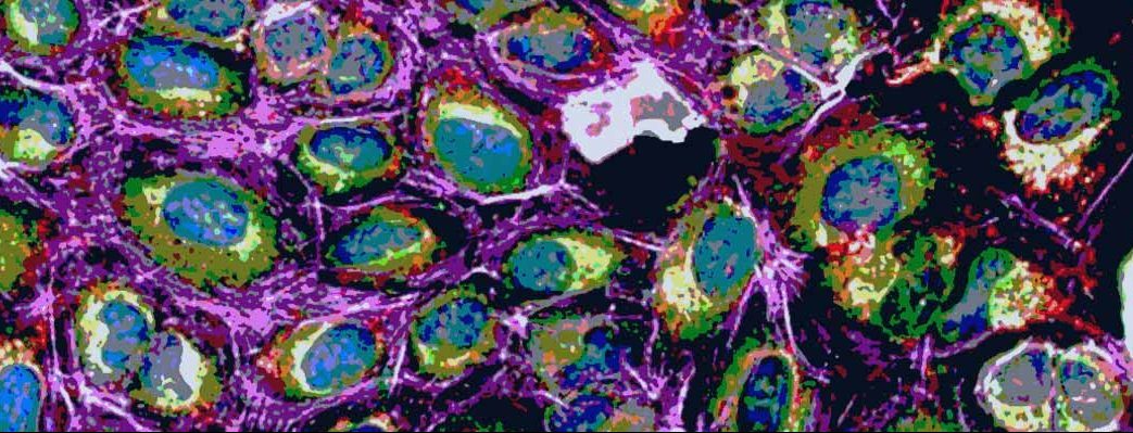
The banner for today’s post was sourced from Perkinelmer.

If the writer of this article can not be bothered to check it for its many (hopefully typographical, not illiterate) errors before publication, how can a reader have confidence in the accuracy of the data reported therein? My wife has PD. I am most interested in advances in understanding and potential therapies, but this article makes me doubt that it is worthwhile reading other PD reports from this source.
LikeLike
Michael Snaith, you reveal your lack of research into trusted commentators on the Parkinson’s research scene. Criticising spelling in the context of Blogs will keep you very busy.
LikeLiked by 1 person
Michael, this site is a fire hose of cutting edge information on Parkinson’s research, and typos are just one small part of the turbulence in that torrent. Just take off your shirt and get a little wet–it’s well worth the minor inconvenience.
LikeLike
The fact that the skin cells of Parkinson’s patients have distinct traits brings to mind the fact that Parkinson’s patients have 3.6 times the likelihood of developing melanoma. Perhaps the differences detected in this study will throw light upon the specific differences that are responsible for that increased susceptibility.
More generally, the notion that the skin cells of Parkinson’s patients could express a phenotype that identifies the illness suggests that the disease itself may be more systemic than its localized manifestations within the nervous system might otherwise suggest.
Certainly, for hereditary PD, it might be surmised that the differences in skin cells could simply derive from the same genetic sources as the susceptibility to the illness. But considering that the majority of patients involved had idiopathic PD, the fact that their status as PD patients was nonetheless detectable, suggests that either
1) There are hereditary factors that have not been identified thus far, OR
2) There could be some systemic state of unheathfulness developed due to lifestyle or environment which affects the quality of skin cells and also increases susceptibility to Parkinson’s, OR
3) The skin cells might have been affected as a *result* of developing the illness (i.e., that these skin cells might not have shown the same traits prior to the development of Parkinson’s).
If (2) is correct, then one might ask what particular form of systemically unhealthful condition would affect both skin cells *and* the likelihood of developing PD. And also, what other effects that condition might have.
And if (3) is correct, then one might ask how the development of PD itself might have an identifiable effect upon skin cells–and what other aspects of the body might also be affected by PD.
From a philosophical standpoint, I have always been interested in the notion that traits that pertain to something very large might have a resonant reflection in the smallest parts of that larger entity–as Blake wrote:
To see a World in a Grain of Sand
And a Heaven in a Wild Flower
Hold Infinity in the palm of your hand
And Eternity in an hour
LikeLike